Arq. Bras. Med. Vet. Zootec., v.66, n.6, p.1623-1630, 2014
Short-term preservation of Pecari tajacu ovarian preantral follicles using phosphate
buffered saline (PBS) or powdered coconut water (ACP
®) media
[Preservação de folículos ovarianos pré-antrais de Pecari tajacu por curtos períodos utilizando meios à base de solução salina fosfatada tamponada (PBS) ou água de coco em pó (ACP®)]
G.L. Lima1, E.A.A. Santos1, L.F. Lima2, V.B. Luz1, A.P.R. Rodrigues2, A.R. Silva1*
1Universidade Federal Rural do Semi-Árido − UFERSA − Mossoró, RN 2Universidade Estadual do Ceará − Fortaleza, CE
ABSTRACT
We compare protocols for the short-term preservation of collared peccarie’s ovarian preantral follicles (PFs) by using phosphate buffered saline- (PBS) or powdered coconut water- (ACP®) based medium. For morphology analysis each pair of ovaries collected from six females was divided into nine fragments. One fragment was destined for morphology analysis (histology and transmission electron microscopy − TEM), constituting the control group and the other fragments were placed in tubes with PBS or ACP®,
packed in 5 L Styrofoam boxes, stored for 4h, 12h, 24h, and 36h, and then analyzed. For viability analysis a pair of ovaries from two additional females was divided into nine fragments; one fragment was immediately destined for viability analysis (Trypan blue test) and the other fragments were stored as previously described, until 24h and then analyzed. After 4h storage in ACP® medium, the follicular
integrity was similar to control (87.8% vs 94.4%, respectively); however, ultrastructural analyses revealed swollen mitochondria as the first signals of PF degeneration. It was observed that ACP® (66.7%) was
more efficient than PBS (49.4%) to preserve the morphological integrity after 36h storage (P<0.05); however, no differences were observed on follicular viability (P>0.05). In conclusion, the use of the ACP® is recommended for the short-term preservation of Pecari tajacu preantral follicles.
Keywords: collared peccaries, preantral follicles, coconut water, ovary, oocytes
RESUMO
Compararam-se protocolos para a preservação por curtos períodos de folículos ovarianos pré-antrais (PFs) de catetos, utilizando meios à base de solução salina tamponada (PBS) ou água de coco em pó (ACP®). Para a análise morfológica, cada par de ovários coletados de seis fêmeas foi dividido em nove fragmentos. Um fragmento foi destinado para a análise da morfologia (histologia e microscopia eletrônica de transmissão – MET), constituindo o grupo controle, e os demais fragmentos foram colocados em tubos contendo PBS ou ACP®, acondicionados em caixas térmicas de poliestireno expandido de 5L, armazenados durante quatro, 12, 24 e 36 horas, e, então, analisados. Para a análise da viabilidade, pares de ovários de duas fêmeas adicionais foram divididos em nove fragmentos; um deles foi imediatamente destinado à análise da viabilidade (teste com azul de Trypan), os outros fragmentos foram armazenados como descrito previamente até 24h e, então, foram analisados. Após quatro horas de armazenamento em meio ACP®, a integridade folicular foi similar ao grupo controle (87,8% vs. 94,4%, respectivamente); contudo, a análise ultraestrutural revelou mitocôndrias edemaciadas como os primeiros sinais de degeneração dos PFs. Foi observado que o ACP® (66,7%) foi mais eficiente do que o PBS (49.4%) em preservar a integridade morfológica após 36h (p<0,05); entretanto, nenhuma diferença foi observada para a viabilidade folicular (p>0,05). Em conclusão, o uso da ACP® é recomendado para a preservação por curtos períodos de folículos pré-antrais de Pecari tajacu.
Palavras-chave: catetos, folículos pré-antrais, água de coco, ovário, oócitos
Recebido em 3 de fevereiro de 2014 Aceito em 10 de junho de 2014
INTRODUCTION
The development of protocols for the preservation of female gametes from collared peccaries (Pecari tajacu), which is amongst the most hunted species in Latin America, has been neglected so far, since researches have only focused on semen preservation (Castelo et al., 2010; Silva et al., 2012). In general, the preservation of female gametes is better achieved by the storage of ovarian tissue that contains innumerous immature oocytes enclosed in preantral follicles (PFs) (Paynter, 1999). In collared peccaries, the PFs represent more than 90% of the total ovarian follicle population (Lima et al., 2011). Since most of these animals are far from the specialized laboratories, especially in Brazil, which presents continental dimensions, the development of protocols for short-term preservation is necessary to maintain PFs viability during the interval between ovary collection and its use in some assisted biotechnology technique.
The phosphate buffered saline solution (PBS) has been commonly used for short-term ovarian tissue preservation in mammals (Andrade et al., 2002; Santos et al., 2002). However, as PBS is a very simple medium comprised of a few constituents, the use of richer substances is suggested for female genetic material preservation. In this context, coconut water based solutions (Cocos nucifera) have proved to be efficient for the transportation of caprine (Silva et al., 2000), ovine (Andrade et al., 2002), and bovine (Lucci et al., 2004) ovaries. Recently, a media based on powdered coconut water (ACP®,
ACP Biotechnology®, Fortaleza, CE, Brazil) has also provided successful preservation of canine PFs in situ (Lima et al., 2010).
The present study aimed to evaluate the use of PBS- or ACP®-based media for the short-term
storage of Pecari tajacu ovarian PFs, by examining the follicular morphology, ultrastructure, and viability.
MATERIALS AND METHODS
The ethics committee of the UFERSA, Mossoró, Brazil, approved the experimental protocols and
animal care practices. (Process nº
23091.000254/11-88). The animals used in this research belonged to the Centre of Multiplication of Wild Animals (IBAMA nº 1478912) – UFERSA, located in the northeast of Brazil (Mossoró, RN, Brazil; 5°10’S, 37°10’W). The climate of that region istypicallysemi-arid, with an average annual temperature of 27°C. This center shelters a population of 200 collared peccaries, and a programmed slaughter is conducted every year for population control; the carcasses are destined for several experiments. In the present research, eight mature females aging 3–4 years and weighting 21.67±2.08kg were used.
Pairs of ovaries were aseptically removed from female collared peccaries following slaughtering. After removal from the bursa ovarica, the ovaries were rinsed once with 70% ethanol for 10s and twice in sterile phosphate-buffered saline (PBS). Initially, six pairs of ovaries from different animals were recovered and each pair was cut into nine fragments of ~ 2mm thickness each, from which one was immediately fixed in Carnoy for 4 hours and submitted to histological analysis, constituting the fresh control group (0h). The other eight ovarian fragments were incubated in polystyrene plastic tubes (2.5mL) containing PBS or ACP® (280 mOsm/L; ACP
Biotecnologia®, Fortaleza, CE, Brazil) at room
temperature (~27ºC); the latter was obtained by the atomization process in a spray dryer and was dissolved in ultrapure water (Salgueiro et al., 2002). The pH of both media was ~ 7.0, and 20mg gentamicin (Gentatec®, Chemitec Agro-Veterinária, São Paulo, SP, Brazil) was added to them. The tubes were stored in 5-L isothermal Styrofoam boxes (15 x 21 x 17cm3; Isoplast®,
Fortaleza, CE, Brazil) containing 3-L biological ice packs (Gelo Eutético®, Campinas, SP, Brazil)
Figure 1. Experimental design – pair of collared peccaries ovaries (n=6) were cut in 9 fragments, being one of them randomly selected as control group while the other were incubated in polystyrene plastic tubes (2.5mL) containing PBS or ACP for 4, 12 24 or 36h, under refrigeration. Then, fragments were submitted to morphological analysis (histological and ultrastructural).
The histological analysis was conducted as described by Lopes et al., (2009). Every 5th section was mounted on glass slides, stained with hematoxylin–eosin, evaluated by light microscopy at 1000× magnification (Zeiss, Germany) (Carl Zeiss Optical Inc., Chester, USA), and images were recorded by microphotographs. The PFs were counted and classified as morphologically normal – when containing an oocyte with regular shape and uniform cytoplasm and organized layers of flattened or cuboidal granulosa cells, without antrum; or as degenerated – when the oocyte exhibited pycnotic nucleus and/or ooplasma shrinkage and occasionally the granulosa cell layers became disorganized and detached from the basement membrane and/or included enlarged cells. To avoid evaluating and counting a follicle more than once, PFs were analyzed only in the sections where oocyte nuclei was observed.
To better examine the follicular morphology, transmission electron microscopy (TEM) was performed for analyzing the PFs ultrastructure of the control group and of treatments that did not differ from control, according to Oliveira et al. (2008). Semi-thin sections (0.5µm) were cut, stained with toluidine blue, and analyzed by light microscopy at 400× magnification. From the PFs classified as morphologically normal through semi-thin sections, ultra-thin sections (60–70nm)
were obtained by using an automatic ultramicrotome (Ultracut R, Leica Microsystems, Germany). Subsequently, the ultra-thin sections were contrasted with uranyl acetate and lead citrate, and examined under a Morgagni 268 D (FEI Company, Hillsboro, USA) transmission electron microscope operating at 80kV.
Data were checked for normality by Cramer von Mises test using the univariate procedure of SAS (Statistical Analysis System Version 6.1, SAS, Cary, USA). Data were submitted to ANOVA, using the subdivided installment design. Differences between the control and treatment groups (combinations of medium and time of storage) in terms of the percentages of morphologically normal PFs were determined by Tukey’s test. The effects of the medium and time of storage on PFs viability were evaluated by the Qui square test. Values were considered statistically significant when P<0.05.
RESULTS
Considering the control group and all the treatments, a total of 1627 PFs were evaluated. Regarding the preservation of morphological integrity, all samples suffered a decrease in the percentage of normal PFs through the time of storage, taking the fresh control group as a reference (Table 1, P<0.05). However, a better morphological preservation was achieved in the use of the ACP® based media that presented no
differences from the control group at 4h storage
(P>0.05). Besides, a significant higher proportion of intact PFs was verified in the use of ACP®
medium when compared with PBS at 36 h storage (P>0.05).
Table 1. Percentage mean values (normal/total) of collared peccaries’ (Pecari tajacu) morphologically normal ovarian preantral follicles stored under refrigeration in phosphate buffered saline solution (PBS) or in powdered coconut water based medium (ACP) until 36h
Storage time
(h) PBS ACP
0 (Fresh control) 94.4A
4 81.5aB 87.8aAB
12 71.9aBC 78.1aBC
24 63.9aC 68.9aC
36 49.4bD 66.7aC
a – b within a row, means without a common superscript differed (P<0.05).
A – C within a column, means without a common superscript differed (P<0.05
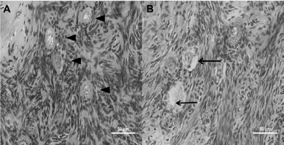
The aspect of morphologically normal or degenerated PFs at histology analysis is shown in Figure 2.
Figure 2. Histological features of collared peccaries’ (Pecari tajacu) ovarian fragments stored under refrigeration. A – Morphologically normal preantral follicles, exhibiting an oocyte with homogenous cytoplasm and a large central nuclei surrounded by well-organized granulosa cells (head arrows). B – Degenerated preantral follicles exhibiting oocyte cytoplasm shrinkage (arrows).
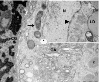
Transmission electron microscopy (TEM) was performed in order to analyze the PFs ultrastructure of the control group, and of those PFs stored in ACP® for 4h, which did not differ
from the control group. In order to compare the
invagination and the nuclear pores were evident. The oocyte cytoplasm showed several mitochondria dispersed through it and lysosomes were often observed as well. Furthermore, few profiles of endoplasmic reticulum were observed, and protein inclusions arranged as crystalloids were frequently observed. Lipid droplets were also common in the cytoplasm (Figure 3).
Follicles stored in ACP® for 4h presented an
unaltered nucleus and an evident nucleolus could be observed. The membrane nucleus remained intact and presented a double membrane, and
several nucleus pores. Lipid droplets were present in the oocyte cytoplasm extension, without evident alterations when compared to the control group and an evident smooth endoplasmic reticulum could be observed. Crystalloids and multilamellar inclusions were also present at ooplasm. By this evaluation, the alterations most commonly found were related to the mitochondrias, as most of them presented membranes and cristae disruption. Follicles stored in PBS presented similar characteristics but some vacuoles between oocyte and granulosa cells were identified in various PFs (Figure 4).
Figure 3. Electron micrograph of collared peccary preantral follicles from the control group (0h). CG, granulosa cell; GA, Golgi apparatus; c, crystalloid; *, vacuole; normal mitochondria (arrow); N, oocyte nucleus; LD, lipid droplet; nuclear pore (head arrow).
Figure 4: Electron micrograph of collared peccary preantral follicles, stored in ACP® based medium (A)
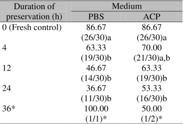
A total of 209 follicles were examined for viability by Trypan blue dye exclusion test (Table 2). It should be mentioned that the ACP®
-based media was efficient in maintaining the PFs viability similar to the fresh control group for 4h storage (P>0.05). No differences were evidenced between the media during the entire storage time for PFs viability (P>0.05). Due to the low number of isolated follicles recovered at 36h, it was not possible to proceed with a statistical analysis to evaluate the effect of medium and time of storage on follicle viability at this time.
Table 2. Percentage mean values (viable/total) of collared peccaries’ (Pecari tajacu) viable preantral follicles preserved under refrigeration in phosphate buffered saline solution (PBS) or in powdered coconut water (ACP) based solution for 24h
Duration of preservation (h)
Medium
PBS ACP
0 (Fresh control) 86.67 (26/30)a
86.67 (26/30)a
4 63.33
(19/30)b (21/30)a,b 70.00
12 46.67
(14/30)b
63.33 (19/30)b
24 36.67
(11/30)b (16/30)b 53.33
36* 100.00
(1/1)*
50.00 (1/2)*
a,b Within a row, means without a common superscript
differed (P < 0.05).
* Values could not be submitted to statistical analysis.
Furthermore, a temperature increase (P<0.05) inside the isothermal boxes was verified after 24h; however, this temperature was always lower than 8.4ºC and the external temperature remained constant (25.2ºC). For both media used, the pH was always closer to 7.0.
DISCUSSION
This study shows for the first time the short-term preservation of collared peccaries’ ovarian tissue in different media. According to the results, peccaries’ PFs seem to be very sensitive to preservation at low temperatures regardless of the media used. In swine, the domestic species more related to the collared peccaries (Cavalcante-Filho et al., 1998), a high quantity of total fatty acids was found in the oocytes, which reflects an abundant store of triglycerides
(McEvoy et al., 2000). It is hypothesized that the exposure of swine oocytes to low temperatures promotes the liberation of fatty acids from triglycerides resulting in high levels of free fatty acids that are reported as causing toxicity in innumerous cell types (Andrade et al., 2005; Cury-Boaventura et al., 2006). This is possibly the reason why swine oocytes are more sensitive to low temperatures (under 15 ºC) than other species (Didion et al., 1990). These facts could also be extrapolated for P. tajacu, once the presence of a large amount of lipid droplets in the oocyte cytoplasm was confirmed through the TEM. However, it is necessary to emphasize that differences regarding the sensitivity to the ovary tissue preservation exist even between collared peccaries and domestic swine. Lucci et al. (2007) demonstrated that the morphology and viability of swine PFs could be efficiently preserved for 18 h at 4 ºC using a saline solution (0.9%), while the present study shows an efficient preservation of peccaries’ PFs for only 4 h in the ACP® media
use. Adictionaly, Brito (2008) showed that swine PFs can be stored in situ at 4 or 20 ºC for up to 18 h without significative morphological alterations in histology and TEM analisis.
The TEM revealed that the swollen mitochondria and endoplasmic reticulum (ER) were the main alterations found during PFs storage both in the use of ACP® or PBS for 4h. In general, such
patterns are described as damages caused by ionic balance modification owing to alterations in the cellular membrane permeability (Borges et al., 2009). It is known that mitochondrial damage is the first sign of degeneration and it is also described for swine (Brito, 2008) PFs during in vitro storage. The analysis of PFs’ morphological integrity has been largely used for the evaluation of treatments applied to ovarian follicles (Lucci et al., 2007). However, the morphology does not always represent the PFs’ viability, and other tests are required for its analysis (Santos et al., 2007). The Trypan blue dye test indicated that the PFs viability of collared peccaries was kept similar to the control group when ACP®-based
In general, when media were compared with each other, a more efficient preservation of the ovarian tissue was verified in the use of ACP®.
These results demonstrate that medium composition is a very important concern for P. tajacu PFs storage. In fact, studies have shown that PFs use a different source of nutrients such as their own endogenous resource and also the contents of the medium used for preservation (Santos et al., 2002). It is known that with increase in time and temperature, depletion of the endogen reserve occurs, and the medium composition becomes the main source of nutrients (Celestino et al., 2007). The efficiency of ACP® as a medium for PFs short-term
preservation could be attributed to its rich contents of amino acids, sugars, vitamins, and minerals (Salgueiro et al., 2002). Also, 3-indol-acetic acid (IAA) is a vegetal hormone present in coconut water that has demonstrated beneficial effects on the preservation of caprine PFs when added to the commercial media used for the preservation of ovarian tissue (Ferreira et al., 2001). Possibly, the IAA interacts with growth factors present on ovarian tissue and thereby promotes its activation, while simultaneously maintaining the patterns of cell permeability and respiration (Ferreira et al., 2001).
It was verified that temperature inside the boxes did not exceed 8.4 ºC, allowing the viability preservation of more than 30% PFs for 24h, regardless of the medium used. In spite of the difference from the control group, these percentages still provide an adequate number of ovarian follicles available for the use in other techniques, such as in vitro culture or cryopreservation, as the PFs population in collared peccaries was estimated in 33 273.45±3 019.30 follicles (Lima et al., 2011).
In conclusion, ACP® provided a more adequate
medium for short-term preservation of P. tajacu preantral follicles under low temperatures when compared to PBS based medium in the same conditions. This study provided important information that will be useful for the exchange of genetic material of the abovementioned species, thereby contributing to its conservation.
ACKNOWLEDGEMENTS
The authors thank Dr. Emer S. Ferro from Instituto de Ciências Biológicas/USP, Dr. Maria Angélica Miglino and Dr. Rose Eli G. Rici from Laboratório de Anatomia/USP, M.Sc. Bruno R.
Simão from Departamento de Ciências
Ambientais e Tecnológicas/UFERSA, and Dr. José R. Figueiredo from LAMOFOPA/UECE for technical assistance. G.L. Lima and A.R. Silva were supported by grants from CNPq (National Research Council).
REFERENCES
ANDRADE, E.R.; AMORIM, C.A.; MATOS, M.H.T. et al. Evaluation of saline and coconut water solutions in the preservation of sheep preantral follicles in situ. Small. Rum. Res., v.43, p.235-243, 2002.
ANDRADE, L.N.S.; LIMA, T.M.; CURI, R.; CARTRUCCI, A.M.L. Toxicity of fatty acids on murine and human melanoma cell lines. ToxicologyIn Vitro, v.19, p.553-560, 2005.
BORGES, E.N.; SILVA, R.C.; FUTINO, D.O. et al. Cryopreservation of swine ovarian tissue: Effect of different cryoprotectants on the structural preservation of preantral follicle oocytes. Cryobiology. v.59, p.195-200, 2009.
BRITO, R.C.B. Morfologia de ovócitos imaturos inclusos em folículos ovarianos primordiais suínos. 2008. 40f. Dissertação (Mestrado em Medicina Veterinária) – Faculdade de Agronomia e Medicina Veterinária, Universidade de Brasília, Brasília. CASTELO, T.S.; BEZERRA, F.S.B.; LIMA, G.L. et al. Effect of centrifugation and sugar supplementation on the sperm cryopreservation of captive collared peccaries (Paccary tajacu). Cryobiology. v.61, p.275-279, 2010.
CAVALCANTE-FILHO, M.F.; MIGLINO, M.A.; MACHADO, G.V. et al. Comparative study of the morphology of the stomach of White lipped peccary (Tayassu pecari) and of the collared peccary (Pecari tajacu). Braz. J. Morph. Sci., v.15, p.206-207, 1998.
CELESTINO, J.J.H.; SANTOS, R.R.; MARTINS, F.S. et al. Conservação de folículos pré-antrais bovinos em solução salina 0,9% ou TCM 199. Arq. Bras. Med. Vet. Zootec., v.59, p.591-599, 2007.
DIDION, B.A.; POMP, D.; MARTIN, M.J. et al. Observations on the cooling and cryopreservation of pig oocytes at germinal vesicle stage. J. Anim. Sci., v.68, p.2803-2820, 1990.
FERREIRA, M.A.L.; BRASIL, A.F.; SILVA, J.R.V. et al. Effects of storage time and temperature on atresia of goat ovarian preantral follicles held in M199 with or without indole-3-acetic acid supplementation. Theriogenology, v.55, p.1607-1617, 2001.
FIGUEIREDO, J.R.; HULSHOF, S.C.J.; VAN DEN HURK, R. et al. Development of a combined new mechanical and enzymatic method for the isolation of intact preantral follicles from fetal, calf and adult bovine ovaries. Theriogenology, v.40, p.789-799, 1993.
JEWGENOW, K.; PENFOLD, K.L.; MEYER, H.H.D.; WILDT, D.E. Viability of small preantral ovarian follicles from domestic cats after cryoprotectant exposure and cryopreservation. J. Reprod. Fertil., v.112, p.39-47, 1998.
LIMA, G.L.; COSTA, L.L.M.; CAVALCANTI, D.M.L.P. et al. Short-term storage of canine preantral ovarian follicles using a powdered coconut water (ACP®)-based medium. Theriogenology, v.74, p.46-152, 2010.
LIMA, G.L.; SANTOS, E.A.A.; LUZ, V.B. et al. Estimativa e caracterização da população folicular ovariana de catetos (Pecari tajacu Linnaeus, 1758) In: CONGRESSO BRASILEIRO DE REPRODUÇÃO ANIMAL, 19., 2011, Recife. Anais... Recife: [s.n.] 2011. p. (Resumo).
LOPES, C.A.P.; SANTOS, R.R.; CELESTINO, J.J.H. et al. Short-term preservation of canine preantral follicles: Effects of temperature, medium and time. Anim. Reprod. Sci., v.115, p.201-214, 2009.
LUCCI, C.M.; KACINSKIS, M.A.; RUMPF, R.; BÁO, S.N. Effects of lowered temperatures and media on short-term preservation of zebu (Bos indicus) preantral ovarian follicles. Theriogenology, v.61, p.461-472, 2004.
LUCCI, C.M.; SCHREIER, L.L.; MACHADO, G.M. et al. Effects of storing pig ovaries at 4 or 20◦C for different periods of time on the morphology and viability of preantral follicles.Reprod. Domest. Anim., v.42, p.76-82, 2007.
LUZ, V.B.; SANTOS, R.R.; PINTO, L.C. et al. Dimethyl sulfoxide perfusion in caprine ovarian tissue and its relationship with follicular viability after cryopreservation. Fertil. Steril., v.91, p.1512-1515, 2009.
McEVOY, T.G.; COULL, G.D.; BROADBENT, P.J. et al. Fatty acid composition of lipids in immature cattle, pig and sheep oocytes with intact zona pellucida. J. Reprod. Fertil., v.118, p.163-170, 2000.
OLIVEIRA, M.F.; MESS, A.; AMBRÓSIO, C.E. et al. Chorioallantoic placentation in Galea spixii (Rodentia, Caviomorpha, Caviidae). Reprod. Biol. and Endocrinol., v.6, p.39-45, 2008.
PAYNTER, S.J.; COOPER, A.; FULLER, B.J.; SHAW, R.W. Cryopreservation of bovine ovarian tissue: structural normality of follicles after thawing and culture in vitro. Cryobiology. v.38, p.301- 309, 1999.
SALGUEIRO, C.C.M.; NUNES, J.F.; OLIVEIRA, K.P.L., et al. Utilização de diluentes à base de água de coco “in natura” e em pó na inseminação artificial programada de cabras. Rev. Bras. Reprod. Ani., v.5, p.96-98, 2002.
SAS Institute Inc. Base SAS® 6.1 Procedures Guide. Cary, NC: SAS Institute Inc. 2011.
SANTOS, R.R.; THARASANIT, T.; VAN HAEFTEN, T. et al. Vitrification of goat preantral follicles enclosed in ovarian tissue by using conventional and solid-surface vitrification methods. Cell Tissue. Res., v.327, p.167-17, 2007.
SANTOS, R.R.; SILVA, J.R.V.; COSTA, S.H.F. et al. Effect of 0.9% saline solution and Phosphat Buffer Saline at different temperatures and incubation times on the morphology of goat preantral follicles. Braz. J. Vet. Res. Anim. Sci., v.39, p.254-259, 2002.
SILVA, M.A.; PEIXOTO, G.C.X.; LIMA, G.L. et al. Cryopreservation of collared peccaries (Tayassu tajacu) semen using a powdered coconut water (ACP-116c) based extender plus various concentrations of egg yolk and glycerol. Theriogenology, v.78, p.605-611, 2012.