1,2-
b
-Oligoglucan Phosphorylase from
Listeria innocua
Masahiro Nakajima1,2., Hiroyuki Toyoizumi1., Koichi Abe1, Hiroyuki Nakai3, Hayao Taguchi1, Motomitsu Kitaoka2*
1Department of Applied Biological Science, Faculty of Science and Technology, Tokyo University of Science, Noda, Chiba, Japan,2National Food Research Institute, National Agriculture and Food Research Organization, Tsukuba, Ibaraki, Japan,3Graduate School of Science & Technology, Niigata University, Niigata, Niigata, Japan
Abstract
We characterized recombinant Lin1839 protein (Lin1839r) belonging to glycoside hydrolase family 94 fromListeria innocua. Lin1839r catalyzed the synthesis of a series of 1,2-b-oligoglucans (Sopn: n denotes degree of polymerization) using
sophorose (Sop2) as the acceptor anda-D-glucose 1-phosphate (Glc1P) as the donor. Lin1839r recognized glucose as a very
weak acceptor substrate to form polymeric 1,2-b-glucan. The degree of polymerization of the 1,2-b-glucan gradually decreased with long-term incubation to generate a series of Sopns. Kinetic analysis of the phosphorolytic reaction towards
sophorotriose revealed that Lin1839r followed a sequential Bi Bi mechanism. The kinetic parameters of the phosphorolysis of sophorotetraose and sophoropentaose were similar to those of sophorotriose, although the enzyme did not exhibit significant phosphorolytic activity on Sop2. These results indicate that the Lin1839 protein is a novel inverting
phosphorylase that catalyzes reversible phosphorolysis of 1,2-b-glucan with a degree of polymerization of$3. We propose 1,2-b-oligoglucan: phosphatea-glucosyltransferase as the systematic name and 1,2-b-oligoglucan phosphorylase as the short name for this Lin1839 protein.
Citation:Nakajima M, Toyoizumi H, Abe K, Nakai H, Taguchi H, et al. (2014) 1,2-b-Oligoglucan Phosphorylase fromListeria innocua. PLoS ONE 9(3): e92353. doi:10. 1371/journal.pone.0092353
Editor:Nancy E. Freitag, University of Illinois at Chicago College of Medicine, United States of America
ReceivedDecember 11, 2013;AcceptedFebruary 20, 2014;PublishedMarch 19, 2014
Copyright:ß2014 Nakajima et al. This is an open-access article distributed under the terms of the Creative Commons Attribution License, which permits unrestricted use, distribution, and reproduction in any medium, provided the original author and source are credited.
Funding:This work was supported by the Science and Technology Research Promotion Program for Agriculture, Forestry, Fisheries and Food Industry, Internal Research Fund of NARO, and Research Fund of Tokyo University of Science. The funders had no role in study design, data collection and analysis, decision to publish, or preparation of the manuscript.
Competing Interests:The authors have declared that no competing interests exist.
* E-mail: mkitaoka@affrc.go.jp
.These authors contributed equally to this work.
Introduction
‘Phosphorylase’ is the general term for enzymes that reversibly phosphorolyze glycosyl linkages to generate sugar 1-phosphates [1,2]. Phosphorylases are generally thought to be involved in metabolism of specific oligosaccharides or storage polysaccharides in cytosol through their phosphorolytic activity. Reverse phospho-rolysis is useful for preparation of specific oligosaccharides because of their strict regioselectivity [1–3].
This reversibility makes it possible to practically produce several oligosaccharides from abundantly available natural resources, such as sucrose and starch, by combining the reactions of two phosphorylases. Based on these features, practical preparative methods of several oligosaccharides and polysaccharides have been developed [2,4–8]. These combinations enable practical production of such compounds without using expensive sugar 1-phosphates. However, the number of phosphorylases based on EC numbers is only 19, which is much smaller than those of glycoside hydrolases, and limits the usage of the enzyme class. Therefore, it would be beneficial to find new phosphorylases showing unreported substrate specificities and regioselectivities.
Phosphorylases belong to one of the glycoside hydrolase families (GH) 13, 65, 94, 112, and 130 or the glycosyl transferase families (GT) 4 and 35 on the Carbohydrate-Active EnZymes database (http://www.cazy.org) on the basis of the sequence similarity [9]. Among them, activities of GH94 members reported are cellobiose phosphorylase (EC 2.4.1.20) [10], N,N9-diacetylchitobiose
phos-phorylase (EC 2.4.1.280) [11], laminaribiose phosphos-phorylase (EC 2.4.1.31) [12,13], cellodextrin phosphorylase (EC 2.4.1.49) [14– 16], cellobionic acid phosphorylase (EC 2.4.1.x) [17] and C-terminal domain of cyclic 1,2-b-glucan synthase (EC 2.4.1.x, CGS) possessing phosphorolytic activity on protein-bound 1,2-b-oligo-glucan [18].
In the phylogenetic tree analysis of GH94, enzymes catalyzing the same reactions appeared in the same cluster, except for cellodextrin phosphorylase. We noticed that strains of genus Listeriagenerally possess a gene encoding a GH94 protein at a cluster in which no enzyme has been characterized. In this study, we describe the GH94 protein from Listeria innocua with phosphorylase activity specific to 1,2-b-oligoglucans that requires a new EC number.
Materials and Methods
Sequence Analysis
ClustalW2 (http://www.ebi.ac.uk/Tools/msa/clustalw2/) was used to perform multiple alignments, and MEGA5.1 was used to construct a phylogenetic tree [19].
Cloning, Expression, and Purification
using KOD plus DNA polymerase (Toyobo, Osaka, Japan) and the genomic DNA as a template. Primer pair was forward primer
59-GTGGATATccaTGgCAATGTTAAAAG-39 and reverse
primer 59-ATACACAAAACAACCctcGAGACGG-39(lower case represents sequences different from original sequence of the genome sequence ofL. innocuaClip 11262) containing additional NcoI and XhoI sites (underlined), respectively. The amplified lin1839 gene was inserted into the NcoI and XhoI sites of pET28a(+) (Novagen, Madison, WI, USA) to encode a His6–tag fusion at the C terminus. The constructed plasmid was used to transformEscherichia coliBL21 (DE3). The transformant was grown in 1 l of Luria–Bertani medium containing 30mg/ml kanamycin at 37uC until the absorbance reached 0.8 at 660 nm, followed by induction using 0.1 mM IPTG at 20uC overnight. The cells were collected by centrifugation at 3,9006g for 5 min and then suspended in 20 mM MOPS–NaOH buffer (pH 7.5) containing 250 mM NaCl (buffer A). The suspended cells were disrupted by sonication and centrifuged at 27,0006gfor 20 min. The obtained supernatant was loaded onto a HisTrap FF crude column (5 ml; GE Healthcare, Buckinghamshire, UK) equilibrated with buffer A containing 10 mM imidazole using a AKTA Prime Plus chroma-tography system (GE Healthcare). After unbound components were washed with buffer A containing 10 mM imidazole, a linear
gradient of 10–250 mM imidazole in buffer A was used to elute Lin1839r (actually eluted between 25 and 100 mM of imidazole). An Amicon Ultra 30,000 molecular weight cut-off (Millipore, Billerica, MA, USA) was used to change the buffer in the pooled protein solution to 20 mM MOPS–NaOH buffer (pH 7.5). A theoretical extinction coefficient of 175,450 M21cm21, based on the amino acid sequence, was used to determine the protein concentration spectrophotometrically at 280 nm [20]. Finally, 88 mg of recombinant Lin1839 protein (Lin1839r) was obtained from 1 l of culture medium.
Size-exclusion Chromatography
Lin1839r (1 ml of 2.0 mg/ml solution) was loaded onto Superdex 200 (Hiload 16/60; GE Healthcare) equilibrated with 50 mM MOPS–NaOH (pH 7.0) containing 150 mM NaCl. Ovalbumin (44 kDa), conalbumin (75 kDa), aldolase (158 kDa), ferritin (440 kDa) and thyroglobulin (669 kDa; GE Healthcare) were used as standard proteins. Blue dextran 2000 (2000 kDa; GE Healthcare) was used to determine the void volume of the column.
Measurement of Phosphorolytic and Synthetic Activity
The phosphorolytic activity was determined by measuring a-glucose 1-phosphate (Glc1P) generated in a reaction mixture on
Figure 1. Phylogenetic tree of GH94 proteins. Genes are represented with the organism names and GenBankTM accession numbers. Characterized enzymes are categorized in boxes framed with broken lines according to their substrate specificities. In the case of CGSs, only GH94 domains are used for the alignment. The gene cloned in this study is represented with a black background and white letters. GH94 proteins from Listeriaare boxed with a bold line.
the basis of the phosphoglucomutase-glucose 6-phosphate dehy-drogenase method [21]. The reaction was performed in a reaction mixture (200ml) placed in a well of a 96-well microplate (EIA/RIA plate, 96-well half area, Corning, NY, USA) containing appropri-ate concentrations of each sugar substrappropri-ate and inorganic phosphate with 5.0mM a-glucose 1,6-bisphosphate (Sigma– Aldrich, St. Louis, MO, USA), 5.0 IU/ml glucose 6-phosphate dehydrogenase from Leuconostoc mesenteroides (Oriental Yeast, Tokyo, Japan), 6.25 IU/ml phosphoglucomutase from rabbit muscle (Sigma–Aldrich), 0.5 mM thio-NAD+ (Oriental Yeast),
and 25 mM MgCl2in 100 mM MOPS–NaOH buffer (pH 7.5) at 30uC. The concentration of Glc1Pwas calculated by determining absorbance at 400 nm continuously on a Spectramax 190 (Molecular Devices, CA, USA).
The method of Lowry and Lopez [22], as described below, was used to determine the reverse phosphorolytic activity by measuring the amount of inorganic phosphate released in a reaction mixture. In brief, the enzymatic reaction was performed in a reaction mixture (160ml) containing 10 mM Glc1P and an appropriate concentration of each acceptor in 100 mM MOPS–NaOH buffer
(pH 7.5) at 30uC. An aliquot (20ml) was mixed with 160ml of 0.2 M sodium acetate buffer (pH 4.0) and 20ml of 1% ammonium molybdate containing 25 mM sulfuric acid to stop the reaction every 3 minutes. Then, 20ml of 1% ascorbic acid and 0.05% potassium bisulfate were mixed with the samples. The mixtures were incubated at 37uC for 1 h, and the concentrations of phosphate released were quantified by measuring absorbance at 700 nm. One unit of the phosphorolytic and reverse phosphoro-lytic activities were defined as the amount of the enzyme producing 1mmol of Glc1P and phosphate, respectively, per minute under the above conditions with the concentrations of all substrates at 10 mM.
Temperature and pH Profile
The effect of pH on the enzymatic activity using 4.0mg/ml Lin1839r was determined by measuring synthetic activities under the standard conditions described above and by substituting 100 mM MOPS–NaOH buffer (pH 7.5) with various 100 mM buffers. Similarly, the effect of temperature on the enzymatic activity of 4.0mg/ml Lin1839r was determined by measuring the reverse phosphorolytic activities at various temperatures for 20 min. The thermal and pH stabilities were evaluated by measuring the residual synthetic activity of 10 mM sophorose (Sop2) and 10 mM Glc1Pafter incubation of Lin1839r (0.5 mg/ ml) at different temperatures in 20 mM MOPS–NaOH buffer (pH 7.5) for 1 h and at different pH at 30uC for 1 h, respectively.
Thin Layer Chromatography (TLC)
An aliquot (1ml) of the reaction mixture for the synthetic reaction was spotted onto a TLC plate (Kieselgel 60 F254; Merck, Darmstadt, Germany), and a solution of acetonitrile:water (3:1, v/ v) was used to elute the samples. The TLC plate was soaked in 5% sulfuric acid:methanol solution and heated in an oven until bands were sufficiently visible.
Structural Analysis of the Reaction Products
Reaction products (oligosaccharides) for structural determina-tion were generated by incubadetermina-tion of a reacdetermina-tion mixture (4 ml) containing 20 mM Glc1P, 10 mM Sop2, and 40mg/ml Lin1839r in 100 mM MOPS–NaOH (pH 7.0) at 30uC for 2 h. After using Amberlite MB4 (Organo, Tokyo, Japan) to desalt the sample, it was concentrated and loaded onto a Toyopearl HW-40F column (5.0 cm Q680 cm; Tosoh, Tokyo, Japan), equilibrated with distilled water and eluted at a flow rate of 5.0 ml/min. Fractions containing trisaccharide and tetrasaccharide were collected, followed by lyophilization. Polysaccharide was synthesised by incubation of a reaction mixture (1 ml) containing 200 mM Glc1P, 50 mM glucose, and 125mg/ml Lin1839r in 100 mM MOPS–NaOH (pH 7.0) at 30uC for 4 days. The products were separated on the same column as described above. Fractions containing polysaccharide were collected and desalted using Amberlite MB4, followed by lyophilization. The amounts of trisaccharide, tetrasacchride, and polysaccharide obtained were 2.3, 1.6 and 7.5 mg, respectively. One-dimensional (1H and13C) and two-dimensional [double-quantum-filtered correlation spec-troscopy (DQF-COSY), totally correlated specspec-troscopy (TOCSY), heteronuclear single-quantum coherence (HSQC), and hetero-nuclear multiple-bond correlation (HMBC)] NMR spectra of the product were acquired in D2O with 2-methyl-2-propanol as an internal standard on a Bruker Avance 500 or Bruker Avance 800 spectrometer (Bruker Biospin, Rheinstetten, Germany). Proton signals were assigned on the basis of the DQF-COSY and TOCSY spectra.13C signals were assigned on the HSQC spectra on the basis of the assignment of proton signals. The linkage position in
Table 1.Substrate specificity of Lin1839r in the synthetic reaction.
Substrate Relative activitya(%)
Acceptorb
D-Glucose N.Dd,e
2-Deoxy-D-glucose N.D
D-Xylose N.D
D-Mannose N.D
D-Galactose N.D
L-Fucose N.D
L-Arabinose N.D
L-Rhammose N.D
D-Glucosamine N.D
N-Acetyl-D-glucosamine N.D
D-Galactosamine N.D
N-Acetyl-D-galactosamine N.D
Sucrose N.D
Maltose N.D
Lactose N.D
Sophorose 100 (43f
)
Laminaribiose 1.1 (0.48)
Cellobiose N.D
Gentiobiose N.D
Donorc
Glc1P 100 (43)
a-D-Galactose 1-phoshate N.D
a-D-Mannose 1-phoshate N.D
N-Acetyl-a-D-glucosamine 1-phoshate N.D
aThe specific activity of Lin1839r in the presence of 10 mM Sop
2and 10 mM
Glc1Pwas defined as 100% relative activity.
bMeasured at 10 mM with 10 mM Glc1Pas the donor. cMeasured at 10 mM with 10 mM Sop
2as the acceptor. dN.D represents not determined owing to,0.2% relative activity. eThe specific activity on glucose at 100 mM was 0.095 s21. fValues in parentheses represent specific activity (s21).
doi:10.1371/journal.pone.0092353.t001
the oligosaccharides was determined by detecting inter-ring cross peaks in each HMBC spectrum.
Preparation of 1,2-b-oligoglucans and 1,2-b-glucan for Assay
1,2-b-Oligoglucans [Sopn, n denotes the degree of polymeriza-tion (DP) of the oligosaccharide] was synthesized without using expensive Sop2 by incubation of a reaction mixture (20 ml)
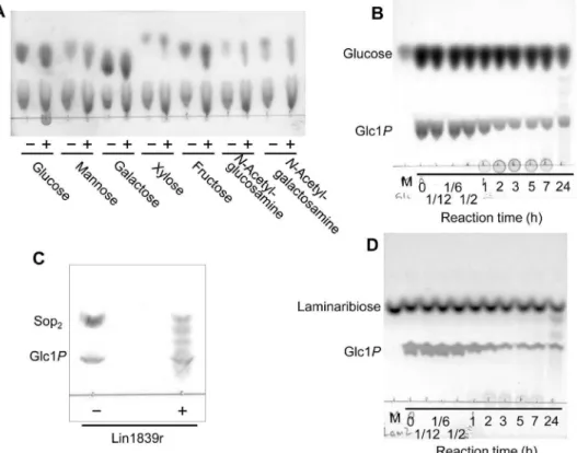
Figure 2. TLC analysis of reaction products from acceptors and Glc1P.(A) Reaction products from monosaccharides and Glc1P. Reactions were performed with 2 mg/ml of Lin1839r in the presence of 50 mM monosaccharides and 50 mM Glc1Pfor 1 h. (B) Time course of the reaction products from glucose and Glc1P. Substrates used were 50 mM glucose and 50 mM Glc1P. The enzyme concentration used was 2 mg/ml. (C) Reaction products after reaction for 2 h using 10 mM Sop2 and 20 mM Glc1P as substrates. (D) Time course of the reaction products from laminaribiose and Glc1P. The substrates used were 10 mM laminaribiose and 10 mM Glc1P. The enzyme concentration used was 0.5 mg/ml. (A, C) Presence and absence of Lin1839r are represented with ‘+’ and ‘2’, respectively. (B, D) M, marker. Numbers under a line represent reaction time. doi:10.1371/journal.pone.0092353.g002
Figure 3. Effects of temperature and pH on enzyme activity and stability of Lin1839r.(A) The temperature optimum and stability are represented by closed and open circles, respectively. (B) The optimum pH and stability are represented by a single line and a dashed line, respectively. The buffers used for reaction and incubation are sodium acetate (pH 4.0–5.5, closed circle), MES–NaOH (pH 5.5–6.5, open circle), BISTRIS (pH 6.0–7.0, closed triangle), MOPS–NaOH (pH 6.5–7.5, open triangle), HEPES–NaOH (pH 7.5–8.5, closed square), 2-hydroxy-3-[4-(2-hydroxyethyl)-1-piperazinyl]propanesulphonic acid–NaOH (pH 8.0–8.5, open square), Tris–HCl (pH 7.5–9.0, open rhombic), and glycine–NaOH (pH 8.6–10, closed rhombic).
containing 500 mM Glc1P, 250 mM glucose, and 2.0 mg/ml Lin1839r (pH 7.0 adjusted with HCl) at 30uC for 14 days. After performing electrodialysis on a Microacylizer S1 with an AC-220-10 cartridge (Astom Corp., Tokyo Japan) to remove the salts contained in the reaction mixture, a Toyopearl HW-40F column, as described above, was used to separate the products. Fractions containing Sop3, Sop4, and Sop5 were collected, followed by lyophilization. 1,2-b-glucan was produced by incubation of a reaction mixture (50 ml) containing 750 mM Glc1P, 180 mM glucose, and 80mg/ml Lin1839r in 375 mM MOPS–NaOH (pH 7.0) at 30uC for 8 days. The product was separated as described above. The collected sample solution was concentrated into approximately 15 ml, and then an equal volume of ethanol was added to the 1,2-b-glucan solution. The first precipitate was separated from the supernatant by centrifugation after incubation of the sample at 20uC for 1 day. The second precipitate generated in the first supernatant after the incubation for an additional 1 day was also collected by centrifugation. The final precipitate was obtained after storage of the second supernatant at 230uC for over 1 year. The precipitates were dried under vacuum. The average DP of 1,2-b-glucan was calculated from the ratio of the peak area of C-6 protons of the internal glucose units appearing near 3.94 ppm against that of the C-2 proton at the non-reducing end appearing near 3.33 ppm.
Kinetic Analysis
The initial velocities of the phosphorolytic reactions with Sopns were determined under the standard conditions with 1.0mg/ml Lin1839r and a combination of initial concentrations of each substrate and Pi. The kinetic parameters for Sop3were calculated by curve-fitting the experimental data to the theoretical equation (1) for a sequential Bi Bi mechanism using GraFit version 7.0.3 (Erithacus Software Ltd., London, UK).
v~
kcat½E 0½A½B
KiAKmBzKmA½BzKmB½Az½A½B
(A~sugar, B~Pi)
ð1Þ
Kinetic analysis of the synthetic reactions with suitable acceptors was performed under the standard conditions with 2.0mg/ml Lin1839r and different concentrations of the acceptor substrate or Glc1P as the donor with 10 mM each opposite substrate. The kinetic parameters were calculated by curve fitting the experimental data to the Michaelis-Menten equation (2) using GraFit version 7.0.3.
v~
kcat½E0½S
Kmz½S ð2Þ
Results
Sequence Analysis
L. innocuaClip11262 possesses a single gene encoding the GH94 protein (Lin1839) in its genome (GenBank accession number: AL592022.1). Alignment of the amino acid sequences of GH94 proteins revealed that the common catalytic nucleophile aspartate residue was conserved as D739 in Lin1839. The amino acid sequence of the Lin1839 protein showed no predicted N-terminal signal peptide on the basis of a SignalP 4.0 analysis (http://www. cbs.dtu.dk/services/SignalP/) [23], suggesting that it is a cytosolic protein. Its activity was not predictable from the phylogenetic tree analysis with other characterized GH94 enzymes (Fig. 1).
Table 2.Kinetic parameters for Sopns in the synthetic reaction.
Substrate kcat(s21) Km(mM) kcat/Km(s21mM21)
Sop2
a
9764 8.560.6 1161
Sop3
a
110610 6.060.9 1861
Sop4
a
9065 6.860.7 1361
Glc1Pb
4362 1.260.2 3464
aUp to 10 mM acceptors were used. 10 mM Glc1Pwas used as a donor. bUp to 10 mM Glc1Pwas used. 10 mM Sop
2was used as an acceptor.
doi:10.1371/journal.pone.0092353.t002
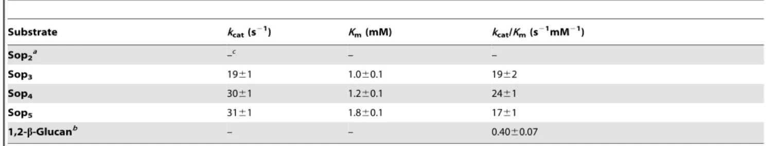
Table 3.Kinetic parameters for phosphorolysis of Sopns and 1,2-b-glucan.
Substrate kcat(s21) Km(mM) kcat/Km(s21mM21)
Sop2a –
c
– –
Sop3 1961 1.060.1 1962
Sop4 3061 1.260.1 2461
Sop5 3161 1.860.1 1761
1,2-b-Glucanb – – 0.40
60.07
aSpecific activity was,0.2 (s21) at 10 mM.
bUp to 4 mg/ml 1,2-b-glucan (average DP = 77) was used. c–, not determined.
doi:10.1371/journal.pone.0092353.t003
Substrate Specificity in the Synthetic Reaction
The acceptor specificity of Lin1839r in the synthetic reaction was examined using 10 mM of various putative carbohydrate acceptors in the presence of 10 mM Glc1Pas the donor. Lin1839r did not utilize any monosaccharides as the acceptor to significantly liberate inorganic phosphate under the reaction conditions described in Table 1. We noticed that a spot of probable polysaccharide appeared at the origin of the TLC for the reactions with 50 mM monosaccharides and 50 mM Glc1P with higher concentrations of Lin1839r (2 mg/ml) only when glucose was used as the acceptor, but no oligosaccharides were detected (Fig. 2A). After a long incubation period, a series of oligosaccharides were detected on TLC (Fig. 2B). Next, we examined the acceptor specificity for disaccharides at 10 mM. The enzyme showed the highest activity with Sop2(43 s21) and a detectable activity with laminaribiose (0.48 s21) but no activity with other disaccharides (Table 1). The reaction with Sop2 produced a series of
oligosaccharides (Fig. 2C). The reaction with laminaribiose as the acceptor produced a polymer initially followed by generation of oligomers (Fig. 2D), a pattern similar to that for glucose. The enzyme exhibited activity only on Glc1P among the sugar 1-phosphates examined (Table 1).
Basic Properties of Lin1839r
Purified Lin1839r was detected at 120 kDa as a single band on SDS-PAGE, which corresponded to the theoretical molecular mass of Lin1839r with His6 tag (123,817 Da). Size-exclusion chromatography of Lin1839r resulted in elution of Lin1839r as a 110-kDa protein, suggesting that Lin1839r is a monomeric protein. Lin1839r was stable up to 37uC. The remaining activity of Lin1839r after incubation for 20 min drastically decreased at temperatures .45uC. The optimal temperature was 37–45uC (Fig. 3A). Lin1839r was stable in the range of pH 4.5–9.5, and its optimal pH was 7.5–8.0 (Fig. 3B).
Analysis of Products of the Synthetic Reaction Catalyzed by Lin1839r
Each oligosaccharide produced in the reaction mixture with Sop2as the acceptor and Glc1Pas the donor was isolated by gel filtration. The trisaccharide and tetrasaccharide produced were identified as Sop3and Sop4, respectively, on the basis of the NMR spectra (Fig. S1–S2, Table S1–2). The polymer product produced from glucose and Glc1Pat the early stage of the reaction was also isolated by gel filtration. Simple1H and13C NMR spectra of the compound indicated that it is a homopolymer containing only a 1,2-b-glucosyl linkage and an average DP of 39 (Fig. S3, Table S3).
Preparation of 1,2-b-glucan and Sopns
High concentrations of glucose and Glc1P as substrates were successfully adapted to production of 1,2-b-glucan (at a low concentration of Lin1839r) and Sopns (at a high concentration of Lin1839r for a long reaction term) at a larger scale. No precipitate was generated during the entire reaction period, suggesting that the generated 1,2-b-glucan was very soluble in water. For the production of the polymer, addition of ethanol to the 1,2-b-glucan solution caused gradual precipitation. The amounts of the first, second and third precipitates were 1.3, 0.90 and 0.25 g, respectively. The average DPs of these precipitates were 77, 64 and 39, respectively. The amounts of Sop2, Sop3, Sop4and Sop5 obtained were 98, 120, 100 and 35 mg, respectively.
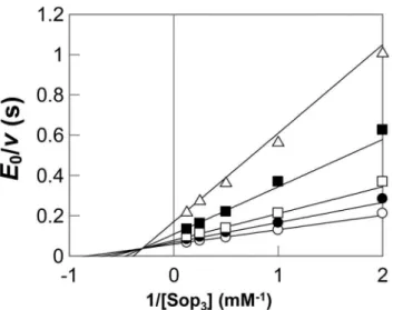
Figure 4. Double reciprocal plot for the phosphorolysis of Sop3
with different concentrations of inorganic phosphate. Concen-trations of inorganic phosphate were 0.5 mM (open triangle), 1.0 mM (filled square), 2.0 mM (open square), 3.0 mM (filled circle), and 5.0 mM (open circle). The kinetic parameters are as follows:kcat= 2161 (s21), KmA= 0.6660.14 (mM), KmB= 1.360.2 (mM), and KiA= 3.360.6 (mM), where A represents Sop3and B is Pi. Grafit version 7.0.3 was used to perform non-linear regression and calculation of values.
doi:10.1371/journal.pone.0092353.g004
Figure 5. Reaction scheme for the Lin1839 protein.
Kinetic Analysis of the Reverse and Forward Phosphorolytic Reactions
In the reverse phosphorolytic reaction, the kinetic parameters of Lin1839r for activity on the Sopns as acceptors were determined. The enzyme showed similar values ofkcatandKmfor Sop2, Sop3, and Sop4, which led to similar kcat/Km values for these oligosaccharides (Table 2). The kinetic parameters for Glc1Pwere in the same range as those of other inverting phosphorylases [13,24,25]. In the phosphorolytic reaction, the kinetic parameters of Lin1839r for the Sopns and 1,2-b-glucan (DP = 77) as a substrate were determined. The enzyme exhibited similarkcatand Kmvalues for Sop3, Sop4, and Sop5(Table 3). Thekcat/Kmvalues for 1,2-b-glucan were much smaller than those for Sop3, Sop4and Sop5, but the activity was significantly detectable. The enzyme showed only a negligible phosphorolytic activity on Sop2(Table 3). These results indicated that the enzyme catalyzed reversible phosphorolysis of Sopns with$DP 3.
Reaction Mechanism of Phosphorolysis
Double reciprocal plots of initial velocities against various initial concentrations of Sop3 and phosphate yielded a series of lines intersecting at a point (Fig. 4). This result indicated that the phosphorolytic reaction on Sop3 followed a sequential Bi Bi mechanism, same as inverting phoshorylases [24,26–28]. The kinetic parameters of Lin1839r determined by regression data are shown in the Fig. 4 legend. The values ofkcat,KmA, andKmBwere
in the same range as those of other inverting phosphorylases [24,28–30].
Discussion
Classification of Lin1839 Protein
We found that Lin1839r has phosphorylase activity highly specific to Sopns with $DP 3 in phosphorolysis (Fig. 5). The specificity on DP is similar with that of cellodextrin phosphorylase and implies the existence of subsites21 to+2 in Lin1839 protein. The structural prediction of the architecture of Lin1839 is not possible due to the poor identities (less than 20%) in the amino acid sequence with other GH94 enzymes whose structures have been solved [10,11,31,32].
The C-terminal GH94 domain of CGS from Brucella abortus possesses phosphorolytic activity on 1,2-b-glucan [18]. However, Lin1839r is clearly different from the CGS in the catalytic reaction with respect to factors described below.
CGS is a fusion enzyme composed of GT84 and GH94 that synthesizes cyclic 1,2-b-glucan (CbG) [33,34]. CGS synthesizes CbGs through the four enzymatic reactions: (i) initiation (transferring glucose to CGS), (ii) elongation of 1,2-b-glucan, (iii) regulation of DP of 1,2-b-glucan and (iv) cyclization [35]. The GH94 domain is involved in (iii) by controlling the length of the polysaccharide chain produced by the GT domain [18]. The GT domain is able to produce 1,2-b-glucan from UDP-glucose without the GH94 domain. Thus, CGS is virtually a glycosyl
Figure 6. Possible mechanism of the production of 1,2-b-glucan and Sopns from glucose and Glc1P.Possible reaction schemes at the beginning of the reaction (A), after Glc1P/Pi ratio reached equilibrium (B), and at the end of the reaction (C) are shown. Open and slashed circles represent the glucose moiety derived from Glc1Pand material glucose, respectively. Lines connecting these circles represent the 1,2-b-glucosyl linkage. Dashed and bold arrows represent reactions with poor and preferential substrates, respectively. Glc1Pis drawn with an open circle, and Pi represents inorganic phosphate. Accumulation of 1,2-b-glucan and Sopns are surrounded by a dashed rounded box.
doi:10.1371/journal.pone.0092353.g006
transferase, and the GH94 domain is enzymologically just an accessory domain. The phosphorolytic activity of the C-terminal domain (1493–2867) of CGS on linearized CbG was 0.725 U/mg [18], whereas the phosphorolytic activity of Lin1839r on Sop3was much higher (19 s21= 9.2 U/mg). Therefore, Lin1839r is a novel enzyme that should be given a new EC number, and we propose 1,2-b-oligoglucan: phosphatea-glucosyltransferase as the system-atic name and 1,2-b-oligoglucan phosphorylase as the short name for this Lin1839 protein. Sophorodextrin phosphorylase is a possible alternative name.
Synthesis of 1,2-b-glucan and Sopns
Lin1839r synthesized a series of Sopns from Sop2and Glc1P (Fig. 2C). It also produced 1,2-b-glucan from glucose and Glc1P without any detectable oligosaccharides at the early stage of the reaction, and then a series of Sopns accumulated (Fig. 2B). These phenomena can be explained as described below.
At the initial stage, glucose acted as a poor acceptor to generate Sop2slowly. The resultant Sop2was preferentially converted into a polymer because the Sop2 and Sopns generated act as actual acceptors much more than glucose (Fig. 6A). Similar generation of polymeric compounds with a monosaccharide as the acceptor has been reported on production of cellulose-like material using cellodextrin phosphorylase [36]. The polymer formation contin-ued until Glc1P/Pi ratio reached nearly equilibrium. Equilibriums between Sopn and Sopn+1 (n$2) were nearly completed at this stage (Fig. 6B). However, most part of glucose still remained in the reaction mixture, meaning that equilibrium between glucose and Sop2was not completed (Fig. 6B). Sop2was still being generated from glucose and Glc1P. The continuous formation of Sop2 increased the number of the Sopnmolecules, causing the decrease in the average DP of Sopns. Finally, small oligosaccharides were concomitantly generated with disappearance of the polymer (Fig. 6C). This finding is in clear contrast to the reaction of cellodextrin phosphorylase in which the products immediately precipitated no longer participated in the reaction [36].
Physiological Role of 1,2-b-oligoglucan Phosphorylase fromL. innocua
Mostb-1,2-linked glucose polymers have been found as CbG, a homopolymer ofb-1,2-linked glucose with DP 17–24 [37]. CbG is distributed in some a-proteobacteria [38–40]. After CbG is synthesized by CGS, it is transported into periplasm by an ABC-transporter system involving Cgt protein. CbG is then modified with anionic molecules, such as succinate, in the case of Brucella. Rhizobium phaseolisecretes CbG as an exopolysaccharide [41]. CbG is involved in adaption to hypo-osmotic conditions for Rhizobium melilotiandAgrobacterium tumefaciens[42]. Defects in thecgs genes of these bacteria cause non-motile phenotypes because of defects in the assembly of flagella [43,44].BrucellaCbG acts in lipid rafts found on host cell membranes to avoid the innate immune system [45]. CbG suppresses plant immune responses in the case of Xanthomonas campestris pv. campestris [46]. However, L. inncoua does not have a CGS homolog or synthetic system of CbG according to genomic information [47], implying thatL. inncoua utilizes exogenous 1,2-b-glucan. A gene cluster is formed around the lin1839 gene. A putative ATP-binding protein ABC-type transporter gene (lin1841–lin1843) may be involved in intake of Sopns and/or 1,2-b-glucan. A putative GH3b-glucosidase gene (lin1840) is thought to degrade the substrates with concerted action of the Lin1839 protein. LacI transcription factor gene (lin1838) may regulate expression of the genes in the gene cluster. Therefore, it is speculated that the gene cluster is involved in
metabolism of Sopns and/or 1,2-b-glucan in a specific environ-mental condition.
Conclusion
Although numerous studies on 1,3- and 1,4-b-glucans have been reported, 1,2-b-glucan has not been further studied. This is probably because of the difficulty in obtaining 1,2-b-glucan and its oligomers. For instance, Sop2 is only available as an expensive reagent, although it has been well known since 1962 as a powerful inducer of cellulase from Trichoderma reesei, the hyper-cellulase-producing fungus [48]. We identified the Lin1839 protein as a novel 1,2-b-oligoglucan phosphorylase requiring a new EC number. This enzyme enables easy preparation of 1,2-b-glucan and its oligomers, including Sop2, and is an important milestone in the development of procedures for study of the functions of 1,2-b-glucan and related compounds.
Supporting Information
Figure S1 NMR spectra of Sop3. (A)1H-NMR, (B)13C-NMR, (C) DQF-COSY, (D) TOCSY, (E) HSQC and (F) HMBC. I, II, and III denote first, second, and third glucose residues from reducing end, respectively. Letters in parenthesis represent position of hydroxyl group on the anomeric carbon. Arabic numbers shown with roman numbers represent positions of carbons and protons in sugar rings.
(PDF)
Figure S2 NMR spectra of Sop4. (A)1H-NMR, (B)13C-NMR, (C) DQF-COSY, (D) TOCSY, (E) HSQC and (F) HMBC. I, II, and III denote first, second, and third glucose residues from reducing end, respectively. Letters in parenthesis represent position of hydroxyl group on the anomeric carbon. Arabic numbers shown with roman numbers represent positions of carbons and protons in sugar rings.
(PDF)
Figure S3 NMR spectra of 1,2-b-glucan. (A)1
H-NMR, (B)13 C-NMR. Numbers under chemical shifts and in parenthesis represent positions of protons (A) and carbons (B). Letters in parenthesis represent position of hydroxyl group on the anomeric carbon.
(PDF)
Table S1 Chemical shifts in13C-NMR and1H-NMR spectra of Sop3.
(PDF)
Table S2 Chemical shifts in13C-NMR and1H-NMR spectra of Sop4.
(PDF)
Table S3 Chemical shifts in13C-NMR and1H-NMR spectra of 1,2-b-glucan.
(PDF)
Acknowledgments
Author Contributions
Conceived and designed the experiments: MN MK. Performed the experiments: MN H. Toyoizumi KA MK. Analyzed the data: MN H.
Toyoizumi HN H. Taguchi MK. Contributed reagents/materials/analysis tools: H. Taguchi MK. Wrote the paper: MN H. Toyoizumi MK.
References
1. Nakai H, Kitaoka M, Svensson B, Ohtsubo K (2013) Recent development of phosphorylases possessing large potential for oligosaccharide synthesis. Curr Opin Chem Biol 17: 301–309.
2. Kitaoka M, Hayashi K (2002) Carbohydrate-processing phosphorolytic enzymes. Trends Glycosci Glycotechnol 14: 35–50.
3. Luley-Goedl C, Nidetzky B (2010) Carbohydrate synthesis by disaccharide phosphorylases: reactions, catalytic mechanisms and application in the glycosciences. Biotechnol J 5: 1324–1338.
4. Nakajima M, Nishimoto M, Kitaoka M (2010) Practical preparation of D-galactosyl-b1R4-L-rhamnose employing the combined action of
phosphorylas-es. Biosci Biotechnol Biochem 74: 1652–1655.
5. Nishimoto M, Kitaoka M (2007) Practical preparation of lacto-N-biose I, a candidate for the bifidus factor in human milk. Biosci Biotechnol Biochem 71: 2101–2104.
6. Ohdan K, Fujii K, Yanase M, Takaha T, Kuriki T (2006) Enzymatic synthesis of amylose. Biocatal Biotransform 24: 77–81.
7. Ohdan K, Fujii K, Yanase M, Takaha T, Kuriki T (2007) Phosphorylase coupling as a tool to convert cellobiose into amylose. J Biotechnol 127: 496–502. 8. Nishimoto M, Kitaoka M (2009) One-pot enzymatic production of b-D-galactopyranosyl-(1R3)-2-acetamido-2-deoxy-D-galactose (galacto-N-biose) from sucrose and 2-acetamido-2-deoxy-D-galactose (N-acetylgalactosamine). Carbohydr Res 344: 2573–2576.
9. Cantarel BL, Coutinho PM, Rancurel C, Bernard T, Lombard V, et al. (2009) The Carbohydrate-Active EnZymes database (CAZy): an expert resource for Glycogenomics. Nucleic Acids Res 37: D233–238.
10. Hidaka M, Kitaoka M, Hayashi K, Wakagi T, Shoun H, et al. (2006) Structural dissection of the reaction mechanism of cellobiose phosphorylase. Biochem J 398: 37–43.
11. Hidaka M, Honda Y, Kitaoka M, Nirasawa S, Hayashi K, et al. (2004) Chitobiose phosphorylase from Vibrio proteolyticus, a member of glycosyl transferase family 36, has a clan GH-L-like (a/a)(6) barrel fold. Structure 12: 937–947.
12. Nihira T, Saito Y, Kitaoka M, Nishimoto M, Otsubo K, et al. (2012) Characterization of a laminaribiose phosphorylase fromAcholeplasma laidlawii
PG-8A and production of 1,3-b-D-glucosyl disaccharides. Carbohydr Res 361: 49–54.
13. Kitaoka M, Matsuoka Y, Mori K, Nishimoto M, Hayashi K (2012) Characterization of a bacterial laminaribiose phosphorylase. Biosci Biotechnol Biochem 76: 343–348.
14. Reichenbecher M, Lottspeich F, Bronnenmeier K (1997) Purification and properties of a cellobiose phosphorylase (CepA) and a cellodextrin phosphorylase (CepB) from the cellulolytic thermophileClostridium stercorarium. Eur J Biochem 247: 262–267.
15. Krishnareddy M, Kim YK, Kitaoka M, Mori Y, Hayashi K (2002) Cellodextrin phosphorylase fromClostridium thermocellumYM4 strain expressed inEscherichia coli. J Appl Glycosci 49: 1–8.
16. Sawano T, Saburi W, Hamura K, Matsui H, Mori H (2013) Characterization of
Ruminococcus albus cellodextrin phosphorylase and identification of a key phenylalanine residue for acceptor specificity and affinity to the phosphate group. FEBS J 280: 4463–4473.
17. Nihira T, Saito Y, Nishimoto M, Kitaoka M, Igarashi K, et al. (2013) Discovery of cellobionic acid phosphorylase in cellulolytic bacteria and fungi. FEBS Lett 587: 3556–3561.
18. Ciocchini AE, Guidolin LS, Casabuono AC, Couto AS, de Iannino NI, et al. (2007) A glycosyltransferase with a length-controlling activity as a mechanism to regulate the size of polysaccharides. Proc Natl Acad Sci USA 104: 16492–16497. 19. Tamura K, Peterson D, Peterson N, Stecher G, Nei M, et al. (2011) MEGA5: molecular evolutionary genetics analysis using maximum likelihood, evolution-ary distance, and maximum parsimony methods. Mol Biol Evol 28: 2731–2739. 20. Pace CN, Vajdos F, Fee L, Grimsley G, Gray T (1995) How to measure and predict the molar absorption coefficient of a protein. Protein Sci 4: 2411–2423. 21. Michal G (1984) D-Glucose 1-phosphate. In: Bergmeyer HU, Bergmeyer J, Grassl M, editors. Methods of enzymatic analysis 3rd edition. Weinheim: Verlag Chemie. 185–191.
22. Lowry OH, Lopez JA (1946) The determination of inorganic phosphate in the presence of labile phosphate esters. J Biol Chem 162:421–428.
23. Petersen TN, Brunak S, von Heijne G, Nielsen H (2011) SignalP 4.0: discriminating signal peptides from transmembrane regions. Nat Methods 8: 785–786.
24. Rajashekhara E, Kitaoka M, Kim YK, Hayashi K (2002) Characterization of a cellobiose phosphorylase from a hyperthermophilic eubacterium,Thermotoga maritimaMSB8. Biosci Biotechnol Biochem 66: 2578–2586.
25. Kim YK, Kitaoka M, Krishnareddy M, Mori Y, Hayashi K (2002) Kinetic studies of a recombinant cellobiose phosphorylase (CBP) of the Clostridium thermocellumYM4 strain expressed inEscherichia coli. J Biochem 132: 197–203. 26. Nakajima M, Nihira T, Nishimoto M, Kitaoka M (2008) Identification of
galacto-N-biose phosphorylase from Clostridium perfringens ATCC13124. Appl Microbiol Biotechnol 78: 465–471.
27. Nakajima M, Kitaoka M (2008) Identification of lacto-N-Biose I phosphorylase fromVibrio vulnificusCMCP6. Appl Environ Microbiol 74: 6333–6337. 28. Tsumuraya Y, Brewer CF, Hehre EJ (1990) Substrate-induced activation of
maltose phosphorylase: interaction with the anomeric hydroxyl group of a-maltose anda-D-glucose controls the enzyme’s glucosyltransferase activity. Arch Biochem Biophys 281: 58–65.
29. Nihira T, Nakai H, Chiku K, Kitaoka M (2012) Discovery of nigerose phosphorylase from Clostridium phytofermentans. Appl Microbiol Biotechnol 93: 1513–1522.
30. Nakajima M, Nishimoto M, Kitaoka M (2009) Characterization of three b-galactoside phosphorylases from Clostridium phytofermentans: discovery of D-galactosyl-b1R4-L-rhamnose phosphorylase. J Biol Chem 284: 19220–19227.
31. Van Hoorebeke A, Stout J, Kyndt J, De Groeve M, Dix I, et al. (2010) Crystallization and X-ray diffraction studies of cellobiose phosphorylase from
Cellulomonas uda. Acta Crystallogr Sect F Struct Biol Cryst Commun 66: 346– 351.
32. Bianchetti CM, Elsen NL, Fox BG, Phillips GN Jr (2011) Structure of cellobiose phosphorylase fromClostridium thermocellum in complex with phosphate. Acta Crystallogr Sect F Struct Biol Cryst Commun 67: 1345–1349.
33. Castro OA, Zorreguieta A, Ielmini V, Vega G, Ielpi L (1996) Cyclic b-(1,2)-glucan synthesis inRhizobiaceae: roles of the 319-kilodalton protein intermediate. J Bacteriol 178: 6043–6048.
34. Cohen JL, Miller KJ (1991) A novel membrane-bound glucosyltransferase from
Bradyrhizobium japonicum. J Bacteriol 173: 4271–4276.
35. In˜o´n de Iannino N, Briones G, Tolmasky M, Ugalde RA (1998) Molecular cloning and characterization of cgs, the Brucella abortuscyclic b(1–2) glucan synthetase gene: genetic complementation of Rhizobium meliloti ndvB and
Agrobacterium tumefacienschvB mutants. J Bacteriol 180: 4392–4400.
36. Hiraishi M, Igarashi K, Kimura S, Wada M, Kitaoka M, et al. (2009) Synthesis of highly ordered cellulose II in vitro using cellodextrin phosphorylase. Carbohydr Res 344: 2468–2473.
37. Koizumi K, Okada Y, Horiyama S, Utamura T, Hisamatsu M (1983) Separation of cyclic (1R2)-b-D-glucans (cyclosophoraoses) produced by
agrobacterium and rhizobium, and determination of their degree of polymer-ization by high-performance liquid chromatography. J Chromatogr A 265: 89– 96.
38. Bohin JP (2000) Osmoregulated periplasmic glucans in Proteobacteria. FEMS Microbiol Lett 186: 11–19.
39. Roset MS, Ciocchini AE, Ugalde RA, In˜o´n de Iannino N (2006) TheBrucella abortus cyclic b-1,2-glucan virulence factor is substituted with O-ester-linked succinyl residues. J Bacteriol 188: 5003–5013.
40. Breedveld MW, Miller KJ (1994) Cyclicb-glucans of members of the family
Rhizobiaceae. Microbiol Rev 58: 145–161.
41. Higashiura T, Ikeda M, Okubo M, Hisamatsu M, Amemura A, et al. (1985) An improved method for preparation of cyclic (1–2)-b-D-glucan using an acidic polysaccharide-negative mutant of Rhizobium phaseoliAHU1 133. Agric Biol Chem 49: 1865–1866.
42. Gay-Fraret J, Ardissone S, Kambara K, Broughton WJ, Deakin WJ, et al. (2012) Cyclic-b-glucans ofRhizobium(Sinorhizobium) sp. strain NGR234 are required for hypo-osmotic adaptation, motility, and efficient symbiosis with host plants. FEMS Microbiol Lett 333: 28–36.
43. Douglas CJ, Halperin W, Nester EW (1982)Agrobacterium tumefaciensmutants affected in attachment to plant cells. J Bacteriol 152: 1265–1275.
44. Geremia RA, Cavaignac S, Zorreguieta A, Toro N, Olivares J, et al. (1987) A
Rhizobium melilotimutant that forms ineffective pseudonodules in alfalfa produces exopolysaccharide but fails to formb-(1R2) glucan. J Bacteriol 169: 880–884.
45. Arellano-Reynoso B, Lapaque N, Salcedo S, Briones G, Ciocchini AE, et al. (2005) Cyclicb-1,2-glucan is aBrucellavirulence factor required for intracellular survival. Nat Immunol 6: 618–625.
46. Rigano LA, Payette C, Brouillard G, Marano MR, Abramowicz L, et al. (2007) Bacterial cyclicb-(1,2)-glucan acts in systemic suppression of plant immune responses. Plant Cell 19: 2077–2089
47. Buchrieser C, Rusniok C, Kunst F, Cossart P, Glaser P, et al. (2003) Comparison of the genome sequences ofListeria monocytogenesandListeria innocua: clues for evolution and pathogenicity. FEMS Immunol Med Microbiol 35: 207–213. 48. Mandels M, Parrish FW, Reese ET (1962) Sophorose as an inducer of cellulase
inTrichoderma viride. J Bacteriol 83: 400–408.