The Oxidative Response of Mouse Hearts is Modulated by Genetic
Background
Marco Aurélio Santos-Silva
1, Akinori Cardozo Nagato
1, Eduardo Tavares Lima Trajano
1,2, Jackson Nogueira Alves
2,
Ana Carla Balthar Bandeira
3, Luís Cristóvão Porto
2, Frank Silva Bezerra
3Universidade Severino Sombra1, Vassouras; Universidade do Estado do Rio de Janeiro2, Rio de Janeiro, RJ; Universidade Federal de Ouro Preto3,Ouro Preto, MG, Brazil
Mailing Address: Frank Silva Bezerra •
Laboratório de Bioquímica Metabólica - Departamento de Ciências Biológicas (DECBI) - Campus Universitário, s/n - Morro do Cruzeiro – Postal Code 35400-000 - Ouro Preto, MG, Brazil
E-mail: [email protected], [email protected]
Manuscript received February 29, 2012, revised manuscript August 14, 2012, accepted September 04, 2012.
Abstract
Background: Smoking plays an important role in cardiovascular diseases. However, the reasons why some individuals develop those diseases and others do not remain to be explained.
Objective: This study aimed at assessing the redox profile of the heart of different mouse strains after exposure to cigarette smoke.
Methods: Male mice of the Swiss (n = 10), C3H (n = 10), BALB/c (n = 10) and C57BL/6 (n = 10) strains were exposed to cigarette smoke (12 cigarettes/day), while their respective controls (n = 10) were exposed to ambient air for 60 days. After being euthanized, their heart was removed for biochemical analyses.
Results: Although the malondialdehyde content did not increase in any of the groups, catalase activity decreased in the Swiss (p < 0.05) and BALB/c (p < 0.05) strain mice as compared with their respective control groups, while myeloperoxidase decreased in the C3H (p < 0.05) and C57BL/6 (p < 0.001) strain mice as compared with their respective control groups. The reduced glutathione content decreased in the Swiss, C3H, C57BL/6 (p < 0.05) and BALB/c (p < 0,001) strain mice as compared with their respective control groups. Regarding reduced glutathione content, an increase was observed in the Swiss strain mice (p < 0.05), while a decrease was observed in the C3H (p < 0.05) and BALB/c (p < 0.001) strain mice as compared with their respective control groups. The reduced glutathione/reduced glutathione ratio showed a reduction in the Swiss and C57BL/6 (p < 0.05) strain mice as compared with their respective control groups.
Conclusion: The genetic background of mice can influence the antioxidant response after exposure to cigarette smoke and seems to be a determinant factor for redox imbalance in Swiss and C57BL/6 strain mice. Understanding antioxidant responses and genetic background of C3H and BALB/c strain mice might provide important information regarding cardiac resistance to cigarette smoke. (Arq Bras Cardiol. 2013;100(2):157-163)
Keywords:Oxidative Stress; Tobacco; Smoke; Genetic Enhancement; Mice.
progression of cellular damage, there is an organized antioxidant and detoxifying system formed by enzymatic antioxidants (superoxide dismutase, catalase and glutathione peroxidase) and non-enzymatic antioxidants (reduced and oxidized glutathione, GSH and GSSG, respectively)8. However, in the
presence of too much oxidants and/or a deficient protective system, a reduction in catalase (CAT) activity and imbalance between the consumption of GSH and the production of GSSG occur, characterizing oxidative stress9. When the
reduction-oxidation (redox) system is intact, GSH recovers. The indirect analysis of oxidative stress performed by measuring the activity of antioxidant enzymes, such as CAT, and the concentration of tripeptides, such as GSH and GSSG, and of peroxidases, such as myeloperoxidase (MPO), can provide important information about redox imbalance and future therapies.
Studies have suggested that exposure of different mouse strains to cigarette smoke can impair cutaneous wound healing10,11. Organs of different mouse strains respond to
oxidative stress induced by cigarette smoke. A study has shown that C57BL/6 and BALB/c mice are the best experimental models to investigate oxidative responses to cigarette smoke
Introduction
Cigarette smoke, a mixture of more than 4,700 substances1,
predisposes to pulmonary2 and cardiovascular3 diseases, such
as peripheral artery disease and myocardial infarction4. The
smoking habit increases the risk of those diseases5. Cigarette
smoke is one of the exogenous sources of free radicals, such as reactive oxygen and nitrogen species (ROS and RNS, respectively). Those oxidants play an important role in the regulation of cell homeostasis, and participate in the pathogenesis of several cardiovascular diseases6. The ROSs originating from
exposure to cigarette smoke(ECS) have the ability to trigger a chemical reaction chain that leads to irreversible changes, resulting in cell dysfunction and cytotoxicity7. To prevent the
in the liver and lungs, while C3H and C57BL/6 mice are the best models to study those responses in the brain12. However,
the oxidative responses to ECS in the heart of different mouse strains remain to be clarified. Thus, knowing that might explain why some human smokers develop cardiovascular diseases and others do not. This study aimed at investigating the redox profile of the heart of Swiss, C3H, BALB/c and C57BL/6 mice in response to ECS.
Methods
Animals
Male Swiss, C3H, BALB/c and C57BL/6 mice (20 animals / strain) were stored in groups of ten animals/box at controlled temperature and humidity (21 ± 2ºC, 50% ± 10%, respectively) for eight weeks. They were submitted to inverted 12-hour light/dark cycles (artificial light, 19 h-7 h) and 15 min/h exhaustion cycles at the Department of Histology and Embryology. For each strain, ten animals were exposed to cigarette smoke (ECS groups) and ten animals were exposed to ambient air (control [CTR] groups) for 60 days.
The management and experimentation protocols followed the Guide for the Care and Use of Laboratory Animals published by the US National Institutes of Health (publication 85-23, reviewed in 1985). This project was approved by the Committee of Ethics for laboratory animals (IBRAG – UERJ).
Exposure to cigarette smoke
For 60 days, each animal of the ECS groups was exposed to the smoke of 12 commercial cigarettes per day (three exposures per day), in a chamber for cigarette smoke inhalation. Each cigarette was coupled to a 60-mL syringe, which, after being filled with smoke, was emptied into the inhalation chamber. That procedure ended with each cigarette burning up to its final third, taking, on average, three minutes. Each cigarette produced approximately one liter of smoke, which was diluted into the 30 liters present in the chamber, generating a 3% concentration for inhalation during a total of six minutes per cigarette. After that period, the chamber was opened for total exhaustion of the smoke, and the animals had contact with ambient air for one minute. Then, the procedure was repeated with the remaining cigarettes.During the entire experiment, the animals received a standard food preparation and water ad libitum.
After the 60 days of exposure to cigarette smoke or ambient air, the animals were euthanized by cervical dislocation, and had their hearts removed through a sagittal incision in their chests.
Tissue processing
The heart was placed inside a hemolysis tube with 1 mL of KPE buffer and was homogenized in a tissue homogenizer (Nova Técnica, SP, Brazil), in which the grinding movement was repeated ten times, enabling differential centrifugation. Then, the homogenized samples were centrifuged at 7,500 rpm for ten minutes (FANEM®, SP, Brazil), the supernate was stored at
4 oC for biochemical analysis, and the pellet was discarded.
Determination of the MPO content
The content of MPO, the enzyme released by neutrophils and that participates in the antioxidant pathway, because it degrades harmful elements, such as hydrogen peroxide, was extracted through the addition of hexadecyltrimethylammonium bromide (HTAB) and 3,3’,5,5’ tetramethylbenzidine (TMB). The tissue (5 to 50 mg) was homogenized with 1 mL of HTAB, or, when the sample originated from body fluids, 100 µl of it were added to 900 µl HTAB in a tube. Then, the samples were centrifuged for 15 minutes at 11,000 rpm (14,000 g) and refrigerated at 4 °C. The supernate was collected and absorbance was determined at 650 nm by using an ELISA plate reader (model 550, Bio-Rad, Hercules, CA, USA).
Analysis of thiobarbituric acid reactive substances (TBARS) The formation of TBARS during an acid-heating reaction as described by Draper was used as an index of oxidative
damage13. The samples of homogenized lung were mixed
with 1 mL of 10% trichloroacetic acid (TCA) and 1 mL of 0.67% thiobarbituric acid (TBA), and heated for one hour in hot water bath. The TBARS levels were determined by absorbance at 532 nm and expressed as malondialdehyde equivalents (MDA - nM/mg of protein).
Assays of CAT, GSH and GSSG
Catalase activity (U/mg of protein) was measured in response to the amount of H2O2 and read at the wave length of 240 nm (14)@[esse número é referência?]@. Aliquots of the homogenate were treated with sulfosalicylic acid at the proportion of 1:1 to remove cellular debris. The supernate was used in the GSH and GSSG assays. The GSH measurement is a cyclic method, because GSH reacts with dithionitrobenzoic acid (DTNB), forming a conjugate (GSH-TNB) and an anion ((GSH-TNB). Then, the conjugate reacts with the remaining GSH, forming GSSG and TNB, which was measured by spectrophotometry (412 nm). All GSSG present in the sample was converted to GSH through the action of glutathione reductase (GSH-Rd) and NADPH consumption. The TNB production rate is then measured by use of a kinetic method, being proportional to the initial GSH concentration in the sample. GSSG was measured in samples treated with vinylpyridine, to prevent the cyclic conversion of GSH to GSSG. The method to measure GSSG is also kinetic and based on NADPH consumption by glutathione reductase in the reduction reaction of GSSG to GSH. Then, GSH reacts with DTNB, generating TNB, which is then measured by use of spectrophotometry (412 nm)14.
Statistical analysis
Results
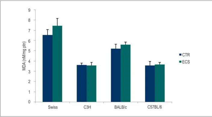
ECS does not change MDA content in the heart
Aiming at characterizing oxidative stress in our ECS model, we analyzed the oxidative damage by measuring TBARS (MDA). Regarding the MDA content in the different mouse strains exposed to cigarette smoke, no increase in MDA equivalent was observed in the ECS groups as compared with the CTR groups (Figure 1).
ECS reduces CAT and MPO activities in the heart
Regarding the activity of antioxidants in the different mouse strains exposed to cigarette smoke, CAT activity decreased in the Swiss (p < 0.05) and BALB/c (p < 0.05) groups as compared with their respective CTR groups, while MPO activity decreased in the C3H (p < 0.05) and C57BL/6 (p < 0.001) groups as compared with their respective CTR groups (Figure 2 A and B).
ECS reduces the GSH/GSSG ratio in the heart of Swiss and C57BL/6 mice
The GSH content decreased in the Swiss, C3H, C57BL/6 (p < 0.05) and BALB/c (p < 0.001) groups after ECS as compared with their respective CTR groups (Figure 3A). After ECS, an increase in the GSSG content was observed in the Swiss group (p < 0.05) and a decrease in the C3H (p < 0.05) and BALB/c (p < 0.001) groups as compared with their respective CTR groups (figure 3B). The GSH/GSSG
ratio decreased in the Swiss and C57BL/6 groups (p < 0.05) as compared with their respective CTR groups (Figure 3C).
Discussion
Cardiac remodeling results from geometric and volumetric cardiac changes in response to myocardial injury15. Oxidative
stress originates from the imbalance between oxidants and antioxidants, and the oxidative damage is caused by that imbalance that modifies cellular macromolecules, leading to cell death due to either apoptosis or necrosis16. An
experimental study has shown that chronic exposure to carbon monoxide increases the gene expression of endothelin-1 and induces cardiac hypertrophy17. Ventricular hypertrophy has
been recognized as an important agent in the remodeling process, as previous studies have confirmed that ECS is followed by left ventricular enlargement in rats18. Thirty days
of ECS are sufficient to impair ventricular function19. However,
ECS for four months causes ventricular dilation associated with systolic function reduction20.
Pressure overload causes oxidative stress and cardiovascular
system inflammation3. The oxidative damage in the
cardiovascular system is assessed by use of lipid peroxidation, protein oxidation, and damage to DNA21. For over 30 years,
TBARS, such as MDA, have been used as important markers of lipid peroxidation22. Although C57BL/6 and BALB/c mice
have been described as animals prone to lung oxidative damage after a long ECS12, oxidative parameters following
ECS observed in the present study showed a trend towards resistance to cardiac oxidative damage in those strains.
Figure 1 – Effect of the exposure to cigarette smoke on the concentration of malondialdehyde (MDA) in the heart of different mouse strains. MDA equivalent (nM/mg protein) was measured by use of the thiobarbituric acid reactive substances (TBARS) method. Results of the group exposed to cigarette smoke (ECS) as compared with the control (CTR) group by strain. The values are expressed as mean ± SEM and analyzed by use of one-way ANOVA followed by Bonferroni post-test (p < 0.05). *p < 0.05; **p < 0.01; and ***p < 0.001.
MDA
(nM/mg ptn)
CTR
Swiss
ECS
C3H
BALB/c
C57BL/6
9
8
7
6
5
4
3
2
1
Figure 2 – Effect of the exposure to cigarette smoke on the catalase (CAT) and myeloperoxidase (MPO) enzymatic activities in the heart of different mouse strains. (A) catalase activity (U/mg protein) and (B) myeloperoxidase activity (mU/mg protein) in the groups exposed to cigarette smoke (ECS) as compared with the control (CTR) group by strain. The values are expressed as mean ± SEM and analyzed by use of one-way ANOVA followed by Bonferroni post-test (p < 0.05). *p < 0.05; **p < 0.01; and ***p < 0.001.
Swiss C3H BALB/c C57BL/6
CA
T
(U/mg ptn)
7
6
5
4
3
2
1
0
CTR
ECS
CTR
ECS
MPO (mU/mg ptn)
0 0,5 1,5 2,5 3,5 4,5
1 2 3 4 5
Swiss C3H BALB/c C57BL/6
A
B
Figure 3 – Effect of the exposure to cigarette smoke on the content of reduced glutathione (GSH) and oxidized glutathione (GSSG) in the heart of different mouse strains. (A) GSH content (nM/mg protein), (B) GSSG content (nM/mg protein), and (C) GSH/GSSG ratio in the groups exposed to cigarette smoke (ECS) as compared with the control (CTR) group by strain. The values are expressed as mean ± SEM and analyzed by use of one-way ANOVA followed by Bonferroni post-test (p < 0.05). *p < 0.05; **p < 0.01; and ***p < 0.001.
Swiss
Swiss
Swiss C3H
C3H
C3H BALB/c
BALB/c
BALB/c C57BL/6
C57BL/6
C57BL/6
GSH (mM/mg ptn)
GSH/GSSG
GSSG (nM/mg ptn)
7
7 6
6 5
5 4
4 3
3 2
2 1
1 0
0
CTR
ECS
CTR
ECS
0 0,5 1,5 2,5
1 2
CTR
Approximately 80% of the studies have found a significant increase in MDA in smokers as compared with non-smokers; however, that result is not unanimous21-24. Investigations regarding ECS
dose-dependency and MDA content have shown a significant increase according to the daily dose of cigarettes. A study with 298 healthy individuals divided into non-smokers, smokers of less than 30 cigarettes/day, and smokers of more than 30 cigarettes/day has shown a strong relation between ECS and MDA plasma content (p < 0.0001)25. Another study with 130 healthy volunteers divided
into mild smokers (≤ 10 cigarettes/day), moderate smokers (11-20
cigarettes/day), and heavy smokers (> 20 cigarettes/day) has shown that MDA plasma content increased in all groups of smokers as compared with those of non-smokers (p < 0.05, p < 0.001 and p < 0.001, respectively)26.
Catalase is a cytoplasm hemoprotein that catalyzes the reduction of H2O2 to H2O and O2
27,being located in the
peroxisomes of cardiomyocytes28. In transgenic mice, in which
there is only catalase isolated from other antioxidants, the enzyme is constitutive and expressed in different amounts in the heart, and, when doxorubicin is administered, catalase activity increases 60 to 100 times, protecting the heart against toxicity29. Increased catalase activity causes resistance to
ischemia and reperfusion30. Our results showed that catalase
decreased in the ECS groups of the Swiss and BALB/c strains. BALB/c mice are resistant to cardiovascular diseases induced by an atherosclerotic diet; however, they are sensitive to immune and inflammatory diseases31.
Nicotine can activate polymorphonuclear cells, inducing neutrophils to produce IL-8. Polymorphonuclear cells significantly contribute to the beginning and progression of vascular inflammation in smokers, in which MPO contributes to the development and progression of cardiovascular diseases32. The
content of MPO, a neutrophil-derived enzyme that catalyzes the formation of innumerous ROS, is significantly high in smokers as compared with non-smokers33. Macrophages use NADPH
oxidase to produce O2
–, which, when undergoing dysmutation,
forms H2O2; thus, MPO catalyzes reactions with H2O2 to generate more potent cytotoxic oxidants, such as hypochlorous acid (HOCl) and the tyrosyl radical, being the only human enzyme that can generate HOCl34. Our results have shown a decrease
in MPO in the ECS groups of the C3H and C57BL/6 strains. C57BL/6 mice are susceptible to diet-induced lesions. When fed an atherogenic diet (1.25% of cholesterol, 0.5% of cholic acid and 15% of fat) for 14 weeks, they develop atherosclerotic lesions in the aorta35.
MPO is related to the activation of the protease cascade. The oxidative inactivation of protease inhibitors, such as α -1-antitripsin and tissue inhibitors of metalloproteinases (TIMPs), in association with the activation of proelastases and matrix metalloproteinases (MMPs) affect cardiac remodeling and the stability of atherosclerotic plaques. In a study of MPO null (MPO -/-) mice with acute myocardial infarction, the animals showed
decreased leukocyte infiltration, reduced ventricular dilation,
and preservation of the systolic function34. Aldehydes derived
from the oxidation of common amino acids, catalyzed by MPO, represent a rapid and relevant mechanism for the generation of cytotoxic species in inflammatory sites33. A study has reported
an increase in aldehydes in tissues after infarction in wild rats as compared with MPO-/- mice, clearly indicating the role of MPO
in the formation of those species36.
GSH detoxifies chemical agents and eliminates products of lipid peroxidation37-39.After GSH exposure to an oxidant, it is
oxidized to GSSG. In the inactivation of an oxidant, GSSG is produced and GSH depleted. GSH is recovered by the GSH-Rd enzyme, an essential step to maintain the integrity of the cell protection system40. We found a reduction in GSH in all strains of
the ECS groups. Our results suggest that GSH-Rd recovered GSH in the C3H and BALB/c strains of the ECS groups as compared with the CTR groups, because a reduction in GSSG was also seen in those groups. However, GSSG in the Swiss strains of the ECS groups remained increased. When the redox system is intact, GSH recovers. Our results showed that the GSH/GSSG ratio decreased in the Swiss and C57BL/6 strains of the ECS groups, indicating that both strains developed oxidative stress.
Conclusions
Our results show that oxidative stress should be considered in different populations with cardiovascular disease, and start a new discussion about the relationship between the genetic profile and redox imbalance in experimental models of ECS.
Author contributions
Conception and design of the research: Santos-Silva MA, Nagato AC, Bezerra FS; Acquisition of data: Santos-Silva MA, Nagato AC, Trajano ETL, Alves JN; Analysis and interpretation of the data: Santos-Silva MA, Nagato AC, Trajano ETL, Alves JN, Bandeira ACB, Bezerra FS; Statistical analysis: Bandeira ACB, Porto LC, Bezerra FS; Obtaining financing: Porto LC; Writing of the manuscript: Trajano ETL, Alves JN, Bandeira ACB, Porto LC, Bezerra FS; Critical revision of the manuscript for intellectual content: Porto LC, Bezerra FS.
Potential Conflict of Interest
No potential conflict of interest relevant to this article was reported.
Sources of Funding
There were no external funding sources for this study.
Study Association
1. Valenca SS, Bezerra FS, Romana-Souza B, Paiva RO, Costa AM, Porto LC. Supplementation with vitamins C and E improves mouse lung repair. J Nutr Biochem. 2008;19(9):604-11.
2. Valenca SS, Castro P, Pimenta WA, Lanzetti M, Silva SV, Barja-Fidalgo C, et al. Light cigarette smoke-induced emphysema and NFkappaB activation in mouse lung. Int J Exp Pathol. 2006;87(5):373-81.
3. Dimitrow PP, Undas A, Wolkow P, Tracz W, Dubiel JS. Enhanced oxidative stress in hypertrophic cardiomyopathy. Pharmacol Rep. 2009;61(3):491-5.
4. Levitzky YS, Guo CY, Rong J, Larson MG, Walter RE, Keaney JF Jr, et al. Relation of smoking status to a panel of inflammatory markers: the Framingham offspring. Atherosclerosis. 2008;201(1):217-24.
5. Hool LC. Reactive oxygen species in cardiac signalling: from mitochondria to plasma membrane ion channels. Clin Exp Pharmacol Physiol. 2006;33(1-2):146-51.
6. Gu L, Pandey V, Geenen DL, Chowdhury SA, Piano MR. Cigarette smoke-induced left ventricular remodelling is associated with activation of mitogen-activated protein kinases. Eur J Heart Fail. 2008;10(11):1057-64.
7. Charniot JC, Bonnefont-Rousselot D, Albertini JP, Dever S, Vignat N, Nataf P, et al. Oxidative stress implication after prolonged storage donor heart with blood versus crystalloid cardioplegia and reperfusion versus static storage. J Surg Res. 2010;160(2):308-14.
8. Panda K, Chattopadhyay R, Chattopadhyay D, Chatterjee IB. Cigarette smoke-induced protein oxidation and proteolysis is exclusively caused by its tar phase: prevention by vitamin C. Toxicol Lett. 2001;123(1):21-32.
9. Lanzetti M, Bezerra FS, Romana-Souza B, Brando-Lima AC, Koatz VL, Porto LC, et al. Mate tea reduced acute lung inflammation in mice exposed to cigarette smoke. Nutrition. 2008;24(4):375-81.
10. Cardoso JF, Mendes FA, Amadeu TP, Romana-Souza B, Valenca SS, Porto LC, et al. Ccn2/Ctgf overexpression induced by cigarette smoke during cutaneous wound healing is strain dependent. Toxicol Pathol. 2009;37(2):175-82.
11. Cardoso JF, Souza BR, Amadeu TP, Valenca SS, Porto LC, Costa AM. Effects of cigarette smoke in mice wound healing is strain dependent. Toxicol Pathol. 2007;35(7):890-6.
12. Rueff-Barroso CR, Trajano ET, Alves JN, Paiva RO, Lanzetti M, Pires KM, et al. Organ-related cigarette smoke-induced oxidative stress is strain-dependent. Med Sci Monit. 2010;16(7):BR218-26.
13. Draper HH, Squires EJ, Mahmoodi H, Wu J, Agarwal S, Hadley M. A comparative evaluation of thiobarbituric acid methods for the determination of malondialdehyde in biological materials. Free Radic Biol Med. 1993;15(4):353-63.
14. Aebi H. [Erythrocyte catalase]. Expos Annu Biochim Med. 1969;29:139-66.
15. Duarte DR, Minicucci MF, Azevedo PS, Matsubara BB, Matsubara LS, Novelli EL, et al. The role of oxidative stress and lipid peroxidation in ventricular remodeling induced by tobacco smoke exposure after myocardial infarction. Clinics (Sao Paulo). 2009;64(7):691-7.
16. Trachtenberg BH, Hare JM. Biomarkers of oxidative stress in heart failure. Heart Fail Clin. 2009;5(4):561-77.
17. Zornoff LA, Matsubara BB, Matsubara LS, Minicucci MF, Azevedo PS, Camapanha AO, et al. [Cigarette smoke exposure intensifies ventricular remodeling process following myocardial infarction]. Arq Bras Cardiol. 2006;86(4):276-82.
18. Castardeli E, Duarte DR, Minicucci MF, Azevedo PS, Matsubara BB, Matsubara LS, et al. Tobacco smoke-induced left ventricular remodelling is not associated with metalloproteinase-2 or -9 activation. Eur J Heart Fail. 2007;9(11):1081-5.
19. Paiva SA, Matsubara LS, Matsubara BB, Minicucci MF, Azevedo PS, Campana AO, et al. Retinoic acid supplementation attenuates ventricular remodeling after myocardial infarction in rats. J Nutr. 2005;135(10):2326-8.
20. Castardeli E, Duarte DR, Minicucci MF, Azevedo PS, Matsubara BB, Matsubara LS, et al. Exposure time and ventricular remodeling induced by tobacco smoke exposure in rats. Med Sci Monit. 2008;14(3):BR62-6.
21. Ozgen N, Guo J, Gertsberg Z, Danilo P, Rosen MR, Steinberg SF. Reactive oxygen species decrease cAMP response element binding protein expression in cardiomyocytes via a protein kinase D1-dependent mechanism that does not require Ser133 phosphorylation. Mol Pharmacol. 2009;76(4):896-902.
22. Polidori MC, Savino K, Alunni G, Freddio M, Senin U, Sies H, et al. Plasma lipophilic antioxidants and malondialdehyde in congestive heart failure patients: relationship to disease severity. Free Radic Biol Med. 2002;32(2):148-52.
23. Berr C, Coudray C, Bonithon-Kopp C, Roussel AM, Mainard F, Alperovitch A. Demographic and cardiovascular risk factors in relation to antioxidant status: the EVA Study. Int J Vitam Nutr Res. 1998;68(1):26-35.
24. Marangon K, Herbeth B, Lecomte E, Paul-Dauphin A, Grolier P, Chancerelle Y, et al. Diet, antioxidant status, and smoking habits in French men. Am J Clin Nutr. 1998;67(2):231-9.
25. Block G, Dietrich M, Norkus EP, Morrow JD, Hudes M, Caan B, et al. Factors associated with oxidative stress in human populations. Am J Epidemiol. 2002;156(3):274-85.
26. Solak ZA, Kabaroglu C, Cok G, Parildar Z, Bayindir U, Ozmen D, et al. Effect of different levels of cigarette smoking on lipid peroxidation, glutathione enzymes and paraoxonase 1 activity in healthy people. Clin Exp Med. 2005;5(3):99-105.
27. Zhou Z, Kang YJ. Cellular and subcellular localization of catalase in the heart of transgenic mice. J Histochem Cytochem. 2000;48(5):585-94.
28. Yokota S, Asayama K. Proliferation of myocardial peroxisomes in experimental rat diabetes: a biochemical and immunocytochemical study. Virchows Arch B Cell Pathol Incl Mol Pathol. 1992;63(1):43-9.
29. Kang YJ, Chen Y, Epstein PN. Suppression of doxorubicin cardiotoxicity by overexpression of catalase in the heart of transgenic mice. J Biol Chem. 1996;271(21):12610-6.
30. Li G, Chen Y, Saari JT, Kang YJ. Catalase-overexpressing transgenic mouse heart is resistant to ischemia-reperfusion injury. Am J Physiol. 1997;273(3 Pt 2):H1090-5.
31. Paigen B, Ishida BY, Verstuyft J, Winters RB, Albee D. Atherosclerosis susceptibility differences among progenitors of recombinant inbred strains of mice. Arteriosclerosis. 1990;10(2):316-23.
32. Iho S, Tanaka Y, Takauji R, Kobayashi C, Muramatsu I, Iwasaki H, et al. Nicotine induces human neutrophils to produce IL-8 through the generation of peroxynitrite and subsequent activation of NF-kappaB. J Leukoc Biol. 2003;74(5):942-51.
33. Roman RM, Wendland AE, Polanczyk CA. Myeloperoxidase and coronary arterial disease: from research to clinical practice. Arq Bras Cardiol. 2008;91(1):e11-9.
34. Askari AT, Brennan ML, Zhou X, Drinko J, Morehead A, Thomas JD, et al. Myeloperoxidase and plasminogen activator inhibitor 1 play a central role in ventricular remodeling after myocardial infarction. J Exp Med. 2003;197(5):615-24.
35. Ishida BY, Blanche PJ, Nichols AV, Yashar M, Paigen B. Effects of atherogenic diet consumption on lipoproteins in mouse strains C57BL/6 and C3H. J Lipid Res. 1991;32(4):559-68.
36. Vasilyev N, Williams T, Brennan ML, Unzek S, Zhou X, Heinecke JW, et al. Myeloperoxidase-generated oxidants modulate left ventricular remodeling but not infarct size after myocardial infarction. Circulation. 2005;112(18):2812-20.
37. Kadouri L, Kote-Jarai Z, Hubert A, Baras M, Abeliovich D, Hamburger T, et al. Glutathione-S-transferase M1, T1 and P1 polymorphisms, and breast cancer risk, in BRCA1/2 mutation carriers. Br J Cancer. 2008;98(12):2006-10.
38. Niemoeller OM, Foller M, Lang C, Huber SM, Lang F. Retinoic acid induced suicidal erythrocyte death. Cell Physiol Biochem. 2008;21(1-3):193-202.
39. Rainer M, Sonderegger H, Bakry R, Huck CW, Morandell S, Huber LA, et al. Analysis of protein phosphorylation by monolithic extraction columns based on poly(divinylbenzene) containing embedded titanium dioxide and zirconium dioxide nano-powders. Proteomics. 2008;8(21):4593-602.