Using computed tomography colonography in
patients at high risk of colorectal cancer – a
prospective study in a university hospital in South
America
Augusto Castelli von Atzingen,IDario Ariel Tiferes,IElizabeth Deak,II De´lcio Matos,II Giuseppe D9IppolitoI IFederal University of Sa˜o Paulo (UNIFESP), Department of Diagnostic Imaging, Division of Abdominal Imaging, Sa˜o Paulo/SP, Brazil.IIFederal University of
Sa˜o Paulo (UNIFESP), Department of Surgery, Section of Colon and Rectum Surgery, Division of Gastroenterology, Sa˜o Paulo/SP, Brazil.
OBJECTIVES: The purpose of our study was to report the results of the implementation of computed
tomography colonography in a university hospital setting serving a Brazilian population at high risk of colorectal cancer.
METHODS:After creating a computed tomography colonography service in our institution, 85 patients at high risk of colorectal cancer underwent computed tomography colonography followed by a same-day optical colonoscopy from September 2010 to May 2012. The overall accuracy of computed tomography colonography in the detection of lesions$6 mm was compared to that of optical colonoscopy (direct comparison). All colonic segments were evaluated using quality imaging (amount of liquid and solid residual feces and luminal distension). To assess patient acceptance and preference, a questionnaire was completed before and after the computed tomography colonography and optical colonoscopy. Fisher’s exact test was used to measure the correlations between colonic distension, discomfort during the exam, exam preference and interpretation confidence.
RESULTS: Thirteen carcinomas and twenty-two lesions$6 mm were characterized. The sensitivity, specificity and accuracy of computed tomography colonography were 100%, 98.2% and 98.6%, respectively. Computed tomography colonography was the preferred method of investigation for 85% of patients. The preparation was reported to cause only mild discomfort for 97.6% of patients. According to the questionnaires, there was no significant relationship between colonic distension and discomfort (p.0.05). Most patients (89%) achieved excellent bowel preparation. There was a statistically significant correlation between the confidence perceived in reading the computed tomography colonography and the quality of the preparation in each colonic segment (p#0.001). The average effective radiation dose per exam was 7.8 mSv.
CONCLUSION: It was possible to institute an efficient computed tomography colonography service at a university hospital that primarily assists patients from the public health system, with high accuracy, good acceptance and effective radiation doses. Our results seem to be comparable to other centers of excellence and fall within acceptable published guidelines, showing that a successful computed tomography colonography program can be reproduced in a South American population screened in a university hospital.
KEYWORDS: Colonography; Computerized Tomography; Colorectal Neoplasm; Colonic Polyps; Virtual
Colonoscopy.
von Atzingen AC, Tiferes DA, Deak E, Matos D, D9Ippolito G. Using computed tomography colonography in patients at high risk of colorectal cancer – a prospective study in a university hospital in South America. Clinics. 2014;69(11):723-730.
Received for publication onMay 4, 2014;First review completed onJune 3, 2014;Accepted for publication onJuly 28, 2014
E-mail: [email protected]
Tel.: 55 11 6842-3822/55 35 99848151
& INTRODUCTION
Colorectal cancer (CRC) is the third most common cancer, accounting for the second highest rate of cancer-related mortality in the Western world (1). Accurate pre-operative detection is crucial for planning curative treatment and increasing survival (2,3); however, the best means of diagnosis among the variety of resources and skills available at a given institution remains up for debate (4). A complete examination and colon screening can be achieved by Copyrightß2014CLINICS– This is an Open Access article distributed under
the terms of the Creative Commons Attribution Non-Commercial License (http:// creativecommons.org/licenses/by-nc/3.0/) which permits unrestricted non-commercial use, distribution, and reproduction in any medium, provided the original work is properly cited.
No potential conflict of interest was reported.
colonoscopy (the current test of choice in many institutions), direct rectal visualization with sigmoidoscopy plus barium enema, or CT colonography (CTC) (5). CTC has improved rapidly since its appearance in the mid-1990s and has been established as an alternative to optical colonoscopy (OC) for colorectal cancer screening, especially for the detection of colonic polyps (6). In addition, CTC has fewer complications than OC, including reduced perforation rates and it can be better tolerated (2,3,7). Still, most of the available data concerning the accuracy of CTC have been taken from the setting of a well-structured CT division with experience in performing CTC studies. Thus, it remains unclear whether the success of evaluating high-risk patients in centers of excellence can be generalized to other places, such as university hospitals of below-average-income countries, since the large studies have been limited to well-developed countries (3,7). Despite the many reports about the technical aspects of CTC, no results have been published on screening CTC in a high-risk South American population.
CTC was introduced in our country in 2003 and, until recently, was only available for patients in the private health system. Since the end of the last decade, several public and university hospitals have become equipped with CT equipment to perform CTCs. Moreover, the number of trained professionals who perform and read CTC exams in diagnostic imaging departments has increased broadly, including at our center. Our ability to offer this exam to our patients has extended this screening to a greater segment of the population that can benefit from a sensitive and well-tolerated approach. In addition, we have instituted a training program for our interns, nurses and technicians. The aim of this study was to show the feasibility of implementing a CTC program at a university hospital that has limited resources and is focused on providing care to patients in the public health system, while obtaining results comparable to those from international institutions (3,6,8-10).
& MATERIALS AND METHODS
Patients
Approval from the Institutional Review Board was obtained. All patients agreed to join the study and provided their signed consent. From September 2010 to May 2012, CTC followed by same-day OC were prospectively per-formed in 85 high-risk patients to screen for CRC. According to published guidelines, the criteria for high-risk colorectal cancer were the following: age over 40 years and
bright/dark red fecal bleed or unexplained anemia and
altered bowel habit/family history of CRC (1,6,11). Subjects were excluded from the study if they had inflammatory bowel disease, history of segmental colectomy or with colostomy, recent optical colonoscopy follow-up within the preceding two years, or any co-morbidity that would make OC unsuitable.
Bowel preparation
The preparation was composed of two days of a soft diet,
two tablets of bisacodyl (DulcolaxH), simethicone (LuftalH),
10 sachets of MuvinlaxH (macrogol, sodium bicarbonate,
sodium chloride, potassium chloride) diluted in 1.5 L of
water and 50 ml of ionic iodinated contrast (TelebrixH) the
day before the CTC.
CT colonography
A well-trained CT technician carefully insufflated the colon using a rectal tube and a manual air delivery system. To ensure sufficient colon distension, a CT digital radio-graph was acquired. Further insufflations were performed when the colonic distension was not sufficient in both positions (supine and prone). Images were acquired first from the supine and then from the prone position. When necessary, the lateral decubitus position was used to ensure sigmoid and left colon distension. CTC was performed in all
patients with a 64-row MDCT (Brilliance 64H, Philips
Medical Systems, Best, The Netherlands) without the use of intravenous contrast medium or peristaltic drugs. The imaging parameters for CTC were detector configuration,
6460.625 mm; pitch, 1.1; gantry rotation time, 0.5 s; slice
thickness, 1 mm; reconstruction increment, 0.7 mm;
120 kVp; 50 effective mAs; matrix, 5126512; and a fitting
field of view.
The effective radiation dose was obtained from the report produced by the device at the end of each exam. Values are
expressed asdose length product(DLP), which represents the
radiation dose of a CT section multiplied by the length of the study. To calculate the effective radiation dose, the DLP was multiplied by a correction factor depending on the anatomical region studied. In CT scans of the abdominal region, the correction factor ranges from 0.015 to 0.018 (12). In this study, we used a correction factor of 0.015 to estimate the effective dose of radiation. The result of this calculation is not an exact value of estimated radiation but can be used as a benchmark (12).
Optical colonoscopy and gold standard
On the same day, optical colonoscopy (OC) was performed after CTC within a 6-h interval, thereby avoiding the need for a second bowel purge. All patients received intravenous sedation (5 mg of midazolam hydrochloride). Optical colonoscopy examinations were performed by board-certified gastroenterologists. The location, shape and size of all identified polyps were described and the polyp sizes were measured using 8-mm-long biopsy forceps. Specimens for lesions larger than 5 mm were obtained by polypectomy, endoscopic mucosal resection, or biopsy. The endoscopists were unaware of the CTC findings.
Polypectomy was the gold standard for comparing CTC results, as assessed by segment. The colon was divided into six segments per patient (cecum, ascending colon, trans-verse colon, descending colon, sigmoid and rectum). To match the polyps seen on CTC and OC, two radiologists in consensus analyzed supine and prone CTC images and matched them with the optical colonoscopy findings based on colonic segment, size, shape and anatomical relationship to the haustral folds. For a given lesion to be recorded as a true positive, it had to be located in the same or an adjacent colonic segment; the two recorded sizes had to be the same within a 50% margin of error; and the lesion had to have similar morphology on both examinations. Polyps were categorized by size based on the optical colonoscopy results.
Interpretation of CTC
American College of Radiology’s (ACR) polyp classification (C1, smaller than 6 mm; C2, 6–9 mm; C3, 10 mm or larger). All examinations, even with poor preparation or poor distension were included in the analysis.
After CTC was performed, both radiologists evaluated the CTC datasets using the primary 3D method on a dedicated 3D workstation in addition to the 2D images. Electronic cleansing was not done for this interpretation. When a polyp was detected on CTC, the size, shape and location were recorded on a study report sheet. The shape of the polyp was described as flat, sessile, pediculed, ulcerated, or vegetating. Flat lesions were defined as mucosal elevations with a height less than 2 mm (13). Both the radiologists and the endoscopists used the same definition for a flat lesion.
Patient preference questionnaire
Patient acceptance of the CTC was evaluated by means of a semi-structured questionnaire applied before and after the CTC and CO that enabled us to evaluate the tolerance of the patients for the exam preparation, to assess any discomfort and to assess their preferences. The bowel preparation was defined as complete in cases of correct use of all medications by patients and as incomplete in those cases of incorrect use of all medications. Statistical correlations among the colonic distension, discomfort (subjectively informed by patients as ‘‘little’’, ‘‘moderate’’ or ‘‘great’’) expected and experienced, and preference for the type of exam (CTC or OC) were measured.
Quality of bowel preparation
The quality of the preparation was evaluated subjectively per segment using the following parameters: the degree of distension of colonic segments, the amount of residual marked feces, the amount of residual fluid and confidence in the diagnostics for each of the six segments of the large bowel. Colonic distension was rated as follows: a) non-distended segment b) proper distension and c) excellent distension (9,12). The amount of residual marked stool was
rated as follows: a) no fecal residue b) only residues,5 mm
c) residues with one to three particles $5 mm and d)
residues with more than three particles$5 mm (8,10). The
amount of residual fluid was rated as follows: a) absent b)
occupied,25% of the intestinal lumen c) occupied between
25% and 50% of the lumen and d) occupied .50% of the
intestinal lumen (8,10). Confidence in the diagnostic evaluation of each segment was rated as follows: a) not confident b) less confident and c) confident (10). In addition to the overall evaluation by radiologists, the tests were
considered excellent (characterizing all lesions .6 mm),
partially adequate (only characterizing lesions.10 mm) or
inappropriate (not characterizing lesions.10 mm).
The statistical analysis was performed in a descriptive manner. Fisher’s exact test was used for inferential analysis to study the associations between patient discomfort, colonic dilatation, patient preference and confidence inter-preting the exams. In all findings obtained through the
inferential analyses, the significance levelawas equal to 5%.
The per-lesion sensitivity, PPV, per-patient sensitivity, specificity, PPV and NPV were calculated with data pooled from all the radiologists. The sensitivities were compared
per lesion and per patient for polyps$6 mm, using the OC
as a reference standard for the 56 patients with complete OC exams. Data were entered into Excel 2010 for Windows spreadsheets for data storage. Statistical analyses were
performed with the Statistical Package for the Social
Sciences (SPSS) software release 19.0 for Windows. A p
-value of less than 0.05 indicated statistical significance.
& RESULTS
Eighty-five patients underwent CTC and were included in the study. The average age of the patients was 61 years, ranging from 31 to 88 years with a standard deviation of 13.0 years. Among the patients, 53 (62%) were female and 32 (38%) were male. Of the 85 patients studied, 78 (92%) underwent OC, of which 56 (72%) were complete and 22 (28%) were incomplete (due to CRC in 13/22 (59%) and conflicting colon issues in 4/22 (18%), among other causes).
CTC performance
Among the seventy-eight patients submitted to OC, 367 colonic segments were evaluated, including 336 segments in 56 patients with complete OC exams (six segments per patient) and 31 segments in 22 patients with incomplete CO exams. Among the fifty-six patients with complete OC, the
exams identified 22 lesions$6 mm in 19 patients (Tables 1
and 2). Considering all 336 of the colonic segments studied and measuring the per-lesion accuracy of the method in detecting polyps greater than or equal to 6 mm and using the complete OC as a gold standard, we achieved an accuracy of 98.6%, sensitivity of 100%, specificity of 98.2%, NPV of 100% and PPV of 94.7%.
Among the 13 patients with incomplete OC due to CRC,C, CTC the lesion was identified by CTC in all cases. The data contributed to a diagnosis of a synchronous tumor in the transverse colon with extra colonic metastasis in one patient
and of eight sessile polyps$6 mm that were non-visualized
after incomplete colonoscopy in four patients. The anato-mopathological analysis after a full colectomy in one of the patients identified three of the polyps as advanced adenomas with a villous component and as villous adenomas (Figures 1 and 2).
Patients’ acceptance and preference
Eighty-three patients (97.6%) developed no adverse events as a consequence of the bowel preparation and only two patients (2.4%) had mild reactions, which were characterized by vomiting in one patient and palpitations after the ingestion of iodinated media contrast in another patient. Seventy-five patients (88.2%) expected a ‘‘moder-ate’’ or ‘‘great’’ level of discomfort and ten patients (11.8%) expected ‘‘little’’ discomfort before the exam. On the other hand, after the CTC exam had been performed, 83 patients (97.6%) reported only mild discomfort and 63.5% of patients experienced less discomfort than originally expected. The
Table 1 -Correlation between the morphology and dimensions of lesions$6 mm identified in computed tomography colonography and complete colonoscopies.
Lesion morphology 6-9 mm [n = 5] $1 cm [n = 11] $3 cm [n = 6]
Sessile 4(80) 6(55)
-Pedunculated 1(20) 5(45)
-Flat Lesion - - 1(17)
Ulcerated - - 2(33)
Vegetating - - 3(50)
results showed that, for all segments, the colonic distension was not associated with the level of discomfort reported by
patients (p.0.05). When asked about their examination
preference, 85% of the 78 patients submitted to both exams preferred the CTC.
Bowel preparation and overall evaluation
Seventy-five patients (88.2%) underwent a complete preparation and ten (11.8%) an incomplete preparation due to inadequate intake of iodinated contrast in eight
patients and of MuvinlaxHin two patients. On average, for
each colonic segment, the preparation used achieved 94.1%
of the colonic segments with no fecal residue and 71.7% of
segments with,25% residual liquid; 71.8% of patients had
an excellent distension; and the confidence level in the overall diagnoses was rated as 86% (Table 3). The physicians’ confidence in their interpretation of CTC was strongly correlated with the amounts of residual fluid, fecal residue and colonic distension in the respective colonic
segments (p#0.001 for each). Therefore, a greater distension
combined with less residual fluid and fecal residue promoted greater confidence in the interpretation of the CTC exam. Regarding the overall evaluation by radiologists, 89% of patients had an excellent bowel preparation, 6.4% an inadequate preparation and 4.6% a partial preparation. The average effective radiation dose in the eighty-five CTC
examinations was 7.8 mSv (3.8-12.0 mSv; DP =¡2.0).
Table 2 -Anatomopathological analysis of lesions$6 mm identified in computed tomography colonography and optical colonoscopy.
Anatomopathological analysis 6-9 mm [n = 5] $1 cm-,3 cm [n = 11] $3 cm [n = 6]
Adenocarcinoma - 3(27) 3(50)
Low grade tubular adenoma 3(60) 1(9)
-Inflammatory 2(40) -
-Hyperplastic - 2(18)
-Low grade tubulovillous adenoma - 1(9)
-Villous adenoma - - 1(16)
High-grade tubulovillous adenoma - - 1(16)
High-grade villous adenoma - - 1(16)
Lipoma - 2(18)
-Leiomyoma - 1(9)
-Chronic Colitis - 1(9)
-Numbers in parentheses are percentages %.
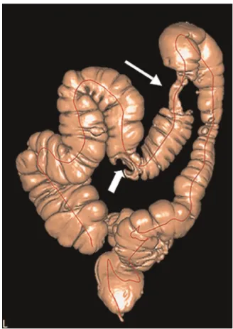
Figure 1 -CTC volume rendering showing a synchronous tumor of the transverse colon (large arrow). In this patient, the colonoscopy was unable to overcome the stenotic and infiltrat-ing lesion located in the splenic angle (thin arrow).
& DISCUSSION
In recent decades, CTC/virtual colonoscopy has emerged as an alternative to OC, not only for the screening of CRC (1-2) but also as an alternative test for investigating sympto-matic patients or those at high risk of developing CRC. Its speed, efficiency (usually lasting less than ten minutes), reproducibility, high level of accuracy, high level of toleration by patients, minimal invasiveness and cost-effectiveness all contribute to the superiority of CTC compared to no screening. For these reasons, the develop-ment of new CTC services for the investigation of sympto-matic and/or high-risk patients is highly desirable. The demand for CTC in a particular country or region is determined by several factors: a) a general demand for colonic investigation; b) OC waiting lists; c) CT scanner capacity; d) the availability of trained radiologists; and e) the unit cost per investigated patient (14,15). In our country, an analysis of these factors has indicated a demand for CTC propagation. In addition, the actual unit cost per CTC is substantially lower in some countries compared to the estimated costs used in previous cost-effectiveness analyses (15), supporting the adoption of this modality in CRC screening programs, even in the context of lower monetary resources. Furthermore, as many hospitals already have CT scanners that can perform CTC (multi-detector row scan-ners), the implementation of CTC requires little, if any, additional investment (16).
Although CTC has been widely accepted as a screening tool for colorectal cancer, even in the United States fewer than 20% of hospitals have adopted this service (16). The use of CTC programs at US hospitals has grown over the last 5 years, increasing from 13% in 2005 to 17% in 2008. The main factors motivating the adoption of CTC include a desire to provide an alternative screening option for elderly patients and for those with failed OC, as well as the often long wait times for OC and strong evidence supporting the use of CTC in the peer-reviewed literature (16). In Brazil, CTC scans are offered in only some private practices and scarce published data are currently available (17,18,19).
Several studies have suggested that most patients prefer CTC to OC or barium enema (5,6). These studies
demon-strate a preference for CTC in 72% of patientsversusOC and
in 97% of patientsversusbarium enema (5). Similar results
were observed in our study, which showed a preference for CTC in 85% of the patients. On the other hand, the prospects of sedation, unconsciousness, discomfort and not witnes-sing the exam were the reasons given by the six patients who preferred the OC.
CTC and OC can effectively identify the adenomatous polyps that are the precursors of CRC (6). However, these methods require the cleaning of the colon with cathartic agents to minimize or eliminate the negative effects of stool or fecal residues on the interpretation of the exam (8). Patients usually report an aversion to laxative preparations, which represents an obstacle for performing these exams
Table 3 -Quality percentages calculated in preparation and confidence in the diagnostic evaluation of each colonic segment.
COLONIC SEGMENT PARAMETER
Cecum Colonic distention Residual marked Residual fluid Confidence
a 9.4% 91.7% 7.1% 11.7%
b 16.5% 2.4% 70.5% 3.5%
c 74.1% 2.4% 16.5% 84.8%
d --- 3.5% 5.9%
---Ascending colon
a 2.4% 89.4% 12.9% 11.7%
b 25.9% 2.4% 68.2% 2.3%
c 71.7% 3.5% 16.5% 86%
d --- 4.7% 2.4%
---Transverse colon
a 1.2% 88.2% 11.8% 11.7%
b 30.6% 2.4% 71.8% 2.3%
c 68.2% 4.7% 12.9% 86%
d --- 4.7% 3.5%
---Descending colon
a 8.2% 88.2% 16.5% 10.5%
b 25.9% 5.9% 76.4% 3.5%
c 65.9% 3.5% 4.7% 86%
d --- 2.4% 2.4%
---Sigmoid colon
a 13% 91.7% 23.5% 10.5%
b 18.8% 2.4% 71.7% 3.5%
c 68.2% 3.5% 2.4% 86%
d --- 2.4% 2.4%
---Rectum
a 8.2% 94.1% 23.5% 10.5%
b 20.0% --- 71.7% 3.5%
c 71.8% 2.4% 2.4% 85.8%
d --- 3.5% 2.4%
---On average 71.8% 94.1% 71.7% 86%
Colonic distention: a) non-distended segment. b) proper distention. and c) excellent distention.Residual marked: a) no fecal residue. b) only residues
and contributes to the relatively low rate of colon cancer screening (9). The ideal cathartic agent for CTC has not been standardized, but phosphor-soda, magnesium citrate and polyethylene glycol (PEG), which was used in the current study, are among the most commonly used agents. PEG is an osmotically balanced wash solution that can be used safely in most patients, unlike phosphor-soda or magne-sium citrate, which are sometimes contraindicated (10). Our selected preparation with PEG was rated excellent in 89% of cases and was accepted by most patients. There was a statistically significant correlation between our level of confidence interpreting the CTC and the residual fluid, fecal residues, and distension in the respective colonic segments that reflected the efficiency of the PEG in the CTC exams performed. Inadequate (6.4%) or partial (4.6%) preparations were due to incorrect use of drugs. The preparation adopted in our study was quite safe, with only two patients experiencing self-controlled sided-effects, such as palpitations and mild vomiting, after ingesting iodinated contrast.
The benefits of CTC screening every 5 years from ages 50 to 80 far exceeded the risks inherent to the exposure to ionizing radiation (20). The American Council and ACR estimate an average recommended dose of radiation for CTC exams of approximately 7 mSv (21). In our study, the CTC exams were performed with a low-dose radiation protocol at a mean effective dose of 7.8 mSv, ranging between 3.8 mSv and 12 mSv, depending on the body types of the patients and the need to change positions to better characterize the entire colon.
That the overall accuracy for detecting polyps larger than 5 mm is above 90% in many studies (22,23) has been used to justify the selection of CTC as a first-option exam in CRC screening, as reinforced by PPVs exceeding 90% for lesions larger than 6 mm, even in high-risk patients (11,24). Our study produced similar results to those previously described, with a sensitivity and specificity above 95% for CTC after standard bowel preparation. This was likely due to the expertise of the readers, one of whom had interpreted more than 600 CTC examinations. An expert radiologist can be defined as a radiologist who has interpreted at least 500 CTC scans (25). In daily practice, greater experience with CTC is often associated with over-sensitivity of polyp detection.
The population included in our study is considered at high risk for the development of CRC. Colorectal cancer occurs in 90% of cases in subjects over 50 years of age. The mid-life risk of 5% (26) increases to 30% in high-risk patients (those with family or personal history, inflammatory disease, or polypoid or non-polypoid syndromes) (27). It is estimated that the
medium prevalence of$6 mm polyps is equal to 13-16% and
that the finding of an advanced neoplasia in polyp varies from 3.3-7.1% of patients (28). In our study group of high-risk patients, including fifty-six patients with complete OC, we
found$6 mm polyps in 19 patients (34%); of those patients,
ten (18% or 10/56) presented with advanced neoplasia (adenocarcinoma, high degree carcinoma and villous com-ponent), a slightly higher frequency than is typically seen in this group of patients. In fact, it is estimated that, among high-risk patients, 10% of polyps larger than 5 mm are advanced adenomas or carcinomas (29); 0.9% of the 6 to 9 mm polyps are adenocarcinomas; and 10% of the 10 to 20 mm polyps constitute CRC (30). The strategies used to approach high-risk patients are often similar to those for
medium-risk patients but should ideally be more careful in nature. In this context, the CTC plays an important role as a method of screening for CRC, generating benefits relevant to clinical, socio-economic and even emotional issues (31).
Another factor to consider when choosing a procedure is the possibility of an incomplete OC. An incomplete OC rate (i.e., the cecum is not reached) is considered acceptable in 10% of cases in daily clinical practice and in 5% of cases for CRC screening (25). However, an incomplete OC can occur in up to 25-30% of patients, especially when infiltrative and stenotic lesions are present. In these cases, it is important to complete further evaluations of the colon. Up to 6.3% of polyps and 4.3% of advanced lesions may be missed if a full assessment of the colon is not performed (32-33). In such cases, CTC has an indisputable role. In our study, for example, CTC proved useful for the pre-operative evalua-tion of a patient diagnosed with left colon carcinoma and incomplete OC, finding a synchronous tumor of the transverse colon that affected the subsequent planned surgery.
The present study has some limitations. Because this prospective clinical investigation was performed amid the daily practice of a general hospital, the sample was small and heterogeneous. The preparation quality was assessed subjectively and without any comparison to the other types of preparations available and described in the literature. The patients were mostly referred by gastroenterologists, increasing the likelihood of true positive findings by CTC. Additionally, the analysis of the images by consensus and the knowledge on the part of radiologists that patients would undergo OC on the same day as the CTC may have increased the sensitivity and specificity of their approach. Many may have accordingly increased their efforts to characterize all possible lesions. Inter-observer agreement was not calculated to determine the reproducibility of CTC in the detection of colonic lesions, which could have contributed to a more comprehensive evaluation of the method at hand, although such data have been previously reported (34).
annual hands-on coursework in the work setting to facilitate the better assimilation of the method, ensuring the permanent inclusion of CTC in the routine CT scans of our department.
However, in this project, we encountered a number of difficulties inherent to the construction of a new care process. Perhaps the greatest barriers were related to the inertia of the system and the lack of familiarity with the screening method on the part the majority of those involved. The persistent pro-active attitude and creativity of the group in overcoming these difficulties were crucial to the success of the project, although we still have not completed the number of exams originally planned. We are currently involved in a study seeking to establish the reasons for the limited number of exam requests and assessing the views of specialists (radiologists, clinicians and surgeons) regarding the value of CTC in our department. The feasibility of implementing the CTC program is undeniable in the setting of genuine interest and appropriate equipment availability. The low cost of the required medical devices (gloves, rectal probe and anesthetic gel) combined with the speed (approximately 10 min) and simplicity of the procedure certainly promote adherence to the CTC examination procedure. In addition, we interfered minimally in the department’s routine.
It is important to observe that the costs of the examination were minimized by using manual insufflation with ambient air (which was met with good acceptance by patients), thereby avoiding the expenses inherent to the use of
mechanical CO2insufflators. Further, by not using
endove-nous antispasmodics, we reduced the costs, made the exam minimally invasive and maintained similar acceptance results with those of other studies (5,8). For a CTC examination performed with iodinated contrast tagging and manual insufflation, the estimated costs (medical devices, drugs and contrast media), including laxative agents taken directly by the patients, were approximately US$ 14.0 per examination. This variable cost per CTC exam is very competitive when considering alternative investiga-tive modalities and the fact that other expensive equipment-investment costs, such as the CT scanner itself, can be minimized by the large quantity of CT exams performed for multiple indications, amounting in our practice to approxi-mately 12,000-15,000 exams per year, per piece of equip-ment. On the other hand, we did not evaluate costs associated with human resources (considering CTC reading and reporting time) or other variables, such as poor bowel cleansing, inadequate tagging, artifacts and poor colon distension. All of these factors may directly or indirectly increase the costs of the examination and should be investigated rigorously. In short, we can claim that the CTC exam is regularly available for those patients treated at our institution and candidates for the screening of CRC.
In conclusion, it was possible to include the CTC among the routine exams performed at a university hospital and to focus on the patients serviced in the public health system at a level of safety, acceptance and effectiveness similar to international standards. In most cases, patients preferred the CTC over the optical colonoscopy. Bowel preparation with PEG provides excellent results and is well accepted by patients. CTC is a fundamental tool in the evaluation of the colon in patients experiencing incomplete optical colono-scopy, especially when the presence of CRC prevents the progression of the device. These data could be beneficial to
public health policymakers and managers, as they consider both a variety of strategies for increasing CRC screening rates and the role that CTC should play in the investigation of high-risk and symptomatic patients.
& AUTHOR CONTRIBUTIONS
von Atzingen AC conceived and designed the study, was responsible for literature research, clinical examinations, experiments/data analysis, statistical analysis, preparation and editing of the manuscript. Tiferes DA conceived and designed the study, was responsible for clinical examina-tions, experiments/data analysis and statistical analysis. Deak E was responsible for clinical examinations. Matos D conceived and designed the study, was responsible for clinical examinations and data analysis. D9Ippolito G conceived and designed the study, was responsible for the manuscript preparation, clinical examinations, experiments/data analysis, manuscript editing and statistical analysis.
& REFERENCES
1. Cummings LC, Cooper GS. Colorectal cancer screening: update for 2011. Semin Oncol. 2011;38(4):483-9, http://dx.doi.org/10.1053/j.seminoncol. 2011.05.002.
2. von Wagner C, Smith S, Halligan S, Ghanouni A, Power E, Liford RJ, et al. Patient acceptability of CT colonography compared to double contrast barium enema: results from a multicentre randomised controlled trial of symptomatic patients. Eur Radiol. 2011;21(10):2046–55, http://dx.doi. org/10.1007/s00330-011-2154-y.
3. Pickhardt PJ, Choi JR, Hwang I, Butler JA, Puckett ML, Hildebrandt HA, et al. Computed tomographic virtual colonoscopy to screen for colorectal neoplasia in asymptomatic adults. N Engl J Med. 2003;349(23):2191-200. 4. Warren JL, Klabunde CN, Mariotto AB, Meekins A, Topor M, Brown ML, et al. Adverse events after outpatient colonoscopy in the Medicare population. Ann Intern Med. 2009;150(12):849–57, http://dx.doi.org/10. 7326/0003-4819-150-12-200906160-00008.
5. von Wagner C, Knight K, Halligan S, Atkin W, Lilford R, Morton D, et al. Patient experiences of colonoscopy, barium enema and CT colonogra-phy: a qualitative study. Br J Radiol. 2009;82(973):13-9.
6. Levin B, Lieberman DA, McFarland B, Smith RA, Brooks D, Andrews KS, et al. Screening and surveillance for the early detection of colorectal cancer and adenomatous polyps, 2008: a joint guideline from the American Cancer Society, the US Multi-Society Task Force on Colorectal Cancer, and the American College of Radiology. CA Cancer J Clin. 2008;58(3):130-60, http://dx.doi.org/10.3322/CA.2007. 0018.
7. Laghi A, Rengo M, Graser A, Iafrate F. Current status on performance of CT colonography and clinical indications. Eur J Radiol. 2013;82(8):1192-200.
8. Pickhardt PJ, Kim DH. Bowel preparation for CT colonography. In: Pickhardt PJ, Kim DH, editors. CT colonography: principles and practice of virtual colonoscopy. Philadelphia: Elsevier; 2010. p.115-30. 9. Zalis ME, Blake MA, Cai W, Hahn PF, Halpern EF, Kazam IG, et al.
Diagnostic accuracy of laxative-free computerized tomographic colono-graphy for detection of adenomatous polyps in asymptomatic adults: a prospective evaluation. Ann Intern Med. 2012;156(10):692-702, http:// dx.doi.org/10.7326/0003-4819-156-10-201205150-00005.
10. Hara AK, Kuo MD, Blevins M, Chen MH, Yee J, Dachman A, et al. National CT colonography trial (ACRIN 6664): comparison of three full-laxative bowel preparations in more than 2500 average-risk patients. AJR Am J Roentgenol. 2011;196(5):1076-82, http://dx.doi.org/10.2214/AJR. 10.4334.
11. White TJ, Avery GR, Kennan N, Syed AM, Hartley JE, Monson JRT. Virtual colonoscopyvsconventional colonoscopy in patients at high risk of colorectal cancer – a prospective trial of 150 patients. Colorectal Dis. 2009;11(2):138–45, http://dx.doi.org/10.1111/j.1463-1318.2008.01554.x. 12. Zalis ME, Barish MA, Choi JR, Dachman AH, Fenlon HM, Ferrucci JT
et al. CT colonography reporting and data system: a consensus proposal. Radiology. 2005;236(1):3-9, http://dx.doi.org/10.1148/radiol. 2361041926.
13. Birnbaum S. Radiation protection in the era of helical CT: practical patient based programs for decreasing patient exposure. Semin Ultrasound CT MR. 2010;31(1):46-52, http://dx.doi.org/10.1053/j.sult. 2009.09.006.
14. Hansmann A, Burling D. Essential requirements of a CT colonography service. Eur J Radiol. 2013;82(8):1187-91.
16. McHugh M, Osei-Anto A, Klabunde CN, Galen BA. Adoption of CT colonography by US hospitals. J Am CollRadiol. 2011;8(3):169-74, http:// dx.doi.org/10.1016/j.jacr.2010.08.008.
17. da Fonte AC, Chojniak R, de Oliveira Ferreira F, Pinto PN, dos Santos Neto PJ, Bitencourt AG. Inclusion of computed tomographic colono-graphy on pre-operative CT for patients with colorectal cancer. Eur J Radiol. 2012;81(3):e298-303.
18. Maia MVAS, von Atzingen AC, Tiferes DA, Sarhan SS, Deak E, Matos D, et al. Patient preferences toward colon cancer screening: a comparison between computerized tomography colonography and conventional colonoscopy. Radiol Bras. 2012;45(1):24-8, http://dx.doi.org/10.1590/ S0100-39842012000100007.
19. von Atzingen AC, Tiferes DA, Matsumoto CA, Nunes TF, Maia MVAS, D9Ippolito G. Common findings and pseudolesions at computerized tomography colonography: pictorial essay. Radiol Bras. 2012;45(3):160-6, http://dx.doi.org/10.1590/S0100-39842012000300008.
20. Berrington de Gonzalez A, Kim KP, Knudsen AB, Lansdorp-Vogelaar I, Rutter CM, Smith-Bindman R, et al. Radiation-related cancer risks from CT colonography screening: a risk-benefit analysis. AJR Am J Roentgenol. 2011;196(4):816-23, http://dx.doi.org/10.2214/AJR.10.4907.
21. American College of Radiology. ACR Practice Guideline for the Performance of Computerized Tomography (CT) Colonography in Adults. Philadelphia: ACR; 2009.
22. Pickhardt PJ, Wise SM, Kim DH. Positive predictive value for polyps detected at screening CT colonography. Eur Radiol. 2010;20(7):1651-6, http://dx.doi.org/10.1007/s00330-009-1704-z.
23. Kim DH, Pooler BD, Weiss JM, Pickhardt PJ. Five year colorectal cancer outcomes in a large negative CT colonography screening cohort. Eur Radiol. 2012;22(7):1488-94, http://dx.doi.org/10.1007/s00330-011-2365-2.
24. Zueco Zueco C, Sobrido Sampedro C, Corroto JD, Rodriguez Fernandez P, Fontanillo Fontanillo M. CT colonography without cathartic prepara-tion: positive predictive value and patient experience in clinical practice. Eur Radiol. 2012;22(6):1195-204, http://dx.doi.org/10.1007/s00330-011-2367-0.
25. Zalis ME, Barish MA, Choi JR, Dachman AH, Fenlon HM, Ferrucci JT, et al. Working Group on Virtual Colonoscopy. CT colonography reporting and data system: a consensus proposal. Radiology. 2005;236(1):3-9.
26. Jemal A, Siegel R, Ward E, Murray T, Xu JQ, Thun MJ. Cancer statistics, 2007. CA Cancer J Clin. 2007;57(1):43-66, http://dx.doi.org/10.3322/ canjclin.57.1.43.
27. Winawer SJ, Fletcher RH, Miller L, Godlee F, Stolar MH, Mulrow CD, et al. Colorectal cancer screening: clinical guidelines and rationale. Gastroenterology.1997;112(2):594-642, http://dx.doi.org/10.1053/gast. 1997.v112.agast970594.
28. Lieberman D, Moravec M, Holub J, Michaels L, Eisen G. Polyp size and advanced histology in patients undergoing colonoscopy screening: implications for CT colonography. Gastroenterology. 2008;135(4):1100-5, http://dx.doi.org/10.1053/j.gastro.2008.06.083.
29. Butterly LF, Chase MP, Pohl H, Fiarman GS. Prevalence of clinically important histology in small adenomas. Clin Gastrentrol Hepatol. 2006;4(3):343-8, http://dx.doi.org/10.1016/j.cgh.2005.12.021.
30. Matek W, Guggenmoos-Holzmann I, Demling L. Follow-up of patients with colorectal adenomas. Endoscopy. 1985;17(5):175-81, http://dx.doi. org/10.1055/s-2007-1018494.
31. Gallo TM, Galatola G, Laudi C, Regge D. CT colonography: screening in individuals at high risk for colorectal cancer. Abdom Imaging. 2006;31(3):297-301, http://dx.doi.org/10.1007/s00261-005-0368-7. 32. Neerincx M, Terhaar sive Droste JS, Mulder CJ, Rakers M, Bartelsman JF,
Loffeld RJ, et al. Colonic work-up after incomplete colonoscopy: significant new findings during follow-up. Endoscopy. 2010;42(9):730-5, http://dx.doi.org/10.1055/s-0030-1255523.
33. Neri E, Giusti P, Battolla L, Vagli P, Boraschi P, Lencioni R, et al. Colorectal cancer: role of CT colonography in preoperative evaluation after incomplete colonoscopy. Radiology. 2002;223(3):615-9, http://dx. doi.org/10.1148/radiol.2233010928.