208
1. Professor of Dentistry, University Salgado Filho. Master in Dentistry (Dental Clinic), Federal University Fluminense (UFF). Niterói, RJ, Brazil.
2. Assistant Professor, University Estácio de Sá, Juiz de Fora (MG) and Master in ENT, Federal University of Rio de Ja-neiro (UFRJ). Rio de JaJa-neiro, RJ, Brazil.
3. Specialist in Stomatology, University of the State of Rio de Janeiro (UERJ), Rio de Janeiro (RJ) and Master Student in Dentistry (Dental Clinic), Federal University Fluminense (UFF). NIterói, RJ, Brazil.
4. Master in Dentistry (Dental Clinic), Federal University Fluminense (UFF). Niterói, RJ, Brazil.
5. Assistant Professor, School of Dentistry, Federal Univer-sity Fluminense (UFF), Nova Friburgo (RJ) and Doctor in Dentistry (Pediatric Dentistry), Federal University of Rio de Janeiro (UFRJ). Rio de Janeiro, RJ, Brazil.
6. Professor, School of Dentistry, Federal University Flumi-nense (UFF). Niterói, RJ, Brazil.
Correspondence to: Roberta Barcelos, M.D.
Rua Sílvio Henrique Braune, 22 – Centro 28625-650 Nova Friburgo, RJ.
E-mail: [email protected]
Impact of supportive therapy for otological changes in patients with
temporomandibular joint disorders*
Impacto da terapia de suporte nas alterações otológicas em pacientes com desordem temporomandibular
Luciana Uemoto
1, Miguel Eduardo Guimarães Macedo
2, Thays Almeida Alfaya
3, Fernanda Nunes
de Souza
4, Roberta Barcelos
5, Cresus Vinícius Depes Gouvêa
6* Received from the Department of Dentistry, Specialization Course in Temporomandibular Disorders and Orofa-cial Pain, School of Dentistry, Federal University of Juiz de Fora. Juiz de Fora, MG.
SUMMARY
BACKGROUND AND OBJECTIVES: Otological
symptoms complaints may be associated to temporo-mandibular joint disorders (TMD). Occlusal splint is an alternative to treat TMD because it provides ideal oc-clusion to patients, acting on painful symptoms. Con-sidering the conservative and reversible characteristics of supportive therapy with occlusal splints, this study aimed at evaluating its impact on the frequency of oto-logical changes in TMD patients.
METHOD: An intervention study was carried out with 35 patients with TMD-associated otological symptoms. TMD patients were considered those with at least three signs and three symptoms of morbidity, being one of them earache, tinnitus, sensation of hypoacusis, ear
fullness or dizziness. Patients were submitted to clini-cal evaluation, orientations and fabrication of maxil-lary occlusal splint in thermoplastic acrylic resin. Splint adjustments and symptoms evaluation were performed fortnightly. Fischer Exact test was used for statistical analysis of the prevalence of signs and symptoms before
(BE) and after (AF) therapy with signiicance level of
5% (p < 0.05).
RESULTS: Nineteen patients have inished the treat -ment. There has been decrease in the prevalence of oto-logical signs and symptoms: tinnitus (BE = 14; AF = 6; p = 0.009), earache (BE = 13; AF = 4; p = 0.003), ear fullness (BE =12; AF = 4; p = 0.006), dizziness (BE = 11; AF = 4; p = 0.009) and hypoacusis (BE = 10; AF = 2; p = 0.001).
CONCLUSION: Occlusal splint is a conservative and reversible therapy able to improve TMD-related otologi-cal symptoms.
Keywords: Ear diseases, Temporomandibular joint
disorders, Therapy.
RESUMO
JUSTIFICATIVA E OBJETIVOS: As queixas de
sin-tomas otológicos podem estar associadas a quadros de disfunção temporomandibular (DTM). A placa mior-relaxante é uma alternativa de tratamento para a DTM, pois oferece oclusão ideal para o paciente, atuando nos sintomas de dor. Considerando as características conser-vadoras e reversíveis da terapia de suporte com placa miorrelaxante, o objetivo deste estudo foi avaliar seu impacto na frequência de alterações otológicas em pa-cientes com DTM.
MÉTODO: Realizou-se estudo de intervenção em 35
apresentassem pelo menos três sinais e três sintomas da morbidade, sendo um deles otalgia, zumbido, sen-sação de hipoacusia, sensen-sação de plenitude auricular ou tontura. Os pacientes foram submetidos a exame clínico, orientações e confecção de placa miorrelaxante maxilar
em resina acrílica termoplastiicável. Ajustes da placa
e avaliação dos sintomas foram realizados quinzenal-mente. Utilizou-se o teste Exato de Fisher para análise estatística da diferença entre a prevalência de sinais e sintomas antes (AN) e após (AP) a terapia, com nível de
signiicância de 5% (p < 0,05).
RESULTADOS: Dezenove pacientes concluíram o
tratamento. Observou-se redução na prevalência de si-nais e sintomas otológicos: zumbido (AN = 14; AP = 6; p = 0,009), otalgia (AN = 13; AP = 4; p = 0,003), sensação de plenitude auricular (AN = 12; AP = 4; p = 0,006), tontura (AN = 11; AP = 4; p = 0,009) e de hipoacusia (AN = 10; AP = 2; p = 0,001).
CONCLUSÃO: A placa miorrelaxante é uma terapia
conservadora e reversível que mostrou ser capaz de mel-horar os sintomas otológicos associados à DTM. Descritores: Otopatias, Terapêutica, Transtornos da ar-ticulação temporomandibular.
INTRODUCTION
Temporomandibular disorder (TMD) includes a group of stomatognathic system abnormalities which cause generally chronic and non progressive pain which im-pacts quality of life (QL). It involves symptoms in tem-poromandibular joint (TMJ), masticatory muscles and associated structures such as ear, with presence of tin-nitus and earache1. Tinnitus has a prevalence of 22% in TMD patients2.
Earache may be TMJ pain perceived more posteriorly, since just a thin part of the temporal bone separates TMJ from external auditory canal and middle ear. In addition, TMJ and part of auditory structures originate
in the irst branchial arch, more precisely in Meckel’s
cartilage and some ear and masticatory system struc-tures have trigeminal with common innervation, thus justifying pain in this region3. So, anatomical proximity together with similar phylogenic heritage may
contrib-ute for the dificulty patients have to locate pain3. An-other manifestation secondary to TMD pain are trigger-points (TP), especially in sternocleidomastoid, masseter and medial pterygoid muscles which, when stimulated, cause local and distant pain4. The formation of algog-enous points in these muscles may cause irradiation to the ear generating tinnitus4.
Occlusal splint is an alternative treatment for TMD. It
has fundamentally an orthopedic function and is alterna-tive to rapidly delete the memory of traumatic occlusion, temporarily changing nociception caused by dental con-tact. Although its action mechanism is not totally clear, the splint supplies ideal occlusion for patients, since
condyles are in a more stable musculoskeletal position5.
Systematic reviews conirm its action on pain6 and on dental wear control in cases of bruxism9.
This study aimed at evaluating the impact of support-ive therapy with occlusal splint in the frequency of otological changes in TMD patients.
METHOD
This was an intervention study in patients with otologi-cal symptoms associated to TMD.
To identify eligible patients, cards of patients who voluntarily attended the Temporomandibular Disorder and Orofacial Pain of a public Dentistry School were screened. Fifty patients were pre-selected and after applying inclusion and exclusion criteria the sample totaled 35 patients.
Inclusion criteria were: (a) adult patients, (b) both gen-ders, (c) without systemic involvement, (d) with TMD and at least one otological symptom (earache, tinnitus, sensation of hypoacusis, sensation of ear fullness or diz-ziness) as primary complaint. TMD patients were consid-ered those with at least three signs and three symptoms of the morbidity, such as TMJ pain, headache, muscle pain, jaw movement limitation, abnormal static and dynamic occlusion, joint noises, muscle fatigue, pain when
chew-ing, open mandibular lockchew-ing, dificulty to chew, dental
wear or mouth opening limitation, and one of them should be earache, tinnitus, dizziness sensation of hypoacusis or of ear fullness. Exclusion criteria were: (a) patients with TMD whose primary complaint was not an otological symptom and (b) presence of systemic involvement. All individuals agreed in participating in the study by signing the Free and Informed Consent Term (FICT).
During evaluation and dental treatment, patients were submitted to history and physical evaluation, orientation about jaw position at rest and neuromuscular relaxation with a maxillary occlusal splint, made of thermoplastic acrylic resin.
Patients were oriented to use the occlusal splint for a minimum period of two months and maximum of six
months, as follows: 24 hours per day in the irst three weeks, 16 hours in the next three weeks, 12 hours for three more weeks and then for 8 hours during sleep
until discharge.
car-bon paper (AccuFilm® (Parkell Inc., Edgewood, New York, USA). Symptoms were evaluated fortnightly using the pain scale (0-3) with the following categories: “0” no pain or discomfort, “1” discomfort, “2” pain and “3” severe pain. Total follow up time was six months. Discharged patients were oriented to interrupt the use of the splint at the mo-ment they would start the second part of the treatmo-ment, consisting in orthodontics, rehabilitation with dental prosthesis, physical therapy or global postural reeduca-tion (RPG). Those not improving were oriented to con-tinue the treatment, which consisted in maintaining ther-apy and/or use alternatives such as physical therther-apy, la-ser therapy or needling. All alternatives were performed
twice a week. Physical therapy used stretching, relaxa -tion and massage techniques; laser therapy was made up of punctual laser applications (Three Light®, Clean Line, São Paulo, Brazil) at TMJ region using 80 mW power, 795 nm wavelength and dose of 4 J/cm2. Dry needling was indicated for patients with PG. The technique was
performed twice a week with short needle (Unoject® Nova DFL, Rio de Janeiro, Brazil) inserted at a depth of 1 to 2 centimeters in acute 30o angle between the
nee-dle and the skin, in different directions, with movements
to inside the tissue. Data were analyzed by the SPSS 17 program (IBM Corp., Chicago, IL, USA), using Fischer Exact test to analyze differences between the prevalence of signs and symptoms before (BE) and after (AF) the
therapy, with signiicance level of 5% (p < 0.05).
This study was approved by the Research Ethics Com-mittee, University Salgado de Oliveira, under opinion 15/2004.
RESULTS
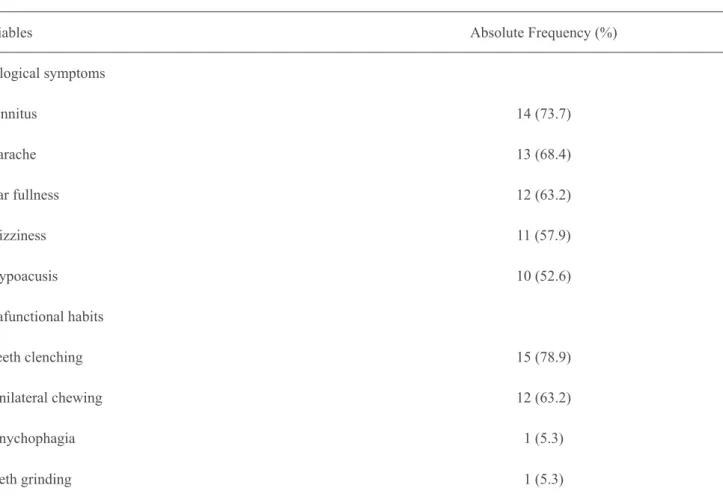
From 35 selected patients, 19 have inished the treat -ment. There has been predominance of females (94.7%) and of the fourth decade of life (36.8%).
With regard to symptoms, among those related to the ear, tinnitus was the most prevalent, predominating unilater-ally to the right, while for parafunctional habits, clench-ing was the most frequent (Table 1).
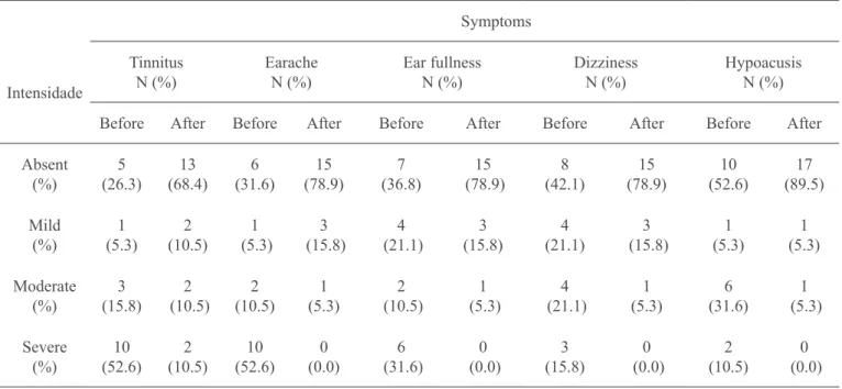
All symptoms were slightly improved (Table 2). Except for tinnitus, all patients reporting pretreatment symptoms as severe had resolution or at least decrease in intensity. In addition, the frequency of patients with no symptoms has increased for all evaluated symptoms.
Table 1 – Clinic characteristics of the sample (n = 19).
Variables Absolute Frequency (%)
Otological symptoms
Tinnitus 14 (73.7)
Earache 13 (68.4)
Ear fullness 12 (63.2)
Dizziness 11 (57.9)
Hypoacusis 10 (52.6)
Parafunctional habits
Teeth clenching 15 (78.9)
Unilateral chewing 12 (63.2)
Onychophagia 1 (5.3)
DISCUSSION
Otological symptoms may be associated to TMD. In our study, tinnitus was the most prevalent otological symp-tom, followed by earache, and in line with previous study10. The relationship between these symptoms and TMD is not totally explained by the literature and there
are controversies as to audiological indings in these pa -tients10. This may be explained by anatomical proximity. So, as from some deep and constant pain site – muscular, visceral, neural or vascular – secondary effects may be present in the ear. Earache in TMD patients may be re-ferred pain, the origin of which may be in some hyperac-tive muscle or in TMJ itself. Myospasm of tensor mus-cle of tympanic membrane may be a secondary effect as response to TMD pain11, and may pull the hammer and the tympanic membrane, changing sound conduction through the middle ear, which would justify the com-plaint of subjective hypoacusis and also of earache12.
Another important inding was related to the presence of parafunctional habits which was also signiicant in
the sample. Masticatory muscles hyperactivity may
lead to relex hyperactivity of tensor muscle of tym -panic membrane, justifying the tinnitus reported by patients. Conversely, muscle relaxation obtained with occlusal splints has improved such symptom13. This
justiies our results because 78.9% of patients report -ed clenching and 73.7% tinnitus; after treatment, only 31.5% have maintained the symptom.
In these cases, the occlusal splint promotes a temporary change in proprioception caused by dental contact and decreases motor activity related to parafunctional hab-its5, justifying the improvement of otological symptoms. Clinical studies and systematic reviews also emphasize its effectiveness for TMD-related pain6,8,14. Al-Ani et al.7
state that in spite of the lack of enough scientiic evidenc -es about the effectiven-ess of occlusal splints for myo-fascial pain, this therapy relieves pain intensity both at rest and at palpation, and improves depression of patients with myofascial pain as compared to untreated patients. Although there are no studies with strong evidences of the effectiveness of occlusal splints to treat otological changes, one may state that this therapy deserves con-sideration for being noninvasive and reversible, that is, there is change in occlusal conditions and dimensions to a more physiological and functional situation with no need for more invasive and irreversible procedures, such as orthodontic treatment or surgery. The splint promotes changes in the occlusal relationship of TMJ and chewing forces, in addition to improving joint and muscle func-tions of the stomatognathic system. Other possible ther-apies would be occlusal adjustment, surgeries and ortho-dontic treatment, but all are invasive and irreversible.
Sample size and lack of a control group may be consid -ered a limiting factor for our study. However,
consider-ing the impact of TMD in QL of patients, our indconsider-ings
support the hypothesis that this less invasive and revers-ible therapy is an effective treatment for this condition, Table 2 – Distribution of otological symptoms by intensity. before and after supportive therapy with myorelaxant splint (n = 19).
Symptoms Intensidade Tinnitus N (%) Earache N (%) Ear fullness N (%) Dizziness N (%) Hypoacusis N (%)
Before After Before After Before After Before After Before After
promoting improvement in patients’ well being. So, we
stress the need for further studies to complement our
indings and to conirm the evidences that supportive
therapy is the treatment of choice for similar cases. Another factor deserving consideration is related to pain evaluation, which is subjective and highly variable
among individuals, representing a dificulty for studies
on the subject. In spite of this variation in pain percep-tion among individuals, we have used a pain scale as the tool for patients to report their level of pain, thus
provid-ing a scale for patients’ discomfort intensity.
The variety of TMD manifestations is, in general, asso-ciated to the number of structures which are part of the stomatognathic system15, and may be reported by patients or be found during physical evaluation. Some symptoms
may also be found in ENT disorders making dificult the task of inding their etiology. So, the interaction between dentists and ENT specialists is needed, taking into con
-sideration early diagnosis and patients’ well being.
CONCLUSION
Occlusal splints were able to improve otological
symp-toms associated to TMD. This treatment should be taken
into consideration since it is a conservative and revers-ible therapy.
REFERENCES
1. Sharma S, Gupta DS, Pal US, et al. Etiological factors of temporomandibular joint disorders. Natl J Maxillofac Surg. 2011;2(2):116-9.
2. Vielsmeier V, Strutz J, Kleinjung T, et al. Temporo-mandibular joint disorder complaints in tinnitus: fur-ther hints for a putative tinnitus subtype. PLoS One. 2012;7(6):e38887.
3. Ash CM, Pinto OF. The TMJ and the middle ear: structural and functional correlates for aural symptoms associated with temporomandibular joint dysfunction. Int J Prosthodont. 1991;4(1):51-7.
4. Bezerra Rocha CA, Sanchez TG, Tesseroli de Siqueira JT. Myofascial trigger point: a possible way of modulat-ing tinnitus. Audiol Neurootol. 2008;13(3):153-60.
5. Almilhatti HJ, Camparis CM, Bönecker G, et al. Como
aumentar o índice de sucesso no tratamento com placas oclusais miorrelaxantes. JBA J Bras Oclusão ATM Dor Orofac. 2002;2(8):340-3.
6. Turp JC, Komine F, Hugger A. Eficacy of stabiliza -tion splints for the management of patients with mastica-tory muscle pain: a qualitative systematic review. Clin Oral Investig. 2004;8(4):179-95.
7. Al-Ani Z, Gray RJ, Davies SJ, et al. Stabilization splint therapy for the treatment of temporomandibu-lar myofascial pain: a systematic review. J Dent Educ. 2005;69(11):1242-50.
8. Forssell H, Kalso E, Koskela P, et al. Occlusal treat -ments in temporomandibular disorders: a qualitative systematic review of randomized controlled trials. Pain. 1999;83(3):549-60.
9. Macedo CR, Silva AB, Machado MA, et al. Occlus-al splints for treating sleep bruxism (tooth grinding). Cochrane Database Syst Rev. 2007;(4):CD005514. 10. Felício CM, Faria TG, Silva MAMR, et al. Desordem Temporomandibular: relações entre sintomas otológicos e orofaciais. Rev Bras Otorrinolaringol. 2004;70(6):786-93. 11. Sicher H. Temporomandibular articulation in man-dibular overclosure. J Am Dent Assoc. 1948;36(2):131-9. 12. Saueressig NS, Kayser FG, Oliveira FL. Disfunções temporomandibulares e sua relação com o zumbido au-ditivo e dorsalgia: relato de caso clínico. JBA J Bras Oclusão ATM Dor Orofac. 2003;3(9):21-5.
13. Felício CM, Mazzetto MO, Bataglion C, et al. De-sordem Temporomandibular: Análise da freqüência e severidade dos sinais e sintomas antes e após a placa de oclusão. J Bras Ortodon Ortop Facial. 2003;8(43):48-57. 14. Sima FT, Gil C. Estudo comparativo do grau de severidade das desordens craniomandibulares em pa-cientes edentados parciais antes e após a utilização de placas interoclusais. Rev Pos Grad. 2005;12(2):179-85. 15. Bretan O, Nogueira EA. Distúrbios temporomandib-ulares e alterações da musculatura mastigatória. Arq Int Otorrinolaringol. 2005;9(2):318.
Submitted in February 02, 2012.
Accepted for publication in August 29, 2012.