(1) Universidade Federal de Pernambuco (UFPE), Recife, PE - Brazil.
Conlict of interest: Nonexistent
Temporomandibular joint dysfunction in
Parkinson’s Disease: an integrative
literature review
Taysa Vannoska de Almeida Silva(1)
Maria das Graças Wanderley de Sales Coriolano(1)
Carla Cabral dos Santos Accioly Lins(1)
Received on: December 14, 2016 Accepted on: August 23, 2017 Mailing address:
Taysa Vannoska de Almeida Silva
ABSTRACT
Temporomandibular joint dysfunction is a set of disorders involving the masticatory muscles, temporomandibular joint and associated structures. It is known that the pro -gression of motor symptoms in Parkinson’s disease is an indication that these peo -ple are more prone to the development of this dysfunction. Thus, this study aims to investigate the signs and symptoms of temporomandibular dysfunction in people with Parkinson’s disease. The search was performed in the databases: MEDLINE/ PubMed, LILACs, CINAHL, SCOPUS, Web of Science and PEDro, without timing or language restriction. Speciic descriptors were used for each database and keywords, evaluated by the instruments: Critical Appraisal Skill Program and Agency for Health care and Research and Quality. A total of 4,209 articles were found but only 5 were included. After critical analysis of the methodology of the articles, one did not reach the mini -mum score required by the evaluation instruments, thus, it was excluded. The selected articles addressed, as signs and symptoms of temporomandibular joint dysfunction, the following: myofascial pain, bruxism, limitation of mouth opening, dislocation of the articular disc and asymmetry in the distribution of occlusal contacts. Further stu -dies are needed in order to determine the relationship between cause and effect of the analyzed variables, so as to contribute to more speciic and effective therapeutic interventions.
Keywords: Parkinson’s Disease; Temporomandibular Joint Disorders;
INTRODUCTION
Parkinson’s disease (PD) is considered one of the most prevalent neurodegenerative disorders in the elderly, affecting 0.3% of the general population, affecting more men than women1. It is a progressive
degenerative clinical syndrome of the central nervous system that causes movement disorders due to
dopamine deiciency in the nigrostriatal pathway of
the brain2,3. The diagnosis is based on clinical features
through motor symptoms that include: bradykinesia, muscle stiffness, postural instability and resting tremors that extend down the neck and face. Non-motor symptoms such as olfactory, gustatory, sleep distur-bance and cognition dysfunction are also present2.
Motor symptoms are related to the development of postural alterations, such as: cervical spine
lexion, thoracic hyperkinesis, scoliosis, abduction of the shoulders and lexion of the arms 4. It has been
demonstrated that changes in neck posture can lead to changes in the biomechanics of the temporoman-dibular joint, affecting the function of the stomato-gnathic system and leading to the development of temporomandibular joint dysfunction (TMD)5,6. A recent
study showed that the prevalence of TMD in the elderly population with PD is 23.08% reaching women more frequently7.
TMD is deined as a set of joint and muscular
disorders of multifactorial and dynamic etiology8. The
main clinical alterations are: headache, neck pain, temporomandibular joint pain, muscle pain, muscle fatigue, limitation of opening of the mouth, opening deviation and joint noises9.
Because it is a multifactorial pathology, there are different instruments for its evaluation, organized in anamnestic10 and clinical11 and diagnostic criteria12.
Among the instruments used for assessment of the condition, the Diagnostic Criteria for TMD Research
(RDC / TMD) has shown the most veriied psychometric
properties and accuracy, characterizing itself as one of the most appropriate instruments for the evaluation of TMD12.
The progression of motor symptoms in PD, such as
deiciency in axial control and mandibular movements,
indicates that these individuals are more likely to the development of this dysfunction2,13,14. In addition,
these functional alterations contribute to generation of pain and poor perception of oral health14,15. Despite
the importance of the subject, there are not published
studies that gather scientiic evidence on the subject
yet. Therefore the purpose of this review is to gather studies that address the signs and symptoms of temporomandibular joint dysfunction in people with PD and its relation with oral health quality.
METHODS
It is an integrative review of the literature, which allows the search, critical evaluation and synthesis of the available evidence on a delimited theme or guiding question based on evidence, which contributes to the practice in health care. The study was performed according to the following steps: elaboration of guiding question, literature research, data collection, critical analysis of included studies, discussion of the results and presentation of the integrative review16.
The guiding question of the present study was:
What published scientiic evidence addresses the signs
and symptoms of temporomandibular joint dysfunction in Parkinson’s disease?
A bibliographic survey was carried out in the databases LILACS (Latin American and Caribbean Literature in Health Sciences), PEDro (Database on Evidence in Physiotherapy), MEDLINE / PubMed (US National Library of Medicine), Scopus Cinahl (Cumulative Index toNursing & Allied Health Literature) and Web of Science. For the research of articles were
used speciic descriptors for each database and
keywords (Figure 1).
The inclusion criteria for obtaining and selecting articles by means of searches performed between June and June 2017 were: (1) Articles that addressed the signs and symptoms of temporomandibular dysfunction in people with Parkinson’s disease; (2) No restriction of language and year of publication; (3) Complete articles published in periodicals. The following were excluded: (1) Repeated studies in databases; (2) Monographs; (3) Review articles; (4) Publications not available in full or whose results have not yet been published.
For a better critical analysis of the methodology of the included articles, two instruments were applied that allowed the evaluation of the different study designs: 1- Critical Appraisal Skill Programme (CASP) (adapted)17,18
The original CASP17 contemplates eight speciic
evaluation tools for different study designs such as reviews, cohorts, clinical trials, cross-sectional studies etc. In this review, an instrument adapted from CASP18 was used, which included 10 items to be scored, including: 1) objective; 2) adequacy of the method; 3) presentation of theoretical and methodological procedures; 4) sample selection criteria; 5) sample detailing; 6) relationship between researchers and respondents (randomization / blinding); 7) respect for ethical aspects; 8) accuracy in data analysis; 9) property to discuss results and 10) research contribu-tions and limitacontribu-tions. For item 8, the appropriateness of data analysis, such as intention-to-treat analysis, was considered methodological analysis rigor. At the end,
studies were classiied in level A (score between 6 and
10 points), being considered as a good methodological quality and reduced bias, or level B (up to 5 points) meaning satisfactory methodological quality, but with risk of considerable bias18.
The AHRQ ranks studies at six levels according to the level of evidence: (1) systematic review or meta-analysis; (2) randomized controlled trials; (3) non-randomized clinical trials; (4) cohort and case-control studies; (5) systematic review of descriptive and qualitative studies and (6) only descriptive or qualitative study20.
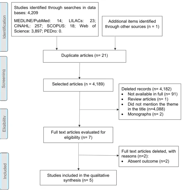
The search by crossing the descriptors and keywords in the pre-established databases resulted in
the identiication and selection of four thousand two
hundred and ten articles. After initial screening, the duplicated articles were excluded and the remaining articles were submitted to a title review for inclusion and exclusion criteria, seven articles were considered eligible. However, of those initially considered eligible,
two published papers did not it in the criteria estab
-lished in this review. After the inal evaluation only ive
articles were eligible for qualitative analysis, as shown in Figure 2.
Data base Search strategy
MEDLINE/PubMed
"temporomandibular joint disorders"[MeSH] OR "temporomandibular joint disorders"[All Fields] OR ("temporomandibular"[All Fields] AND "joint"[All Fields] AND "disorder"[All Fields]) OR "temporomandibular disorder"[All Fields])) AND ("parkinson disease"[MeSH Terms] OR ("parkinson"[All Fields] AND "disease"[All Fields]) OR "parkinson disease"[All Fields] OR ("parkinson's"[All Fields] AND "disease"[All Fields]) OR "parkinson's disease"[All Fields])
PEDro “Temporomandibular joint disorders” AND “Parkinson’s disease” “Temporomandibular disorder” AND “Parkinson’s disease”
LILACS
(tw:(Transtornos da articulação temporomandibular)) OR (tw:(Disfunção temporomandibular)) AND (tw:(Doença de Parkinson))
(tw:(temporomandibular joint disorder)) OR (tw:(temporomandibular disorder)) AND (tw:(Parkinson's Disease))
Scopus ((temporomandibular joint disorder) OR (temporomandibular disorder) AND (parkinson's disease))
Cinahl “Temporomandibular joint disorder” OR “temporomandibular disorders” AND “Parkinson's disease” Web of Science “Temporomandibular joint disorder” OR “Temporomandibular disorder AND “Parkinson's disease”
means satisfactory methodological quality, but with a considerable risk of bias18, this study was taken from
the articles that will be discussed.
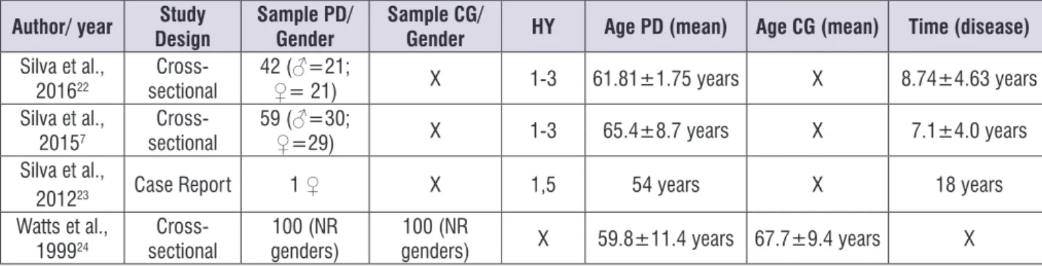
The four studies included a total of 302 people of both sexes, with clinical diagnosis of Parkinson’s disease and similar mean age (between 50 and 70 years). Only three of the analyzed articles showed the time of diagnosis (ranging from 2 to 18 years) and
the classiication of signs and symptoms of disease
severity (mild to moderate) according to the Hoenhn & Yahr scale, as shown in Figure 3.
When analyzing the selected publications one of them scored three according to the instrument, CASP18. This is a case report21 carried out at Okayama
University, Japan, this study showed the usage of an apparatus for pain management in temporomandibular joint disorder, which is a complication of Parkinson’s disease. The article also mentioned in the body of the text: the objective explains the results and presents their contribution to clinical practice. Because it does
not it the score of the instrument used by the research, which deines ive points as a minimum score, which
Scre
en
in
g
In
cl
ud
ed
El
ig
ib
ilit
y
Id
en
tif
ica
tio
n
Duplicate articles (n= 21)
Selected articles (n = 4,189)
Full text articles evaluated for eligibility (n= 7)
Full text articles deleted, with reasons (n=2):
Absent outcome (n=2)
Studies included in the qualitative synthesis (n= 5)
Deleted records (n= 4,182)
Not available in full (n= 91)
Review articles (n= 1)
Did not mention the theme in the title (n=4,088)
Monographs (n= 2) Studies identified through searches in data
bases: 4,209
MEDLINE/PubMed: 14; LILACs: 23; CINAHL: 257; SCOPUS: 18; Web of Science: 3,897; PEDro: 0.
Additional items identified through other sources (n = 1)
criteria for sample selection, randomization / blinding and accuracy in data analysis ; (Iii) Another analyzed study24 reached score 8 because it did not explain
the selection criteria of the sample neither the ethical aspects of the research. The main information of the articles of this review is presented in Figure 4.
After a full reading and analyzing the methodological quality of the studies involved: (i) Two articles7,22
reached score nine, because the researchers did not perform randomization/blinding; (Ii) The article of 201223 is a case report, it score seven points, because
some of the aspects evaluated by the instrument were not suitable for the type of study, such as: to present
Author/ year Study
Design
Sample PD/ Gender
Sample CG/
Gender HY Age PD (mean) Age CG (mean) Time (disease)
Silva et al.,
201622 sectionalCross- 42 (♀= 21)♂=21; X 1-3 61.81±1.75 years X 8.74±4.63 years Silva et al.,
20157 sectionalCross- 59 (♀=29)♂=30; X 1-3 65.4±8.7 years X 7.1±4.0 years Silva et al.,
201223 Case Report 1 ♀ X 1,5 54 years X 18 years Watts et al.,
199924
Cross-sectional genders)100 (NR genders)100 (NR X 59.8±11.4 years 67.7±9.4 years X
PD: Parkinson’s disease; CG: Control Group; HY: Hoehn & Yahr Parkinson disease escalation scale; Gender: ♂= male and ♀= female; NR: Not reported.
Figure 3. General characteristics of the articles included in the review
Author/
Year Goals Evaluations Tools Analyzed Variables Results
Silva et al.,
201622
To investigate the prevalence of TMD in people with PD and to analyze the distribution
of occlusal contacts.
RDC/TMD – assesses the
signs and symptoms of
TMD12;
T-Scan III Portable Occlusal Analysis System.
- Signs and symptoms of TMD;
- occlusal asymmetry
- Prevalence of 23.8% TMD signals in patients with PD;
- The TMD affects more women; - People with PD showed high frequency of
oclusal asymmetry.
Silva et al.,
20157
To analyze the impact of TMD on oral health in people with PD
according to the severity of the disease.
FIM – degree of functional
independence29;
RDC/TMD – assesses the
signs and symptoms of TMD;
OHIP-14 – assesses the
impact of oral health27.
- Functional independence Degree; - Signs and symptoms of
TMD; - Oral health.
- There was a signiicant difference between the groups with and without TMD domain
with respect to psychological disability evaluated by OHIP-14.
Silva et
al.,201223
Trace the presence of signs and symptoms indicative of TMD in a
person with PD.
RDC/TMD - assesses the
signs and symptoms of TMD. - Signs and symptoms of TMD;
- TMD, displacing the disk and limiting the amplitude of mouth opening had a negative impact ADL’s related stomatognathic system.
Watts et al.,
199924
To characterize the relationship between bruxism and
Parkinson’s disease.
Questionnaire structured by the authors.
- Bruxism - DTM
- involuntary jaw movements / mouth
- People with PD show 4.2 times higher risk (RR = 1.42 ; 95% CI 2.3 = 0.88) of
developing bruxism;
- 25% less risk (RR = 0.75 ; 95% CI = 0.17 3.26) having TMD;
- Involuntary movements of the jaw / mouth are speciic to the DP.
PD= Parkinson’s disease; TMD = temporomandibular dysfunction; FIM = Functional Independence Measure; RDC / TMD = Diagnostic Criteria for Research of Temporomandibular Disorders; OHIP-14 = Oral Health Impact Proile
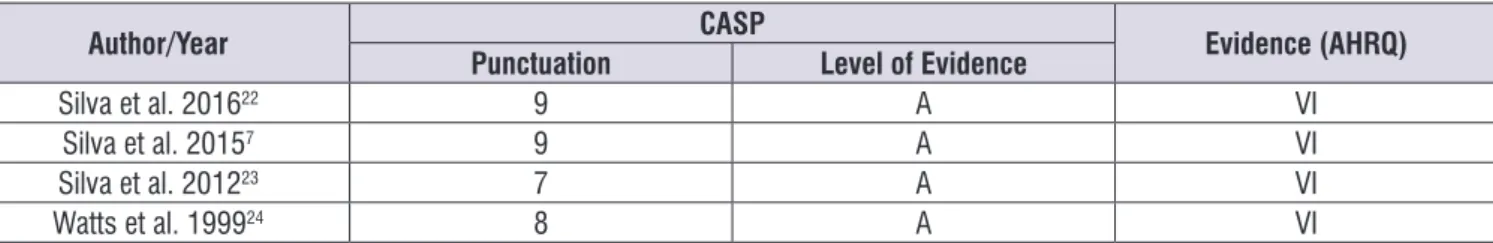
Regarding the evaluation by using the AHRQ20 all
the articles obtained six points fot the level of evidence because they are cross-sectional observational studies and case report. Both the description of the
methodology used as well as the levels of evidence
of the studies, classiied by the adapted CASP18 and
AHRQ20 are represented in Figure 5.
Author/Year CASP Evidence (AHRQ)
Punctuation Level of Evidence
Silva et al. 201622 9 A VI
Silva et al. 20157 9 A VI
Silva et al. 201223 7 A VI
Watts et al. 199924 8 A VI
CASP: Critical Appraisal Skill Programme17; AHRQ: Agency for Healthcare and Research and Quality20
Figure 5. Description of evidence levels of articles according to adapted CASP18 and AHRQ20
LITERATURE REVIEW
In the present review, four articles were found7,22-24,
those aimed to investigate the presence of signs and symptoms of temporomandibular dysfunction in people with Parkinson’s disease (PD) and its relationship with oral health7.
Parafunctional activities can be diurnal or nocturnal (occurring during sleep). The diurnal ones include the clenching of teeth, lip biting, chewing or other objects, digital sucking, improper posture habits, as well as other habits that the individual performs, most of the times, unconsciously. Nocturnal parafunctional habit is very common and is called bruxism. This term consists of a rhythmic attrition of the teeth in non-masticatory movements of the mandible, especially during sleep25.
In the study conducted by Watts et al.24 one hundred
people with Parkinson’s disease were compared to 100 healthy people, using a questionnaire structured by the authors that evaluated the signs and symptoms of TMD. The results indicated that people with Parkinson’s disease present a 4,2 fold greater risk (RR = 1.42, 95% CI = 0.88, 2.3) for developing bruxism, this result corroborates the research carried out by Alencar et al.26 who observed a correlation between bruxism and
reduction in dopamine levels. As well as the group with Parkinson’s disease presented a 25% lower risk (RR = 0.75, 95% CI = 0.17 3.26) for having TMD, this data, however, diverged evidences that show that the progression of motor symptoms in PD: cervical spine
lexion, thoracic hyperkinesis, scoliosis, abduction
these characteristics are an indication that these people are more prone to the development of this dysfunction2,13,14.
In the case report23 presented in this review, a
woman with PD was diagnosed with TMD because of the dislocation of the disc of the temporomandibular joint and limitation of the opening amplitude. This condition has a clinical importance, since studies have shown that these aspects are one of the main factors involved in the development of TMD.
Silva et al. 7,22,23 used as instrument of assessment
for TMD, gold standard; the questionnaire Diagnostic Criteria for Temporomandibular Disorders Research (RDC/TMD)12 . The study aimed to evaluate the
presence of signs and symptoms of this disorder. In the study published in 20157, the authors described that there was no signiicant association between TMD and
the severity of PD, and that the impact of oral health conditions on social aspects was weak. In the studied sample, the most observed characteristic of TMD was joint disc displacement with reduction. In addition, a negative correlation was observed between the severity of PD symptoms and the impact on oral health (r = -0.167, p = 0.207). However, comparing the impact of oral health between the groups with and without TMD,
signiicant differences were observed in the sub-items
of the OHIP-1427 scale: functional limitation”,
“psycho-logical discomfort”, “physical incapacity” and “ with
TMD. A similar inding could be observed in the case
report23, in which the authors reported that the clinical
Bakke et al.14, stated that the oral functions of
individuals with PD are more compromised according to the severity of the motor symptoms, and this could
inluence the occurrence of TMD in this population, however, no signiicant associations were found
between the impairment motor and TMD diagnosis in a study of 20157. The authors explained this result
because the sample was composed of individuals in the PD stage, and propose new studies evaluating other factors such as changes in posture and muscle tone to better evaluate the correlation.
Silva et al. 20157 observed in their study the impact
of oral problems on the quality of life; and the question-naire of choice by the researchers was OHIP-1427,28. In the study, the authors found no signiicant correlation
between motor impairment and impact on oral health. This result is in disagreement with the data described by Bakke et al.14 who evaluated the impact of oral
health on subjects in the moderate to severe stages of PD. The authors explain this divergence by the fact that their sample is made up of individuals in less advanced stages of PD, are semi-dependent individuals with a good perception of their overall health and have no impact on the performance of daily living activities according to the high score of FIM29.
A comparison performed in the same study7 of
the impact of oral health among individuals with and without TMD, higher OHIP-14 scores were found among those with TMD, despite the poor impact indicated by the different sub-scales. The researchers
explain this inding by the fact that only two individuals were classiied with myofascial pain, which is reported
in the literature as the main factor with the greatest negative impact on oral health. The difference was
signiicant in the sub-item Psychological Inability. It has
been demonstrated that all diagnoses resulting from
RDC / TMD have a signiicant impact on oral health.
The authors7 still address the fact that most
evalu-ations and interventions performed in people with PD observe only motor and cognitive aspects which might lead them to ignore symptoms of equal
impor-tance to their health and quality of life, inluencing the
measurement of symptoms DTM and the perception of oral health, since those aspects are based on self-report.
Among the goals of the articles7,22 is the investigation
of the prevalence of temporomandibular dysfunction in
therefore the prevalence used will be the highest value, 23.8% for a sample of 42 individuals.
Silva et al. (2016)22 highlighted the relationship
between TMD and dental occlusion using the T-Scan III version 8.0 occlusal analysis system to determine the occlusal contact points. They observed that a large part of the total sample exhibited asymmetry in the distri-bution of contacts occlusal. Such asymmetry occurred in people with and without TMD, and according to the authors, this may have occurred due to the fact that individuals with PD have altered muscle and postural
tonus that inluence muscle activation and as a conse -quence of the position of the mandible22. Because
constant contractions of the facial and neck muscles
relect on the number of occlusal contacts predomi -nantly on the right side30,31. However, no signiicant
difference was found in the distribution of right-to-left occlusal contacts in relation to the left between the groups with and without TMD (p = 0.883).
In the literature this correlation has divergent opinions32-34. In some researches, the authors says
that this may be partially due to the different evaluation instruments used. Carbon paper has traditionally been used to determine occlusal contact points, however other studies have shown that this material can lead
to false-positive results due to the inluence of factors
such as salivation and occlusal morphologyl32. And that
the analysis with the T-Scan system has shown satis-factory reliability, since its readings are more precise and less subject to external interferences, besides the ability to analyze the symmetry on both sides33,34.
Previous studies have shown that changes in occlusal contacts may lead to functional disharmony between the masticatory muscles and the temporo-mandibular joint, resulting in excessive intra-articular pressure, a set of micro and macro traumas and subse-quent joint degeneration, characteristic of TMD35.
In the scientiic literature, there are a number of
reasons for the lack of consensus about changes in dental occlusion being an etiological factor for TMD, one of the possible reasons is that the studies use different methods of analysis36. In the described study22 there was no signiicant association between TMD and
occlusal contact asymmetry (p=0.466). Silva et al. 22 explain this inding, by showing that the asymmetry
individuals with and without TMD was found, it was observed that this condition is more frequent among people with dysfunction37.
Despite the characteristics present in Parkinson’s disease, they raise the hypothesis about a higher presence of TMD in this population. The case report23
was useful to stimulate new research and to present the importance of knowing new affections with the intention of designing therapeutic strategies claiming the mainte-nance of quality of life38. Two analyzed articles7,22, highlighted the scarcity in scientiic production that
addresses the presence of TMD and its correlation with oral health. In addition, three of the cited articles7,22,24
presented limitation in the research, because they are a cross-sectional study that only allows the creation of associations and does not allow conclusions on causality. Therefore, new studies that address other factors associated with temporomandibular joint dysfunction, such as pain in the stomatognathic system and articular noise, should be performed. In addition longitudinal studies that determine the cause and effect relationships of the analyzed variables.
CONCLUSION
The analyzed articles in this literature review indicated the main signs and symptoms of TMD in people with PD: myofascial pain, bruxism, limitation of the opening of the mouth, displacement of the articular disc with reduction and asymmetry in the distribution of occlusal contacts.
Regarding the impact on oral health, the studies shown a negative correlation between the moderate and severe stages of the disease and that
psycho-logical impairment inluences on the activities of daily
life of those people.
Such information may help guiding future studies that aims to investigate other factors associated with temporomandibular joint dysfunction, since there is not enough data regarding this topic. Consequently, it would be possible to provide valuable information for the application of therapeutic and prevention strategies for this population.
REFERENCES
1. Wirdefeldt K, Adami H, Cole P, Trichopoulos D, Mandel J. Epidemiology and etiology of Parkinson’s
2. Reichmann H. Clinical Criteria for the Diagnosis of Parkinson’s Disease. Neurodegener Dis. 2010;7(5):284-90.
3. Jankovic J. Parkinson’s disease: clinical features and diagnosis. J Neurol Neurosurg Psychiatry. 2008;79(4):368-76.
4. Doherty KM, Warrenburg BP van, Peralta MC, Silveira-Moriayama L. Postural deformities in Parkinson’s disease. Lancet Neurol. 2011;10(6):538-49.
5. Pereira LJ, Gavião MBD, Bonjardim LR, Castelo PM, Bilt van der A. Muscle thickness, bite force, and cranio-facial dimensions in adolescents with signs and symptoms of temporomandibular dysfunction. Eur J Orthod. 2007;29(1):72-8.
6. Olmos SR, Kritz-Silverteins D, Halligan W, Silverstein ST. The effect of condyle fossa relationships on head posture. Cranio. 2005;23(1):48-52.
7. Silva PF da C, Biasotto-Gonzalez DA, Motta LJ, Silva SM, Ferrari RAM, Fernandes KPS et al. Impact in oral health and the prevalence of temporomandibular disorder in individuals with Parkinson’s disease. J Phys Ther Sci. 2015;27(3):887-91.
8. Ingawalé S, Goswami T. Temporomandibular joint: disorders, treatments, and biomechanics. Ann Biomed Eng. 2009;37(5):976-96.
9. Torres F, Campos L, Fillipini H. Efeitos dos
tratamentos isioterapêuticos e odontológicos em
pacientes com disfunção temporomandibular. Rev Fisioter Mov. 2012;25(1):117-25.
10. Fonseca DM, Bonfate G, Valle AL, Freitas SFT.
Diagnóstico pela anamnese da disfunção
craniomandibular. Rev Gauch Odontol. 1994;42(1):23-8.
11. Pehling J, Schiffman E, Look J, Shaefer J, Fricton J. Interexaminer reliability and clinical validity of the temporomandibular index: a new outcome measure for temporomandibular disorders. J Orofac Pain. 2002;16(4):296-304.
12. Dworkin S, LeResche L. Research diagnostic criteria for temporomandibular disorders: review,
criteria, examinations and speciications, critique.
Craniomandib Disord. 1992;6(4):301-55.
with Parkinson’s disease. Eur J Oral Sci. 2011;119(1):27-32.
15. Dworkin SF, Huggins KH, LeResche L, Koff M Von, Howard J, Truelove E. Epidemiology of signs and symptoms in temporomandibular disorders: clinical signs in cases and controls. J Am Dent Assoc. 1990;120(3):273-81.
16. Mendes KDS, Silveira RC de CP, Galvão CM. Revisão integrativa: Método de pesquisa
para a incorporação de evidências na saúde
e na enfermagem. Texto Context Enferm. 2008;17(4):758-64.
17. Critical Appraisal Skills Programme. CASP checklist. Oxford: CASP. 2014. [cited 2017 Jun. 06]. Available from: http://www.casp- net/#!casp-tools-checklists/ c1.
18. Alencar DL, Marques AP de O, Leal MCC, Vieira J de CM. Fatores que interferem na sexualidade de
idosos: uma revisão integrativa. Ciência Saúde
Coletiva. 2014;19(8):3533-42.
19. Gondim ITG de O, Lins CC dos SA, Coriolano
MGWS. Exercícios terapêuticos domiciliares na
doença de Parkinson: uma revisão integrativa. Rev Bras Geriatr e Gerontol [serial on the Internet]. 2016 Apr [cited 2017 Jun. 06];19(2):349-64. Available from: http://www.scielo.br/scielo.php?script=sci_ arttext&pid=S1809-98232016000200349&lng=pt& nrm=iso&tlng=en
20. Stillwell SB, Fineout-Overholt E, Melnyk BM, Williamson K. Evidence-Based Practice: Step by Step. Am J Nurs. 2010;110(1):51-3.
21. Minagi S, Matsunaga T, Shibata T, Sato T. An appliance for management of TMJ pain as a complication of Parkinson’s disease. Cranio. 1998;16(1):57-9.
22. Silva PF da C, Motta LJ, Silva SM, Ferrari RAM, Fernandes KPS, Bussadori SK. Computerized analysis of the distribution of occlusal contacts in individuals with Parkinson’s disease and temporomandibular disorder. Cranio® [serial on the Internet]. 2016 Aug [cited 2017 Jun. 06] 34(6):358-62. Available from: https://www. tandfonline.com/doi/full/10.1080/08869634.2015.10 97315
23. Silva P, Silva S, Ferrari R, Fernandes K, Correa F, Bussadori S. Disfunção temporomandibular em
24. Watts MW, Tan EK, Jankovic J. Bruxism and cranial-cervical dystonia: is there a relationship? Cranio. 1999;17(3):196-201.
25. Cauás M, Alves IF, Tenório K, Filho JBHC, Guerra
CMF. Incidências de hábitos parafuncionais e
posturais em pacientes portadores de disfunção da articulação craniomandibular. Rev Cirur e Traumat Buco-Maxilo-Facial. 2004;4(2):121-9.
26. Alencar M, Martins B, Vieira B. A relação do bruxismo com a dopamina. J Can Dent Assoc. 2006;71(1):62-6.
27. Slade GD. Derivation and validation of a short-form
oral health impact proile. Community Dent Oral
Epidemiol. 1997;25(4):284-90.
28. Dahlström L, Carlsson G. Temporomandibular disorders and oral healthrelated quality of life. A systematic review. Acta Odontol Scand. 2010;68(2):80-5.
29. Linacre J, Heinemann A, Wright D. The structure and stability of the functional independence measure. Arch Phys Med Rehabil. 1994;75(2):127-32.
30. Haralur SB. Digital evaluation of functional occlusion parameters and their association with temporomandibular disorders. J Clin Diagn Res. 2013;7(8):1772-5.
31. Learreta JA, Beas J, Durst A. Muscular activity in relation to intentional occlusal interferences. Cranio. 2007;25(3):193-9.
32. Dworkin S, LeResche L. Research diagnostic criteria for temporomandibular disorders: review,
criteria, examinations and speciications, critique. J
Craniomandib Disord. 1992;6(4):301-16.
33. Koos B, Godt A, Shille C, Göz G. Precision of an instrumentation- based method of analyzing occlusion and its resulting distribution of forces in the dental arch. J Orofac Orthop. 2010;71(6):403-10.
34. Kerstein R. Articulating paper mark misconceptions and computerized occlusal analysis technology. Dent Implant Updat. 2008;19(6):41-6.
35. Iwasaki LR, Crosby MJ, Gonzalez Y, Mccall WD, Marx DB. Temporomandibular joint loads in subjects with and without displacement. Orthop Rev. 2009;1(2):90–3.
37. Munhoz WC, Marques AP, de Siqueira JT. Evaluation of body posture in individuals with internal temporomandibular joint derangement. Cranio. 2005;23(4):269-77.
38. Marques A, Peccin M. Pesquisa em isioterapia:
a prática baseada em evidências e modelos de