Original Ar
tic
Study carried out at the Lato Sensu Course in Speech-Language Pathology and Orthognathic Surgery, Universidade do Sagrado Coração – USC – Bauru (SP), Brazil.
(1) Postgraduate Program (PhD) in Applied Dental Sciences, Faculdade de Odontologia de Bauru, Universidade de São Paulo – USP – Bauru (SP), Brazil. (2) Postgraduate Program (Masters degree) in Speech-Language Pathology and Audiology, Speech-Language Pathology and Audiology Department, Faculdade de Odontologia de Bauru, Universidade de São Paulo – USP – Bauru (SP), Brazil.
(3) Postgraduate Program (Masters degree) in Speech-Language Pathology and Audiology, Speech-Language Pathology and Audiology Department, Faculdade de Odontologia de Bauru, Universidade de São Paulo – USP – Bauru (SP), Brazil.
(4) Universidade do Sagrado Coração – USC – Bauru (SP), Brazil. (5) Speech-Language Pathology and Audiology Department, Faculdade de Odontologia de Bauru, Universidade de São Paulo – USP – Bauru (SP), Brazil.
Correspondence address: Giédre Berretin-Felix. Al. Dr. Octavio Pinheiro Brisolla, 9-75, Bauru (SP), Brazil, CEP: 17012-901. E-mail: [email protected]
Received: 11/5/2009; Accepted: 5/4/2010
symptoms of temporomandibular dysfunction in subjects
with dentofacial deformities
Influência do tratamento ortodôntico-cirúrgico nos sinais e
sintomas de disfunção temporomandibular em indivíduos com
deformidades dentofaciais
Marcela Maria Alves da Silva
1, Amanda Tragueta Ferreira
2, Renata Resina Migliorucci
3,
Hugo Nari Filho
4, Giédre Berretin-Felix
5ABSTRACT
Purpose: To verifying whether orthodontic-surgical treatment results in modiication on the signs and symptoms of temporomandi-bular dysfunction in individuals with dentofacial deformities. Methods: Twenty patients of both genders (ages between 15 and 44 years) with different dental-occlusion and facial characteristics, submitted to mandibular and/or maxillary osteotomy, participated in this study. All patients presented at least one sign or symptom of temporomandibular dysfunction, veriied through interview and clinical evaluation carried out before and after 30 to 60 days of the orthognathic surgery. The inal evaluation investigated the presence of pain on palpation of masticatory and cervical muscles, and temporomandibular joint pain; auscultation of the temporomandibular joints was performed, and jaw movements were measured. Results: All symptoms investigated in the interview decreased after the orthognathic surgery. Regarding mandibular movements, there was a signiicant decrease in postoperative mandibular opening, and little variation was observed in measures of lateral excursions. It was also noticed a decrease of pain in cervical muscles. Conclusion:
The orthodontic-surgical treatment resulted in short term decrease of the investigated symptoms and clinical signs of pain in cervical muscles, and reduction of mandibular opening in patients with dentofacial deformities. Clinical trials register: 083578.
Keywords: Maxillofacial development; Surgery, oral; Temporomandibular joint/pathology; Malocclusion; Tooth abnormalities
dibular movements, triggered for masticatory muscles
(1)and
is considered the most complex joint in the human body
(2).
The occurrence of unfavorable situations that affect TMJ
is frequently because that joint should be adapted to the
oc-clusion, muscle and cervical changes
(3). Imbalance conditions
may result in a dysfunction, which is the generic term for
temporomandibular dysfunction (TMD), related to clinical
signs and symptoms involving the masticatory muscles,
tem-poromandibular joint and associate structures
(4).
TMD’s cause is multifactorial, involving occlusion
chan-ging, dysfunctional habits (like bruxism and clenching), stress,
anxiety or abnormalities in intra-articular disc. This factors
may be related to joint inlammatory occurrence, muscular
damage and pain or throes
(5).
Recently, many theories about TMD’s development has
been questioned
(5). Some authors say that abnormal dental
occlusion occurs equally in people with or without TMD’s
symptoms
(6,7); other authors say that normal occlusion doesn’t
improve TMD’s signs and symptoms
(8,9).
Therefore, some studies have checked the impact of
INTRODUCTION
man-orthodontic surgery treatment in dentofacial deformities in
modiication of signs and symptoms of TMD
(10-12).
So, it is important to consider that there are contradictions
on the literature about TMD and occlusion which dificult
for professional to understand about the rehabilitation of this
dysfunction. The object of this study was verifying if the
or-thodontic surgery treatment may modify signs and symptoms
of temporomandibular dysfunction (TMD) in individuals with
dentofacial deformities.
METHODS
The study was approved by the Ethics in Research Comitte
of the Universidade do Sagrado Coração (number 108/2005).
Twenty adults patients were included in this study (with
age ranging between 15 and 44 years, mean of 25,5 years);
17 were female and three were male, with different occlusion
(Angle molar relationship class II for 11 patients and class
III for nine) and facial features (medium facial type for 11
patients and dolichofacial for nine), submitted to mandibular
and/or maxillary osteotomy responsible for advance or retreat
mandibular procedures.
All selected patients presented at least on sign or symptom
of TMD, veriied through interview and clinical evaluation
made by an interdisciplinary group, consisting of a dentist,
physiotherapist and speech and language pathologist expert
in the area, before the orthognathic surgery. The aspects were
evaluated again 30 and 60 days after the surgery.
It was investigated the following symptoms in the
inter-view: headache; pain in facial muscles and/or in TMJ; pain
during chewing; auditory or vestibular aspects (earache,
ver-tigo, dizziness, tinnitus, ear fullness and hearing loss).
On the stomatognathic system evaluation were measured
the mandibular movements (mouth opening and laterality);
realized auscultation from TMJ during opening, closing,
laterality and mandibular protrusion and registered the
presence of articular reproducible noise (pops and cracks),
when it was found in one or more mandibular movement
evaluated; investigated pain during palpation on supericial
and profound masseter muscles, lateral and medial pterygoid,
anterior, medium and posterior temporalis, superior, lateral
and posterior TMJ.
The data were submitted to statistical analysis using
McNemar test for qualitative data and T test from Student for
the quantitative data. The 5% level was considered signiicant
(p<0,05).
RESULTS
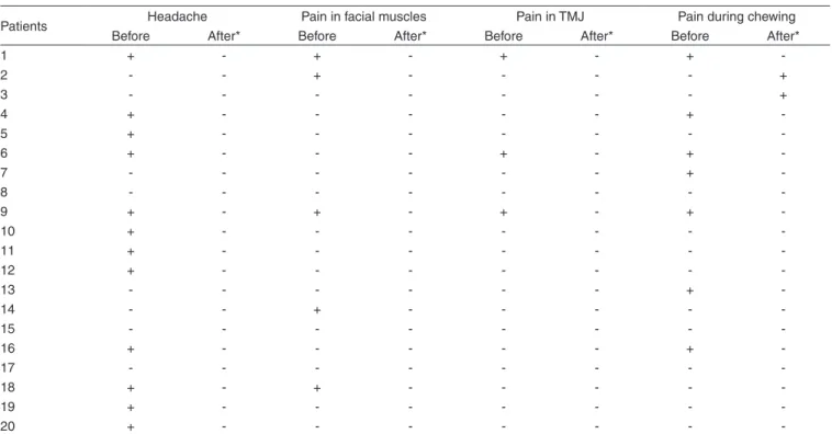
The symptoms investigated through interview are in the
Table 1. After orthognathic surgery all symptoms decreased.
Twelve patients complained about headache before surgery and
did not complain about it after surgery (p=0.007). Five patients
complained about pain in facial muscle only before surgery
(p=0.00006). Three patients complained about pain in TMJ
only before surgery (p=0.00001). Seven patients complained
about pain during chewing before surgery and did not complain
about it after surgery, but other two patients had pain during
chewing after surgery (p=0.007).
About auditory and vestibular symptoms, ive patients
complained about ear fullness before surgery and one patient
reported about earache after surgery.
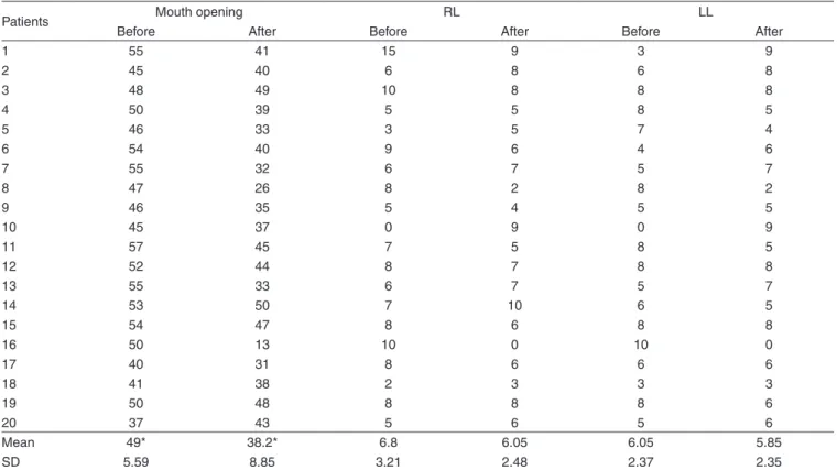
Related to mandibular movements, when compared before
and after surgery, the results showed decrease in the mouth
Table 1. Symptoms investigated considering the different periods (before and after othognathic surgery)
Patients Headache Pain in facial muscles Pain in TMJ Pain during chewing
Before After* Before After* Before After* Before After*
1 + - + - + - +
-2 - - + - - - - +
3 - - - +
4 + - - - +
-5 + - - -
-6 + - - - + - +
-7 - - - +
-8 - - -
-9 + - + - + - +
-10 + - - -
-11 + - - -
-12 + - - -
-13 - - - +
-14 - - + - - - -
-15 - - -
-16 + - - - +
-17 - - -
-18 + - + - - - -
-19 + - - -
-20 + - - -
-* signiicant difference (p<0,05) when comparing the presence of symptoms before and after surgery
Table 2. Individual results, mean and standart deviation (SD) of mouth opening and mandibular laterality movement before and after orthognathic surgery
Patients Mouth opening RL LL
Before After Before After Before After
1 55 41 15 9 3 9
2 45 40 6 8 6 8
3 48 49 10 8 8 8
4 50 39 5 5 8 5
5 46 33 3 5 7 4
6 54 40 9 6 4 6
7 55 32 6 7 5 7
8 47 26 8 2 8 2
9 46 35 5 4 5 5
10 45 37 0 9 0 9
11 57 45 7 5 8 5
12 52 44 8 7 8 8
13 55 33 6 7 5 7
14 53 50 7 10 6 5
15 54 47 8 6 8 8
16 50 13 10 0 10 0
17 40 31 8 6 6 6
18 41 38 2 3 3 3
19 50 48 8 8 8 6
20 37 43 5 6 5 6
Mean 49* 38.2* 6.8 6.05 6.05 5.85
SD 5.59 8.85 3.21 2.48 2.37 2.35
*Signiicant difference (p<0.01). Values expressed in milimeters
Legend: RL = right laterality; LL = left laterality; SD = standart deviation
opening after surgery (p=0.00004) and little variation on
la-terality movements (Table 2).
When compared the data about pain during palpation on
masticatory and cervical muscles and TMJ, before and after
orthognathic surgery, it was noticed pain relief on
trape-zius muscles (p=0.00006) and sternocleidomastoid muscle
(p=0.0005) (Figure 1).
About articulation reproducible noise, it was noticed
presence of articulation noise in eight patients before surgery
(40%) while seven patients presented it after surgery (35%),
result without difference.
DISCUSSION
The examination of TMD in individuals with occlusion
changes can be explained by the high incidence of functional
and anatomic problems of temporomandibular joint in these
cases
(13); however studies that related this condition are
con-tradictory. So, the present study investigated the occurrence of
signs and symptoms os TMD in patients submitted to occlusion
correction using the orthodonthic surgery treatment.
The highest percentage of patients investigated (85%) was
female, agreeing with other studies that demonstrate higher
prevalence of TMD in women
(14,15). Studies that relate hormonal
inluences on the appearance of signs and symptoms of TMD
have found association between this aspects
(16,17).
At this study was observed considerable presence of signs
and symptoms of TMD before surgery, condition justiied by
the decrease of stomatognathic system adaptative capacity
* Signiicant difference (p<0.005)
Legend: SCM = sternocleidomastoid; TMJ = temporomandibular joint
Figure 1. Sign of pain during palpation in masseter muscle, lateral and/ or medial pterygoid, anterior temporalis, trapezius, sternocleidomastoid (SCM) and temporomandibular joint (TMJ), investigated before and after orthognathic surgery
caused by occlusion changes and maxillomandibular
discre-pancy
(5,18).
For patients in this study, after the orthodonthic surgery
treatment was observed signiicant decrease on TMD
symp-toms, agreeing with studies in the literature
(11,12). However,
authors have found the appearance of TMD symptoms after
orthodonthic surgery treatment
(10,19). This disagreement can
irst one investigated cases submitted to maxillary osteotomy
and the second author evaluated cases submitted to mandibular
advancement surgery. At this study, the patients were submitted
to different types of mandibular movement with or without
maxillary intervention.
About auditory and vestibular symptoms, at this study ive
patients complained about ear fullness before surgery, agreeing
with a study that demonstrated relevant occurrence of this
symptom in individuals with TMD
(20). One patient complained
about earache after surgery, what can be related to pain during
mandibular condyle palpation in subjects with dysfunction
(21).
Studies that make an association between earache, tunnitus,
hearing loss, ear fullness and vertigo with DTM are common
in the literature
(22-24).
At this study was found an important decrease of mouth
opening after surgery. According to the literature, after
sur-gery the mandibular movements tend to be reduced, assuming
normal values from three to 14 months after surgery
(25,26). A
study presented some cases that the injury in orofacial myology
improved in three months after orthognathic surgery, with a
signiicant increase of mandibular protrusion and laterality
(27).
A short-term evaluation realized in this study (between 30 and
60 days after surgery) can explain the differences with studies
published before.
With orthognathic surgery, a decrease in pain during
pal-pation in cervical muscles was observed. None studies in the
literature were found relating this aspect, but bad occlusion
can modify the posture as a compensation mechanism. It may
justify the decrease of pain during palpation in cervical muscles
after orthodonthic surgery correction.
On the patients evaluated at this study, it was observed
that, even with the structural correction resultant from the
orthodonthic surgery treatment, the stomatognathic system
reorganization did not occur naturally; and speech and
langua-ge pathology intervention was necessary in order to adapt the
orofacial functions. We should pay attention on masticatory
function, in which the default is inluenced by
temporoman-dibular dysfunction and presence of changes in mantemporoman-dibular
movement
(18-28).
Another aspect not discussed in this study that should be
considered is the change in thermal and tactile sensitivity result
from mandibular osteotomy procedure. It may be transitory
but causing drooling, accumulation of saliva and food on lips
comissure and dificult with lips movement
(29).
So, improvement on signs and symptoms of TMD after
orthodonthic surgery treatment occur because of
morphofunc-tional balance established by the treatment and monitoring of
the patient by interdisciplinary group. The interdisciplinary
group, in these cases, was extremely important considering
the etiology of this dysfunction is multifactorial and need a
management of all causal aspects to have a good prognosis.
CONCLUSION
The orthodontic surgery treatment caused, in a short
term, decrease in the symptoms: headache, pain in the facial
muscle, pain in TMJ, pain during chewing and in the clinical
signs of pain during palpation on the cervical muscle; still
result in decrease of mouth opening in patients with detofacial
deformities.
RESUMO
Objetivo: Veriicar se o tratamento ortodôntico-cirúrgico acarreta modiicações nos sinais e sintomas de disfunção temporomandibular em indivíduos com deformidades dentofaciais. Métodos: Participaram do estudo 20 pacientes de ambos os gêneros (idades entre 15 e 44 anos), com diferentes características dento-oclusais e faciais, submetidos a osteotomias mandibulares e/ou maxilares. Todos os pacientes apresentavam pelo menos um sinal ou sintoma de disfunção temporomandibular, veriicado por meio de entrevista e de avaliação clínica realizada pré e após 30 a 60 dias da cirurgia ortognática. Nesta última buscou-se dor à palpação dos músculos mastigatórios, cervicais e da articulação temporomandibular; foi realizada ausculta das articulações temporomandibulares e mediu-se movimentos mandibulares. Resultados: Quanto aos sintomas investigados na entrevista,os resultados demonstraram que após a cirurgia ortognática houve redução de todos os sintomas. No que diz respeito aos movimentos mandibulares, observou-se signiicante diminuição da abertura da boca no pós-operatório e pouca variação nas medidas de lateralidade mandibular. Em relação à presença de dor à palpação, notou-se diminuição da dor para os músculos cervicais. Conclusão: O tratamento ortodôntico-cirúrgico acarretou, em curto prazo, diminuição dos sintomas investigados e dos sinais clínicos de dor à palpação na musculatura cervical e redução da abertura da boca em pacientescom deformidades dentofaciais. Registro de ensaio clínico: 083578.
Descritores: Desenvolvimento maxilofacial; Cirurgia bucal; Articulação temporomandibular/patologia; Má oclusão; Anormalidades dentárias
REFERENCES
1. Anelli-Bastos W, Oliveira MFR. Atuação fonoaudiológica na disfunção temporomandibular. In: Lopes Filho O, editor. Tratado de fonoaudiologia. 2a ed. Ribeirão Preto: Tecmedd; 2005. p. 755-65.
3. Bianchini EMG. Articulação temporomandibular e fonoaudiologia. In: Ferreira LP, Bei-Lopes DM, Limongi SCO, organizadores. Tratado de fonoaudiologia. São Paulo: Roca; 2004. p. 315-29.
4. de Oliveira AS, Dias EM, Contato RG, Berzin F. Prevalence study of signs and symptoms of temporomandibular disorder in Brazilian college students. Braz Oral Res. 2006;20(1):3-7.
5. Buescher JJ. Temporomandibular joint disorders. Am Fam Physician. 2007;76(10):1477-82. Review.
6. Dworkin SF, Huggins KH, LeResche L, Von Korff M, Howard J, Truelove E, Sommers E. Epidemiology of signs and symptoms in temporomandibular disorders: clinical signs in cases and controls. J Am Dent Assoc. 1990;120(3):273-81.
7. McNamara JA Jr, Seligman DA, Okeson JP. Occlusion, orthodontic treatment, and temporomandibular disorders: a review. J Orofac Pain. 1995;9(1):73-90.
8. Al-Ani MZ, Davies SJ, Gray RJ, Sloan P, Glenny AM. Stabilisation splint therapy for temporomandibular pain dysfunction syndrome. Cochrane Database Syst Rev. 2004;(1):CD002778. Review.
9. Koh H, Robinson PG. Occlusal adjustment for treating and preventing temporomandibular joint disorders. J Oral Rehabil. 2004;31(4):287-92. Review.
10. Aoyama S, Kino K, Kobayashi J, Yoshimasu H, Amagasa T. Clinical evaluation of the temporomandibular joint following orthognathic surgery--multiple logistic regression analysis. J Med Dent Sci. 2005;52(2):109-14. 11. Pahkala RH, Kellokoski JK. Surgical-orthodontic treatment and patients’
functional and psychosocial well-being. Am J Orthod Dentofacial Orthop. 2007;132(2):158-64.
12. Panula K, Somppi M, Finne K, Oikarinen K. Effects of orthognathic surgery on temporomandibular joint dysfunction. A controlled prospective 4-year follow-up study. Int J Oral Maxillofac Surg. 2000;29(3):183-7. 13. Altman EB. Myofunctional therapy and orthognathic surgery. Int J
Orofacial Myology. 1987;13(3):2-12.
14. Barbosa CMR, Queluz DP, Barbosa JRA, Di Hipólito Júnior O. Correlação entre aparelho ortodôntico, sexo e presença de desordens temporomandibulares. J Bras Ortodon Ortop Facial. 2002;7(39):185-92. 15. Conti A, Freitas M, Conti P, Henriques J, Janson G. Relationship between
signs and symptoms of temporomandibular disorders and orthodontic treatment: a cross-sectional study. Angle Orthod. 2003;73(4):411-7. 16. Fischer L, Clemente JT, Tambeli CH. The protective role of testosterone in
the development of temporomandibular joint pain. J Pain. 2007;8(5):437-42.
17. Fischer L, Torres-Chávez KE, Clemente-Napimoga JT, Jorge D, Arsati F, de Arruda Veiga MC, Tambeli CH. The inluence of sex and ovarian hormones on temporomandibular joint nociception in rats. J Pain. 2008;9(7):630-8.
18. Berretin-Felix G, Jorge TM, Genaro KF. Intervenção fonoaudiológica em pacientes submetidos a cirurgia ortognática. In: Ferreira LP, Bei-Lopes DM, Limongi SCO, organizadores. Tratado de fonoaudiologia. São Paulo: Roca; 2004. p. 494-511.
19. Wolford LM, Reiche-Fischel O, Mehra P. Changes in temporomandibular joint diysfunction after orthognathic surgery. J Oral Maxillofac Surg. 2003;61(6):655-60; discussion 661.
20. Kuttila S, Kuttila M, Le Bell Y, Alanen P, Jouko S. Aural symptoms and signs of temporomandibular disorder in association with treatment need and visits to a physician. Laryngoscope. 1999;109(10):1669-73. 21. dos Reis AC, Hotta TH, Ferreira-Jeronymo RR, Felício CM, Ribeiro RF.
Ear symptomatology and occlusal factors: a clinical report. J Prosthet Dent. 2000;83(1):21-4.
22. Pereira KNF, Andrade LLS, Costa MLG, Portal TF. Sinais e sintomas de pacientes com disfunção temporomandibular. Rev CEFAC. 2005;7(2):221-8.
23. Silveira AM, Feltrin PP, Zanetti RV, Mautoni MC. Prevalência de portadores de DTM em pacientes avaliados no setor de otorrinolaringologia. Rev Bras Otorrinolaringol. 2007;73(4):528-32. 24. Zeigelboim BS, Jurkiewicz AL, Martins-Bassetto J, Klagenberg KF.
Avaliação vestibular em mulheres com disfunção temporomandibular. Rev CEFAC. 2007;9(2):255-62.25. Boyd SB, Karas ND, Sinn DP. Recovery of mandibular mobility following orthognathic surgery. J Oral Maxillofac Surg. 1991;49(9):924-31.
26. Zarrinkelk HM, Throckmorton G, Ellis E 3rd, Sinn DP. A longitudinal study of changes in masticatory performance of patients undergoing orthognathic surgery. J Oral Maxillofac Surg. 1995;53(7):777-82; discussion 782-3.
27. Milosevic A, Samuels RH. Mandibular mobility and occlusal relationships after orthognathic surgery. Int J Adult Orthodon Orthognath Surg. 1997;12(2):122-8.
28. rawitzki LVV, Dantas RO, Mello-Filho FV, Marques W Jr. Effect of treatment of dentofacial deformities on the electromyographic activity of masticatory muscles. Int J Oral Maxillofac Surg. 2006;35(2):170-3. 29. Ylikontiola L, Vesala J, Oikarinen K. Repeatability of 5 clinical