IMAGINGASPECTSOF CAMURATI-ENGELMANNDISEASE
REV ASSOC MED BRAS 2016; 62(9):825-827 825
IMAGE IN MEDICINE
Imaging aspects of Camurati-Engelmann disease
SIMONE UEZATO1*, GUSTAVO DIAS2, JULIANA INADA3, MARCELO VALENTE4, ELOY FERNANDES5
1Resident Physician at Hospital Estadual Vila Alpina/Diagnostic Imaging Service II, associated with the Luiz Roberto Barata Barradas Specialist Medical Outpatient Clinic (AME), São Paulo, SP, Brazil
2Resident Physician at Hospital A.C.Camargo Cancer Center, São Paulo, SP, Brazil
3Orthopedist, Prefeitura Municipal de Barueri, Barueri, SP, Brazil
4Supervising Physician at the Continuing Education/Medical Residency in Radiology and Imaging Diagnosis, Hospital Estadual Vila Alpina, São Paulo, SP, Brazil
5Afiliate Professor of the Department of Imaging Diagnosis, Universidade Federal de São Paulo. Radiologist, Diagnostic Imaging Service II, associated with the Luiz Roberto Barradas Barata Specialist Medical Outpatient
Clinic (AME), São Paulo, SP, Brazil
Study conducted at Hospital Estadual Vila Alpina/Diagnostic
Imaging Service II, São Paulo, SP, Brazil
Article received: 1/20/2016
Accepted for publication: 1/30/2016
*Correspondence:
Address: Av. Almirante Delamare, 1534 São Paulo, SP – Brazil
Postal code: 04230-000 [email protected]
http://dx.doi.org/10.1590/1806-9282.62.09.825
C
ASE REPORTA 35-year-old female patient undergoing outpatient mon-itoring at the Specialist Medical Outpatient Clinic (AME, in portuguese acronym) in Mogi Guaçu, São Paulo, Brazil.
Since childhood the patient has presented generalized pain in the body, mainly in the lower limbs, which has been more intense for the last 2 years, with no relation to physical effort, and which gets worse with changes in ambient tem-perature. Generalized weakness, reduced muscle strength and discreetly staggering gait have also been reported, as well the use of hearing aids. Faced with these symptoms, the patient sought successive health services, where she only received symptomatic treatment. Due to the persistence of the complaint, she underwent an X-ray of the lower limbs.
Based on the radiographic changes found, bone scintigraphy and magnetic resonance imaging (MRI) examinations were requested. There are no known cases in the family.
R
ESULTSThe MRI revealed exuberant diffuse cortical thickening in the bone diaphyses, increased diameter of both femurs, intramedullary and peripheral femoral hyperintensity focal areas in the T2 weighted sequences, with enhance-ment after infusion of the intravenous contrast medium, which may correspond to areas of edema or inflamma-tory activity. The contracting mass of the muscle and subcutaneous plane had a preserved signal (Figure 1).
UEZATO S ETAL.
826 REV ASSOC MED BRAS 2016; 62(9):825-827
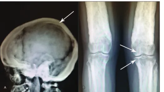
The X-ray of upper limbs, lower limbs and skull also showed diffuse cortical thickening, notably in the bone diaphyses and flat bones, while the bone scintigraphy re-vealed diffuse increase in osteogenic activity at these sites.
D
ISCUSSIONIt is noteworthy that the involvement of the femurs occurs mainly in the cortical region, restricting diagnostic hy-potheses to basically hereditary sclerosing bone dysplasias, particularly those resulting from defects in intramembra-nous ossification.2,3 Intramembranous ossification occurs
in the cortex of tubular bones (such as femurs) and the flat bones of the skullcap, the upper facial bones, tym-panic temporal bones, vomer and medial pterygoid plate.4
The hereditary disorders related to this type of ossification are: generalized cortical hyperostosis (van Buchem’s dis-ease and variants), hereditary multiple diaphyseal sclero-sis (Ribbing disease) and progressive diaphyseal dysplasia (Camurati-Engelmann’s disease).4 Although
Erdheim-Chester disease is considered part of an acquired (and not inherited) syndrome that simulates sclerosing bone dys-plasia,4 it is also part of the differential diagnosis.3
Even-tually, the possibility of osteopetrosis could be suggested; however, this disorder derives from an endochondral ossification defect (which gives rise to bone marrow)4 and
is therefore an ossification condition of the medullary and not the cortical region.
According to the phenotypic presentation, some of these pathologies can be excluded. In generalized cortical hyper-ostosis (van Buchem’s disease and variants),4 facial
abnor-malities occur, such as flattened forehead, elongated jaw and
syndactyly of the second and third fingers. Meanwhile, in hereditary multiple diaphyseal sclerosis (Ribbing disease)4
there is unilateral or bilateral asymmetric/asynchronous involvement of the long bones, typically the tibia and femur,4
as well as non-involvement of skullcap.4 Since the patient in
this study has atypical fascicles, bilateral and symmetrical bone involvement and cortical thickening in the skull radi-ography, other diagnostic possibilities would be more likely. These characteristics4 are seen both in Erdheim-Chester
disease (non-Langerhans cell histiocytosis), and in progres-sive diaphyseal dysplasia (Camurati-Engelmann disease). However, the extra-osseous manifestations of Erdheim-Ches-ter disease (diabetes insipidus, painless bilaErdheim-Ches-teral exophthal-mos, chronic renal failure, hydronephrosis, pulmonary fi-brosis, and heart failure) and associated bone infarcts4 were
not found, as the only comorbidity presented by the patient was dysacusis.
Dysacusis, in turn, is described as one of the symptoms of progressive diaphyseal dysplasia (Camurati-Engelmann disease). Bone involvement in this disease begins in the tibial and femoral diaphyses,3,5 in a bilateral and
symmet-rical manner1,6 progressing to the other long bones with
progressive bone deformity. In descending order of fre-quency, it affects the tibia, femur, fibula, humerus, ulna and radius3,5 and, more rarely, the middle segment of the
clavicles.4,7 In severe cases, isolated sclerosis in the
poste-rior region of the vertebral body can also be observed.3,4
The metaphysis and epiphysis are typically not in-volved, as these regions are formed by endochondral os-sification4 (Figure 2), although during disease progression
they may become involved secondarily.5
IMAGINGASPECTSOF CAMURATI-ENGELMANNDISEASE
REV ASSOC MED BRAS 2016; 62(9):825-827 827
R
EFERENCES1. Bellew N, Wagener G. Camurati-Engelmann disease. SA J Radiol. 2011; 15(1):20-4.
2. Applegate LJ, Applegate GR, Kemp SS. MR of multiple cranial neuropathies in a patient with Camurati-Engelmann disease: case report. AJNR Am J Neuroradiol. 1991; 12(3):557-9.
3. da Fonte AC, Gonçalves CM, Catalan J, Justino Junior RO. Doença de Camurati-Engelmann: relato de caso com acometimento ósseo grave. Radiol Bras. 2005; 38(6):471-2.
4. Ihde LL, Forrester DM, Gottsegen CF, Masih S, Patel DB, Vachon LA, et al. Sclerosing bone dysplasias: review and differentiation from other causes of osteosclerosis. RadioGraphics. 2011; 31(7):1865-82.
5. Bogas M, Bogas V, Pinto F. Doença de Camurati-Engelmann: manifestações típicas de uma doença rara. Rev Bras Reumatol 2009; 49(3): 308-14. 6. Shier CK, Thrall JH, Kottamasu SR, Krasicky GA, Ellis BI. Diaphyseal bone disease:
scintigraphic-radiographic correlation. Radiographics 1989; 9(1):129-51. 7. Antunes ML, Testa JRG, Frazatto R, Barbieri JAF, Silva RFND. Osteodistrofias
raras do osso temporal. Rev Bras Otorrinolaringol. 2005; 71(2):228-32. 8. Janssens K, Vanhoenacker F, Bonduelle M, Verbruggen L, Van Maldergem
L, Ralston S, et al. Camurati-Engelmann disease: review of the clinical, radiological, and molecular data of 24 families and implications for diagnosis and treatment. J Med Genet. 2006; 43(1):1-11.
9. Fuller H, Fuller R, Pereira RMR. Tomografia computadorizada quantitativa periférica de alta resolução para avaliação de parâmetros morfológicos e funcionais ósseos. Rev Bras Reumatol. 2015; 55(4):352-62.
FIGURE 3 Bone scintigraphy after intravenous administration of 99mTc- MDP. Diffuse increase of osteogenic activity in the skull cap and long bones of the upper and lower limbs is observed, suggesting hereditary disease of the bone metabolism.
In rare cases, sclerosis can be found at the base of the cranium4 due to an endochondral ossification defect,
sug-gesting the possibility of two forms of progressive diaph-yseal dysplasia: a pure one, with exclusive disturbance in intramembranous ossification, and a mixed one, in which there is also the endochondral component.3 Cranial nerve
palsy may develop in such cases. Hearing loss occurs in 18% of cases and may be conductive, through fixation of the staples in the oval window, mixed or sensorineural, due to stenosis of the internal auditory meatus.7
Unlike other bone metabolism disorders, in Camu-rati-Engelman disease low-impact fractures are rare, and there is controversy about neuromuscular impairment1,5
probably due to the wide variety of phenotypic expressions described.8
MRI is as effective as computerized tomography for demonstrating the hyperostotic bone, and the compres-sive effect on specific cranial nerves (mainly II, VII and VIII) can be characterized well.2 Perhaps in the near future
techniques such as high resolution peripheral quantitative computed tomography (HR-pQCT)9 could provide
addi-tional information.
Increased osteoblast activity can be detected early through skeletal scintigraphy5 even before the radiographic
changes7 or be normal in some cases.5 In bone scintigraphy
with 99m Tc-MDP, heterogeneous abnormal uptake can be seen in the affected bones3 in a bilateral and symmetrical
manner1,6 similarly to the findings of our study3 (Figure 3).
C
ONCLUSIONWe were able to guide the diagnostic reasoning based on the image findings and clinical data. Knowledge of the different types of ossification was also fundamental to narrow down the range of differential diagnoses. MRI helps to improve the evaluation of the neuromuscular component in these cases and complements the radiographic images in the analysis of bone changes, in relation to their inflam-matory activity and cortical thickening. Despite this being a rare disease whose treatment has been palliative through use of corticosteroids,2 bisphosphonates (with
controver-sial usage),5 decompressive surgeries2 and physiotherapy,5