Evaluation of inter-rater reliability of subjective and objective
criteria for diagnosis of lymphedema in upper and lower limbs
Avaliação da confiabilidade entre os critérios subjetivos e objetivos utilizados para o
diagnóstico de linfedema nos membros superiores e inferiores
Larissa Louise Campanholi1, João Pedreira Duprat Neto1, José Humberto Tavares Guerreiro Fregnani2
Abstract
Background: he diagnosis of lymphedema can be obtained objectively by measurement methods, and also by subjective methods, based on the patient’s complaint. Objective: To evaluate inter-rater reliability of objective and subjective criteria used for diagnosis of lymphedema and to propose a lymphedema cut-of for diferences in volume between afected and control limbs. Methods: We studied 84 patients who had undergone lymphadenectomy for treatment of cutaneous melanoma. Physical measures were obtained by manual perimetry (MP). he subjective criteria analyzed were clinical diagnosis of lymphedema in patients’ medical records and self-report of feelings of heaviness and/or increase in volume in the afected limb. Results: For upper limbs, the subjective criteria clinical observation (k 0.754, P<0.001) and heaviness and swelling (k 0.689, P<0.001) both exhibited strong agreement with MP results and there was moderate agreement between MP results and swelling (k 0.483 P<0.001), heaviness (k 0.576, P<0.001) and heaviness or swelling (k 0.412, P=0.001). For lower limbs there was moderate agreement between MP results and clinical observation (k 0.423, P=0.003) and regular agreement between MP and self-report of swelling (k 0.383, P=0.003). Cut-of values for diagnosing lymphedema were deined as a 9.7% diference between an afected upper limb and control upper limb and a 5.7% diference between lower limbs. Conclusion: Manual perimetry, medical criteria, and self-report of heaviness and/or swelling exhibited better agreement for upper limbs than for lower limbs for diagnosis of lymphedema.
Keywords: lymphedema; diagnosis; methods.
Resumo
Contexto: O diagnóstico de linfedema pode ser obtido tanto de forma objetiva, por métodos de mensuração, quanto por métodos subjetivos, através da queixa do paciente. Objetivo: Examinar a coniabilidade entre critérios objetivos e subjetivos utilizados para o diagnóstico de linfedema e propor um ponto de corte para linfedema de membros superiores e inferiores. Métodos: Foram estudados 84 pacientes submetidos à linfonodectomias para o tratamento do melanoma cutâneo. As mensurações dos membros foram feitas utilizando a perimetria manual. Os critérios subjetivos foram obtidos através do diagnóstico de linfedema nos prontuários dos pacientes (observação clínica) e de auto-relato de sensação de peso e/ou aumento de volume no membro afetado. Resultados: Nos membros superiores, houve uma forte correlação entre a perimetria manual e cada um dos critérios subjetivos: observação clínica (k 0,754, P<0,001) e sensação de peso eaumento de volume (k 0,689, P<0,001); concordância moderada no aumento de volume (k 0,483, P<0,001), peso (k 0,576, P<0,001) e sensação de peso ou aumento de volume (k 0,412, P=0,001). Nos membros inferiores, houve concordância moderada entre a perimetria e observação clínica (k 0,423, P=0,003) e regular no aumento de volume (k 0,383, P=0,003). O ponto de corte para deinir linfedema foi uma diferença de 9,7% entre o membro afetado e o controle, e 5,7% de diferença para membros inferiores. Conclusão: Perimetria, observação clínica e auto-relato de sensação de peso e/ou aumento de volume, apresentaram melhor concordância para membros superiores que para inferiores no diagnóstico de linfedema.
Palavras-chave: linfedema; diagnóstico; métodos.
1A. C. Camargo Cancer Center, São Paulo, SP, Brazil. 2Hospital do Câncer de Barretos, Barretos, SP, Brazil.
Financial support: FAPESP and CNPq.
Conlicts of interest: No conlicts of interest declared concerning the publication of this article. Submitted: May 14, 2014. Accepted: July 12, 2014.
INTRODUCTION
Lymphadenectomy conducted to treat cutaneous melanoma causes lymphedema. Other risk factors for lymphedema are melanoma thickness >4 mm, infection and graft reconstruction, and a combination of these risk factors increases the chances of developing this chronic condition.1
Lymphedema can be diagnosed using several different objective methods, including manual perimetry (MP), water displacement, tonometry, optoelectronic volumetry and bioimpedance.2
However many studies have diagnosed lymphedema subjectively on the basis of patients’ responses to questions about their symptoms, such as heaviness and/or swelling in the limb.3-5 Several prospective
and retrospective studies3,4,6-10 have diagnosed
upper and lower limb lymphedema secondary to treatment of melanoma using combinations of objective or subjective methods, for example, MP and optoelectronic volumetry; patient history and physical examination; self-report and medical records (Table 1).
Manual perimetry offers the advantages of low cost, requiring only a tape measure, and ease of use in clinical practice. It is a simple method that can be used regardless of skin condition and requires minimal technology or training.11 Circumference
measurements are taken at 7 or 10 cm intervals and then it is possible to calculate limb volume from the sum of each truncated cone,12-14 using free online
calculators such as www.armvolume.com and www. legvolume.com. Distances between measurements vary from 4 to 15 cm in different studies.12-16 Studies
comparing limb volume measurements calculated from water displacement with the results of geometric formulas using input values obtained by MP show excellent correlation, indicating that they are equally valid for diagnosis of lymphedema.17,18
The objective of this study was to examine the inter-rater reliability of objective and subjective criteria used for diagnosis of lymphedema. It is also interesting to propose cut-offs for differences in volume between affected and control limbs that could be used to diagnose lymphedema.
METHODS
This study enrolled patients who underwent lymphadenectomy from 1990 to 2008 at our institution. The exclusion criteria were patients with limb amputation or bilateral lymph node dissection and patients who refused to participate. All patients were requested to have an interview and to be examined. During the period, 364 inguinal, ilioinguinal and axillary lymphadenectomies were conducted on patients with melanoma. From these 364 procedures, 186 patients had died from diseases or other causes during follow-up, 63 could not be
located, ive could not be evaluated because they
were bedridden, 17 were excluded because of limb amputation, and seven were excluded because of bilateral dissection. Only two patients refused to participate in the study and so the final sample included 84 patients who had been diagnosed with cutaneous melanoma and had undergone axillary, groin, or ilioinguinal lymph node dissection with a minimum of six months’ follow-up. The project was approved in advance by the Research Ethics Committee at the A.C.Camargo Cancer Center.
Manual perimetry was performed using a regular tape measure. For upper limbs, measurements were taken at 7 cm intervals; 7 and 14 cm above the interarticular line through the elbow and at 7, 14 and 21 cm below the line. For lower limbs, measurements were taken every 10 cm from the sole up to the seventh measurement. Measurements for all patients were obtained by a single researcher to prevent
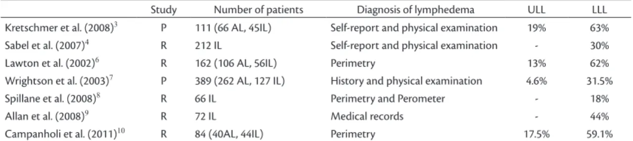
Table 1. Prospective and retrospective studies of diagnosis of lymphedema in upper and lower limbs in patients given axillary, inguinal or ilioinguinal lymphadenectomies for cutaneous melanoma.
Study Number of patients Diagnosis of lymphedema ULL LLL Kretschmer et al. (2008)3 P 111 (66 AL, 45IL) Self-report and physical examination 19% 63%
Sabel et al. (2007)4 R 212 IL Self-report and physical examination - 30%
Lawton et al. (2002)6 R 162 (106 AL, 56IL) Perimetry 13% 62%
Wrightson et al. (2003)7 P 389 (262 AL, 127 IL) History and physical examination 4.6% 31.5%
Spillane et al. (2008)8 R 66 IL Perimetry and Perometer - 18%
Allan et al. (2008)9 R 72 IL Medical records - 44%
Campanholi et al. (2011)10 R 84 (40AL, 44IL) Perimetry 17.5% 59.1%
differences caused by varying tension in the tape. These data were fed into the truncated cone formula:
(
)
= 12+ 1× 2+ 22
12
V h C C C C
π
where: V = volume of the inal segment of the limb,
C1 and C2 = circumference measured between the points, and h = distance between the circles (C1 and C2 in each segment) in centimeters.
Differences between limb volumes measured by
MP were calculated. Lymphedema was deined as
a difference greater than 10% between upper limb volumes12,19 or greater than 6.5% between lower limb
volumes.20,21 Lymphedema deined by MP was also
compared to lymphedema diagnosed subjectively in physicians’ reports (presence of lymphedema in the medical record, when there was a considerable visual difference between limbs) and patients’ complaints (self-report of heaviness and/or swelling in the limb at the time of physical assessment, where the patient notices that his/her shirt sleeve or pants are tighter in the ipsilateral lymphadenectomy limb).
Inter-rater agreement between MP and patient/ medical criteria for diagnosing lymphedema was calculated using the Kappa (k) index. Receiver operator characteristics (ROC) curve analysis was used to establish cut-off values for the difference between limb volumes measured by MP according to the patient and medical criteria. In all statistical
tests, signiicance was accepted at the 5% level. The
Statistical Package for the Social Sciences (SPSS) version 15.0 (Chicago, IL) was used for statistical analyses.
RESULTS
Forty-eight patients were women (57.1%). The average age of patients at surgery was 47.2 years
(sd: 16.7 years), ranging from ive to 80 years and
on the day of assessment, 52.5 years (sd: 16 years),
ranging from 10 to 81 years. There were only three people (3.6%) under 18 years old.
Eighty-four patients were evaluated, 40 (47.6%) had had axillary lymph node dissection, 21 (25%) inguinal and 23 (27.4%) had undergone ilioinguinal lymphadenectomy. The mean time elapsed since lymphadenectomy was 62.5 months (sd: 56.1 months; median: 44 months), ranging from six months thru 17.6 years. The prevalence rates of lymphedema (according to MP) were 17.5% in upper limbs and 59.1% in lower limbs, while prevalence rates of lymphedema according to subjective methods (self-report of heaviness and/or swelling and medical record) were 32.5% in upper and 66% in lower limbs.
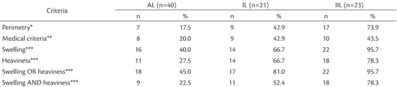
Table 2 shows prevalence rates of lymphedema according to type of lymphadenectomy. Lymphedema was more common after ilioinguinal than after axillary or inguinal lymphadenectomy. Self-report of swelling in the affected limb was the most common complaint, mainly in ilioinguinal lymphadenectomy patients (95.7%). Some patients without lymphedema (according to MP) complained of heaviness and/ or swelling in the limb, irrespective of type of lymphadenectomy.
Comparison of the different diagnostic methods
for lymphedema of upper limbs revealed signiicant
and moderate agreement between MP and subjective patient criteria as follows: self-report of swelling (k=0.483; P<0.001), heaviness (k=0.576; P<0.001) and heaviness or swelling (k=0.412; P=0.001). Self-report of heaviness and swelling (k=0.689; P<0.001) and medical criteria (k=0.754; P<0.001) exhibited strong agreement. When results for lymphedema of
lower limbs were compared, signiicant and regular
agreement was detected between MP result and swelling (k=0.383; P=0.003), and there was moderate agreement with medical records (k=0.423; P=0.003) (Table 3).
Cut-off values for differences between limb volumes measured by MP calculated from patient
Table 2. Prevalence of lymphedema according to type of lymphadenectomy and several criteria for diagnosis of lymphedema.
Criteria AL (n=40) IL (n=21) IIL (n=23)
n % n % n %
Perimetry* 7 17.5 9 42.9 17 73.9
Medical criteria** 8 20.0 9 42.9 10 43.5
Swelling*** 16 40.0 14 66.7 22 95.7
Heaviness*** 11 27.5 14 66.7 18 78.3
Swelling OR heaviness*** 18 45.0 17 81.0 22 95.7
Swelling AND heaviness*** 9 22.5 11 52.4 18 78.3
and medical criteria for deining lymphedema were
as follows: a 9.7% difference between an affected upper limb and control upper limb, and a 5.7% difference for lower limbs. The subjective criteria were also analyzed individually according to MP results, as shown in Table 4. Heaviness, swelling and heaviness and medical records all resulted in the same cut-off (9.6%) for upper limb, while swelling and swelling or heaviness gave a cutoff of 4.7%. The cut-off points for lower limbs were more divergent: 9.5% for medical records and swelling or heaviness, 13.8% for heaviness, 4.4% for swelling and 4.8% for swelling and heaviness.
DISCUSSION
There is a relatively low number of patients with melanoma, compared to other tumors, and because of this most studies of melanoma have restricted
numbers of participants and this is also a limitation of this study. It would have been interesting if these patients could have been studied prospectively.
Some studies have correlated the incidence of morbidity from lymphedema with axillary, inguinal or ilioinguinal lymphadenectomies used to treat cutaneous melanoma. In most cases, studies that mention lymphedema as the primary complication
deine it on the basis of subjective methods only. It
is of interest to investigate lymphedema based on numerical values, using formulas or equipment to provide the data. A comparison of MP with medical diagnosis of lymphedema and self-report measures is interesting because the results can be used to compare different diagnostic techniques.
Kretschmer et al.3 and Sabel et al.4 analyzed
lymphedema using self-report of swelling and heaviness of affected vs. control limbs. The rate of
lymphedema was 19% (mentioned only in the irst
study) in upper limbs and 63% and 30%, respectively, for lower limbs. In contrast, the values obtained in our analysis were higher at 34% for upper limbs and 76% for lower limbs. Both of the studies cited above had larger numbers of participants than this one.
McLaughlin et al.22 reported a significant
discrepancy between self-report and measured lymphedema. Hayes et al.23 concluded that self-report
measures offered good sensitivity for diagnosis real
lymphedemas, but bad speciicity for patients who
did not have lymphedema according to clinical examination. In our study, some patients without lymphedema, according to MP, complained of swelling and/or heaviness, while most patients who did not have heaviness and/or swelling did not have lymphedema.
Armer and Stewart24 studied breast cancer
survivors, comparing four diagnostic criteria: optoelectronic volumetry using a Perometer 350 S (lymphedema was considered as 200 ml and 10% changes in limb volume); MP (2 cm between affected and control upper limb) and self-report of heaviness or swelling during the assessment and/or in the past. It was concluded that there is no gold standard, but that the best criteria was using perometry with 10% limb volume changes. Circumferences using 2 cm
was the worse deinition. The criteria self-report of
heaviness and swelling were better than perometry with 200 ml.
Recently, another study also compared four diagnostic methods in 295 patients post breast cancer to observe the effect of weight lifting on
lymphedema. They deined lymphedema as interlimb change ≥10% for water displacement and MP
results and also used bioimpedance and self-report.
Table 3. Agreement between perimetry and several subjective criteria for diagnosing lymphedema*.
Limb Criteria for deinition of lymphedema
Kappa P value
Upper Heaviness** 0.576 <0.001 Swelling** 0.483 <0.001 Swelling OR heaviness** 0.412 0.001 Swelling AND heaviness** 0.689 <0.001 Medical criteria*** 0.754 <0.001
Lower Heaviness** 0.108 0.453
Swelling** 0.383 0.003
Swelling OR heaviness** 0.207 0.059 Swelling AND heaviness** 0.276 0.064 Medical criteria*** 0.423 0.003 *Lymphedema was deined as: > 10% diference in upper limb volumes or > 6.5% diference in lower limb volumes; **Patient self-report; ***Data retrieved from medical records.
Table 4. Cut-of values for diferences between limb volumes (%) measured by perimetry according to several subjective cri-teria for diagnosing lymphedema.
Limb Criteria for deinition of lymphedema
Diferences in limb vol-umes (Cut-of values - %)*
Upper Heaviness** >9.6
Swelling** >4.7
Swelling OR heaviness** >4.7 Swelling AND heaviness** >9.6 Medical criteria*** >9.6
Lower Heaviness** >13.8
Swelling** >4.4
Swelling OR heaviness** >9.5 Swelling AND heaviness** >4.8 Medical criteria*** >9.5
None of these methods were considered a gold standard. It was concluded that it is important to use multiple methods to evaluate patients with axillary lymphadenectomy.25
In another study that started with 511 women and completed with 176, the prevalence of lymphedema following cancer breast treatment ranged from 0.6 to 27.8%. It was observed that prevalence of lymphedema was higher when determined by self-report (arm swelling) and bioimpedance than when sum of arm circumferences was used. The cut-offs for lymphedema according to sum of circumferences were greater than 5 cm or greater than 10% (as in our paper). Lymphedema was most prevalent according to self-report measures and lowest according to circumferences. Bioimpedance was considered the best method and the use of circumferences was questioned because it exhibited limitations.23
Smoot et al.26 analyzed 144 women with breast
cancer using bioimpedance, truncated cone MP and a self-report questionnaire and concluded that patients with lymphedema must be evaluated using both self-report and objective methods.
Early lymphedemas can be detected when patients notice changes causing sensations such as heaviness and swelling in the upper limb. The ideal would be to relate limb volume measurements to symptoms.27
Patients who did not have lymphedema according to measurements did report swelling and/or heaviness in the affected limb and this should lead us to monitor these individuals closely, since they could have a greater chance of developing lymphedema. Tiwari et al.28 reported that the irst symptom of
lymphedema is complaining of a feeling of heaviness in the limb, especially at the end of the day and on days with higher temperatures.
Most studies of lymphedema are with breast cancer patients and the cut-off value for diagnosing lymphedema is >10% interlimb change for upper limbs.23-25 The problem is to define a cut-off
value for lower limbs. Using optoelectronic volumetry, Spillane et al.8 studied 66 patients who
had undergone inguinal or ilioinguinal dissection and considered lymphedema to be a volume difference
≥15% between affected and control lower limbs using Perometer or ≥7% difference using sum of
circumferences. Katz et al.29 considered lymphedema
to be a volume difference between limbs greater than 6% using optoelectronic volumetry.
The cut-off points for diagnosis of lymphedema
identiied in this study were a 9.7% difference in
volume in the affected upper limb vs. the control upper limb and a 5.7% difference in volume
between lower limbs. These indings were similar
to results from several other studies19,23-25 that deined
lymphedema as a difference of >10% in upper limbs, and a mean of 6.5% difference for lower limbs.8,21,29
In the analysis of results for upper limbs, self-report of heaviness in the affected limb, swelling and heaviness and diagnosis of lymphedema by a physician all had very similar cut-off values (9.6%). For lower limbs, subjective diagnosis of lymphedema was varied. In severe lymphedema, simple observation and palpation of the limb is sufficient for diagnosis, but mild or moderate lymphedema can go unnoticed. For this reason, it is essential to ask patients about their limb complaints (swelling and heaviness). It is also essential for health professionals to take accurate, focused medical histories with regards to the affected limb and to use MP or other objective methods to arrive at more precise diagnoses. Manual perimetry is also useful for observing a limb’s progress, to monitor whether it remains normal or develops lymphedema and to follow the response to physical therapy.
CONCLUSION
Manual perimetry, medical records and self-report of heaviness and swelling in the affected limb exhibited better agreement for upper limbs than for lower limbs when used to diagnose lymphedema. Cut-off values for diagnosing lymphedema were
deined as a 9.7% difference between an affected
upper limb and a control upper limb, and a 5.7% difference between lower limbs. More studies must be conducted using different objective methods and correlating the results with subjective criteria, particularly for lower limbs.
REFERENCES
1. Campanholi LL, Duprat Neto JP, Fregnani JHTG. Mathematical model to predict risk for lymphoedema after treatment of cutaneous melanoma. Int J Surg. 2011;9(4):306-9. http://dx.doi. org/10.1016/j.ijsu.2011.01.007. PMid:21276878
2. Warren AG, Brorson H, Borud LJ, Slavin SA. Lymphedema: a comprehensive review. Ann Plast Surg. 2007;59(4):464-72. http:// dx.doi.org/10.1097/01.sap.0000257149.42922.7e. PMid:17901744 3. Kretschmer L, Thoms KM, Peeters S, Haenssle H, Bertsch HP, Emmert S. Postoperative morbidity of lymph node excision for cutaneous melanoma-sentinel lymphonodectomy versus complete regional lymph node dissection. Melanoma Res. 2008;18(1):16-21. http:// dx.doi.org/10.1097/CMR.0b013e3282f2017d. PMid:18227703 4. Sabel MS, Griffith KA, Arora A, et al. Inguinal node dissection
for melanoma in the era of sentinel lymph node biopsy. Surgery. 2007;141(6):728-35. http://dx.doi.org/10.1016/j.surg.2006.12.018. PMid:17560249
5. van Akkooi AC, Bouwhuis MG, van Geel AN, et al. Morbidity and prognosis after therapeutic lymph node dissections for malignant melanoma. Eur J Surg Oncol. 2007;33(1):102-8. http://dx.doi. org/10.1016/j.ejso.2006.10.032. PMid:17161577
ilioinguinofemoral lymphadenectomy for melanoma. J Am Coll Surg. 2002;195(3):339-51. http://dx.doi.org/10.1016/S1072-7515(02)01230-9. PMid:12229941
7. Wrightson WR, Wong SL, Edwards MJ, et al, and the Sunbelt Melanoma Trial Study Group. Complications associated with sentinel lymph node biopsy for melanoma. Ann Surg Oncol. 2003;10(6):676-80. http://dx.doi.org/10.1245/ASO.2003.10.001. PMid:12839853
8. Spillane AJ, Saw RPM, Tucker M, Byth K, Thompson JF. Defining lower limb lymphedema after inguinal or ilio-inguinal dissection in patients with melanoma using classification and regression tree analysis. Ann Surg. 2008;248(2):286-93. http://dx.doi.org/10.1097/ SLA.0b013e31817ed7c3. PMid:18650640
9. Allan CP, Hayes AJ, Thomas JM. Ilioinguinal lymph node dissection for palpable metastatic melanoma to the groin. ANZ J Surg. 2008;78(11):982-6. http://dx.doi.org/10.1111/j.1445-2197.2008.04716.x. PMid:18959697
10. Campanholi LL, Duprat JP, Fregnani JHTG. Incidence of LE due to treating cutaneous melanoma. J Lymphoedema. 2011;6(1):30-4. 11. Deltombe T, Jamart J, Recloux S, et al. Reliability and limits
of agreement of circumferential, water displacement, and optoelectronic volumetry in the measurement of upper limb lymphedema. Lymphology. 2007;40(1):26-34. PMid:17539462. 12. Kosir MA, Rymal C, Koppolu P, et al. Surgical outcomes after
breast cancer surgery: measuring acute lymphedema. J Surg Res. 2001;95(2):147-51. http://dx.doi.org/10.1006/jsre.2000.6021. PMid:11162038
13. Duff M, Hill AD, McGreal G, Walsh S, McDermott EW, O’Higgins NJ. Prospective evaluation of the morbidity of axillary clearance for breast cancer. Br J Surg. 2001;88(1):114-7. http://dx.doi. org/10.1046/j.1365-2168.2001.01620.x. PMid:11136322 14. Megens AM, Harris SR, Kim-Sing C, McKenzie DC. Measurement
of upper extremity volume in women after axillary dissection for breast cancer. Arch Phys Med Rehabil. 2001;82(12):1639-44. http://dx.doi.org/10.1053/apmr.2001.26822. PMid:11733875 15. Karges JR, Mark BE, Stikeleather SJ, Worrell TW. Concurrent
validity of upper-extremity volume estimates: comparison of calculated volume derived from girth measurements and water displacement volume. Phys Ther. 2003;83(2):134-45. PMid:12564949.
16. Mansel RE, Fallowfield L, Kissin M, et al. Randomized multicenter trial of sentinel node biopsy versus standard axillary treatment in operable breast cancer: the ALMANAC Trial. J Natl Cancer Inst. 2006;98(9):599-609. http://dx.doi.org/10.1093/jnci/djj158. PMid:16670385
17. Chen YW, Tsai HJ, Hung HC, Tsauo JY. Reliability study of measurements for lymphedema in breast cancer patients. Am J Phys Med Rehabil. 2008;87(1):33-8. http://dx.doi.org/10.1097/ PHM.0b013e31815b6199. PMid:17993983
18. Sander AP, Hajer NM, Hemenway K, Miller AC. Upper-extremity volume measurements in women with lymphedema: a comparison of measurements obtained via water displacement with geometrically determined volume. Phys Ther. 2002;82(12):1201-12. PMid:12444879.
19. de Vries M, Vonkeman WG, van Ginkel RJ, Hoekstra HJ. Morbidity after axillary sentinel lymph node biopsy in patients with cutaneous melanoma. Eur J Surg Oncol. 2005;31(7):778-83. http:// dx.doi.org/10.1016/j.ejso.2005.05.003. PMid:15993029 20. de Vries M, Vonkeman WG, van Ginkel RJ, Hoekstra HJ. Morbidity
after inguinal sentinel lymph node biopsy and completion lymph node dissection in patients with cutaneous melanoma. Eur J Surg Oncol. 2006;32(7):785-9. http://dx.doi.org/10.1016/j. ejso.2006.05.003. PMid:16806794
21. Baas PC, Schraffordt Koops H, Hoekstra HJ, van Bruggen JJ, van der Weele LT, Oldhoff J. Groin dissection in the treatment of lower-extremity melanoma. Short-term and long-term morbidity. Arch Surg. 1992;127(3):281-6. http://dx.doi.org/10.1001/ archsurg.1992.01420030043008. PMid:1550473
22. McLaughlin SA, Wright MJ, Morris KT, et al. Prevalence of lymphedema in women with breast cancer 5 years after sentinel lymph node biopsy or axillary dissection: objective measurements. J Clin Oncol. 2008;26(32):5213-9. http://dx.doi. org/10.1200/JCO.2008.16.3725. PMid:18838709
23. Hayes S, Cornish B, Newman B. Comparison of methods to diagnose lymphoedema among breast cancer survivors: 6-month follow-up. Breast Cancer Res Treat. 2005;89(3):221-6. http:// dx.doi.org/10.1007/s10549-004-2045-x. PMid:15754119 24. Armer JM, Stewart BR. A comparison of four diagnostic criteria
for lymphedema in a post-breast cancer population. Lymphat Res Biol. 2005;3(4):208-17. http://dx.doi.org/10.1089/lrb.2005.3.208. PMid:16379589
25. Hayes SC, Speck RM, Reimet E, Stark A, Schmitz KH. Does the effect of weight lifting on lymphedema following breast cancer differ by diagnostic method: results from a randomized controlled trial. Breast Cancer Res Treat. 2011;130(1):227-34. http://dx.doi. org/10.1007/s10549-011-1547-6. PMid:21562712
26. Smoot B, Wong J, Cooper B, et al. Upper extremity impairments in women with or without lymphedema following breast cancer treatment. J Cancer Surviv. 2010;4(2):167-78. http://dx.doi. org/10.1007/s11764-010-0118-x. PMid:20373044
27. Armer JM, Radina ME, Porock D, Culbertson SD. Predicting breast cancer-related lymphedema using self-reported symptoms. Nurs Res. 2003;52(6):370-9. http://dx.doi.org/10.1097/00006199-200311000-00004. PMid:14639083
28. Tiwari A, Cheng KS, Button M, Myint F, Hamilton G. Differential diagnosis, investigation, and current treatment of lower limb lymphedema. Arch Surg. 2003;138(2):152-61. http://dx.doi. org/10.1001/archsurg.138.2.152. PMid:12578410
29. Katz E, Dugan NL, Cohn JC, Chu C, Smith RG, Schmitz KH. Weight lifting in patients with lower-extremity lymphedema secondary to cancer: a pilot and feasibility study. Arch Phys Med Rehabil. 2010;91(7):1070-6. http://dx.doi.org/10.1016/j.apmr.2010.03.021. PMid:20599045
Correspondence Larissa Louise Campanholi Rua Professor Antonio Prudente, 211 – Liberdade CEP 01509-001 – São Paulo (SP), Brazil E-mail: [email protected]
Author information LLC - MD, PhD, Physiotherapist, Skin Cancer Department, A. C. Camargo Cancer Center. JPDN - PhD, Surgical Oncologist, Skin Cancer Department, A. C. Camargo Cancer Center. JHTGF - MD, PhD, Surgical Oncologist, Gynecologic Oncology Department, Hospital do Câncer de Barretos.
Author contributions Conception and design: LLC, JPDN, JHTGF Analysis and interpretation: LLC, JPDN, JHTGF Data collection: LLC Writing the article: LLC Critical revision of the article: LLC, JPDN, JHTGF Final approval of the article*: LLC, JPDN, JHTGF Statistical analysis: LLC, JHTGF Overall responsibility: LLC