Aluísio Henrique Rodrigues de Andrade Lima
1, Cláudia Lúcia de Moraes Forjaz
2, Gleyson Queiroz de Moraes
Silva
1, Annelise Lins Menêses
1, Anderson José Melo Rodrigues Silva
1, Raphael Mendes Ritti-Dias
1Escola Superior de Educação Física - Universidade de Pernambuco1, Recife, PE; Escola de Educação Física e Esportes - Universidade de São Paulo2, São Paulo, SP - Brazil
Mailing address: Raphael Mendes Ritti Dias •
Rua Arníbio Marques, 310 - Santo Amaro - 50100-130 - Recife, PE - Brazil E-mail: [email protected]
Manuscript received September 04, 2010; revised manuscript received November 03, 2010; accepted on November 25, 2010.
Abstract
Background: Cardiac sympathovagal balance is altered after resistance exercise. However, the impact of the characteristics of resistance training in this response remains unclear.
Objective: Analyze the acute effect of resistance exercise intensity for trunk and upper limbs in cardiac autonomic modulation after exercise.
Methods: Fifteen young men performed three experimental sessions in random order: control (C), resistance exercise with 50% of 1-RM (E50%) and resistance exercise with 70% of 1-RM (E70%). The sessions included 05 exercises for the trunk and upper limbs performed in three sets of 12, 9 and 6 repetitions, respectively. Before and at 20 and 50 minutes after the interventions, the heart rate was measured for spectral analysis of variability.
Results: In comparison to the values before the intervention, the RR interval and the band of high frequency (HF) increased (major changes: + 112 ± 83 ms; +10 ± 11 un, respectively, p < 0.01), while the low frequency band (LF) and LF/HF ratio decreased (major changes: -10 ± 11 pc; -2 ± 2, respectively, p < 0.01) after the session C. None of these variables changed significantly after the E50% session (p> 0.05). Compared to pre-exercise values, the RR interval and the HF band decreased (major changes: -69 ± 105 ms; -13 ± 14 un, respectively, p < 0.01), while the LF band and the LF/HF ratio increased (major changes: -13 ± 14 un, 13 ± 3 14 ± 3 and un, respectively, p < 0.01) after E70%.
Conclusion: The higher intensity of resistance exercise for trunk and upper limbs promoted, in an acute manner, greater increase in cardiac sympathovagal balance after exercise. (Arq Bras Cardiol. 2011; [online].ahead print, PP.0-0)
Keywords: Exercise; physical exertion; autonomic nervous system; sympathetic nervous system.
the cardiovascular response to resistance exercise is dependent on the amount of muscle mass involved13, it is possible that
the autonomic responses after trunk and upper limb exercise be different, which requires further analysis.
Another important aspect concerns the overload during exercise. A previous study6 reported that the two protocols
of resistance exercise (10 reps at 80% of 1RM and 20 reps at 40% of 1RM) cause similar alterations in cardiac autonomic modulation. However, since the protocols differed in the volume (number of repetitions) and intensity (overload), it was not possible to establish clearly the isolated impact of exercise intensity in cardiac autonomic modulation after exercise. As the intensity of resistance exercise affects the metabolic and mechanical responses in the vasculature, and consequently in the reflex mechanisms of cardiovascular control, it is possible that different intensities of resistance exercise promote different responses in cardiac autonomic modulation after exercise.
Hence, the purpose of this study is to analyze the acute effect of resistance exercise intensity for trunk and upper limbs in cardiac autonomic modulation after exercise. Our hypothesis is that the sympathovagal balance increases after exercise regardless of the intensity used, but a greater
Introduction
Resistance exercise is recommended for healthy individuals1,2 and those with heart diseases3 due to benefits
on fitness4, health4 and quality of life5. However, after the
completion of the resistance exercise, the cardiac sympathetic modulation remains high, while the parasympathetic modulation remains reduced6-8, which may increase the risk
of acute cardiovascular events9,10.
Health institutions, such as the American College of Sports Medicine, recommend that the program of resistance training be divided into two sessions: one with exercises for the lower limbs and another with exercises for the upper limbs11. A
recent study12 showed that performing exercises only for the
magnitude and longer duration of this response will be observed after an exercise session with increased intensity.
Methods
Sample
The sample included 15 men aged 18 to 25 years, recruited at the university and local communities. Before entering the study, the individuals were informed about the procedures they would be exposed, and those who agreed to participate signed a consent form. This study was approved by the Ethics Committee of the institution to which the authors belong (223/08). As inclusion criteria for this study, the individuals should be well nourished, not present any cardiovascular disease, not on medication and in the 06 months before the study, they could not be practicing any kind of physical exercise.
Clinical data
To obtain the clinical data of individuals, we raised a medical history that included questions on cardiovascular risk factors. In possession of the data, the individuals were stratified with respect to cardiovascular risk at high, moderate or low level1.
Besides the medical history, anthropometric measurements and blood pressure were assessed. Body weight and height were measured using a digital scale with 0.1 kg precision (Filizola, Brazil) and a wooden stadiometer with accuracy of 0.1 cm, respectively. Blood pressure was measured by auscultation with the volunteers sitting and at rest, three consecutive times after 05 minutes of rest, in two separate visits14. To remain in the
study, the individuals were required to be normotensive, thereby presenting (and to present) systolic blood pressure smaller than or equal to 120 mmHg and diastolic blood pressure smaller than or equal to 80 mmHg14. This level of pressure was chosen
because individuals with values above those already present changes in cardiac sympathovagal balance15.
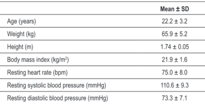
The clinical characteristics of the individuals are presented in Table 1.
Adaptation sessions and test with one maximum repetition (1-RM)
Before the experimental sessions, each individual participated in two sessions to adapt to resistance exercises,
performed on different days. In each adaptation session, the individuals performed three sets of 12 repetitions with the minimum permissible load in the following exercises: i) bench press, ii) barbell rows, iii) front lifting, iv) curl and v) triceps curl. All exercises included in the study were performed with free weights.
Two days after the last adaptation session, the individuals performed the maximum repetition test (1-RM)16. The test
began with a warm-up (10 reps), with approximately 50% of the estimated load for the first attempt, based on the previous experience of participants. Then, there were three attempts with progressive to load until the load of 1-RM was identified. Between the attempts, and between the exercises, we maintained a recovery interval of two minutes. In order to familiarize the individuals with the test17, we performed 04
test sessions of 1-RM, with a minimum interval of 48 hours between sessions, where the highest load found during the tests was used to calculate the exercise intensity in the experimental sessions.
Experimental protocol
After determining the loads of 1-RM, the individuals underwent three experimental sessions: control (C), resistance exercise with 50% of 1-RM (E50%) and resistance exercise with 70% of 1-RM (E70%). The order of sessions was randomly determined. Between sessions, there was an interval of at least three days.
The sessions started at the same time of the day. The individuals were instructed to have a light meal two hours before the experiments, avoid exercise and alcohol intake for at least 48 hours in advance, and caffeine in the last 12 hours, as well as keep similar sleep schedules and daily activities in all three sessions. Moreover, the experimental sessions were scheduled on days on which the individuals had similar routines.
In each experimental session, each individual remained sitting at rest for 10 minutes by the time they got to the laboratory. Then, the heart rate was measured for 10 continuous minutes, using a frequency meter (Polar, RS 800 CX, USA). After resting measurements, the individuals would move to the weight-lifting room where they performed the sessions of resistance or control exercise.
The three sessions were composed of the 05 exercises described above, performed in three consecutive sets of 12, 9 and 06 repetitions. In all sessions (C, E50% and E70%) before each exercise, a warm-up series of 10 repetitions was performed using 25% of 1-RM. In session C, the individuals performed the exercises with a plastic bar (0.1 kg) to ensure that the differences found in comparison with exercise sessions (E50% and E70%) were due to the intensity used other than the motion or changes in posture. In sessions E50% and E70%, the load of the exercises was 50% and 70% of 1-RM, respectively. The resting interval between the series and between the exercises in every session lasted two minutes.
After completion of the interventions, the individuals returned to the laboratory, where they remained sitting at rest for more 60 minutes (post-intervention period). Heart rate was obtained at intervals of 10 minutes, between 20 and 30 and 50 and 60 minutes of recovery.
Table 1 - Characteristics of individuals
Mean ± SD
Age (years) 22.2 ± 3.2
Weight (kg) 65.9 ± 5.2
Height (m) 1.74 ± 0.05
Body mass index (kg/m2) 21.9 ± 1.6
Resting heart rate (bpm) 75.0 ± 8.0
Resting systolic blood pressure (mmHg) 110.6 ± 9.3
Analysis of heart rate variability
The autonomic modulation of the cardiovascular system was obtained by the technique of spectral analysis of heart rate variability. For this purpose, we used the RR intervals obtained from the frequency meter (Polar, RS 800 CX, USA). Hence, stationary periods of the tachogram, with at least 500 beats, were broken down into bands of low (LF) and high (HF) frequencies using the autoregressive method, using the software Kubios HRV (Finland), following the recommendations of the Task Force of Spectral Analysis18.
Frequencies between 0.04 and 0.4 Hz were considered as physiologically significant, whereas the LF component was represented by oscillations between 0.04 and 0.15 Hz and HF was represented by oscillations between 0.15 and 0.4 Hz. The power of each spectral component was normalized by dividing the power of each spectrum band by the total variance, minus the value of very low frequency band (<0.04 Hz), and multiplying the result by 100. For interpreting the results, the LF and HF normalized components of heart rate variability were considered, respectively, as representative of predominantly sympathetic and parasympathetic modulation of the heart, and the ratio between these bands (LF/HF) as the cardiac sympathovagal balance18.
Statistical analysis
The normality and homogeneity of variance of data were confirmed by the Shapiro Wilks and Levene tests, respectively. Data from the pre-intervention period in the three experimental sessions were compared by the analysis of variance (ANOVA) of one factor for repeated measures. Autonomic responses in each experimental session were calculated as the difference between the values measured after and before the intervention (D= after - before). The responses of heart rate variability after the three experimental sessions
were compared by two-factor ANOVA for repeated measures, considering the session (C, E50% and E70%) and time (before, 20 to 30 and 50 to 60 minutes) as major factors. When F value was significant, we performed post hoc Newman-Keuls test. The alpha value of p < 0.05 was considered significant and data are presented as mean ± standard deviation.
Results
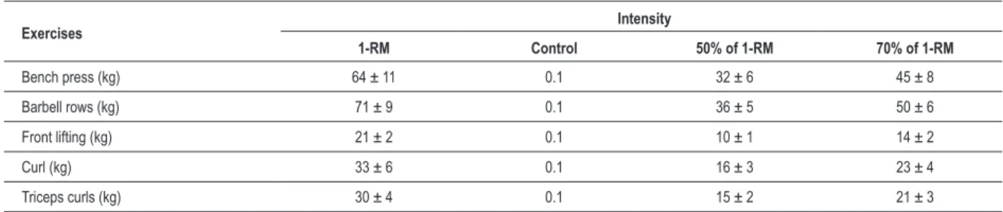
All individuals included in the study had low cardiovascular risk. Out of the 15 individuals, three of them started the protocol with session C, 07 with E50% and 05 with E70%. Tests of 1-RM and the loads used during the sessions of resistance exercise are presented in Table 2.
The data of heart rate variability before the interventions in the three experimental sessions (C, E50% and E70%) are presented in Table 3. The pre-exercise values were similar between sessions C, E50% and E70% (p > 0.05).
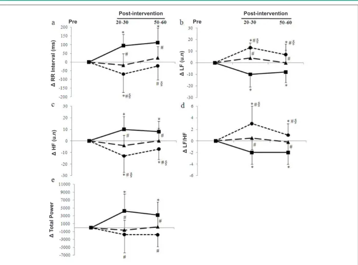
Changes in cardiac autonomic modulation after the three experimental sessions are shown in Figure 1.
Compared with pre-intervention values, the RR interval increased after the C session, throughout the recovery period (greatest increase: +112 ± 83 ms, p < 0,01), remained steady after the session E50%, throughout the recovery period (p > 0.05) and decreased over the first 30 minutes of recovery at session E70% (greatest drop: -105 ± 69 ms, p < 0.01). Thus, the behavior of the RR interval throughout the recovery period differed significantly between the three experimental sessions (p < 0.05).
Compared to the pre-intervention values, the BF band and LF/HF ratio decreased in session C (greatest drop: -10 ± 11 un., -2 ± 2, respectively, p < 0.01), remained steady in session E50% (p > 0.05) and increased in session E70% (greatest increase: +13 ± 14 un.; +3 ± 3, respectively, p
Table 2 - Overloads of resistance exercises used in the control sessions (C), with 50% of 1-RM (E50%) and 70% of 1-RM (E70%)
Exercises Intensity
1-RM Control 50% of 1-RM 70% of 1-RM
Bench press (kg) 64 ± 11 0.1 32 ± 6 45 ± 8
Barbell rows (kg) 71 ± 9 0.1 36 ± 5 50 ± 6
Front lifting (kg) 21 ± 2 0.1 10 ± 1 14 ± 2
Curl (kg) 33 ± 6 0.1 16 ± 3 23 ± 4
Triceps curls (kg) 30 ± 4 0.1 15 ± 2 21 ± 3
Table 3 - Cardiac autonomic modulation at rest before the sessions control (C), 50% of 1-RM (E50%) and 70% of 1-RM (E70%)
Control E50% E70% p
RR interval (ms) 794 ± 104 806 ± 84 809 ± 116 ns
Low frequency band (un) 77 ± 9 74 ± 12 68 ± 14 ns
High frequency band (un) 23 ± 9 26 ± 12 32 ± 14 ns
Low frequency/high frequency ratio 4.1 ± 2.0 3.7 ± 2.1 2.8 ± 1.7 ns
Total power (ms2) 4,227 ± 2,627 5,368 ± 2,844 4,320 ± 3,731 ns
Fig. 1 - Changes in RR interval (a), low frequency band (LF) (b), high frequency band (HF) (c), LF/HF ratio (d) and total power (e) observed in 15 individuals after the sessions C (squares), E50% (triangles) and E70% (circles). * Signiicantly different from pre-intervention (p < 0.05); # Signiicantly different from session C (p < 0.05); Signiicantly different from session E50% (p < 0.05).
Δ
R
R
In
te
rv
al
(m
s)
Δ
T
ota
l Po
w
er
Post-intervention Post-intervention
Pre Pre
Δ
L
F/
H
F
Δ
L
F
(u
.n
)
Δ
H
F
(u
.n
)
< 0.01). Thus, the behavior of these variables throughout the recovery period differed significantly between the three experimental sessions (p < 0.05).
Comparing the pre-intervention values, the HF band increased in session C (greatest increase: +10 ± 11 un., p < 0.01), remained steady in session E50% (p > 0.05) and decreased in session E70% throughout the recovery period (greatest drop: -13 ± 14 un., p < 0.01). Thus, throughout the recovery period, the behavior of HF band differed significantly between the three experimental sessions (p < 0.05).
Comparing the pre-intervention values, the total power increased only in session C (greatest increase: +4,227 ± 4,328 ms2, p < 0.01) and remained steady in sessions E50%
and E70% throughout the recovery period. Thus, the behavior of this variable throughout the recovery period was different between the session C and the sessions E50% and E70%.
Discussion
The results of this study demonstrate that the resistance exercise for trunk and upper limbs led to an increased cardiac sympathetic modulation and reduced cardiac parasympathetic modulation. Moreover, this change in the
cardiac autonomic modulation was higher after a more intense resistance exercise.
Over 60 minutes after the session E70%, we saw an increased sympathetic modulation and reduced cardiac parasympathetic modulation. These results are similar to those observed in previous studies that used resistance exercise for key muscle groups6,8 or for lower limbs12. These results
suggest that regardless of the exercises employed (major muscle groups, lower limbs or trunk and upper limbs), the completion of resistance exercise causes changes in cardiac autonomic modulation, which remain for a long period after the completion of the session.
The mechanisms involved in this response were not investigated in this study. Nevertheless, it is possible that in the control session, the orthostatic stress caused by prolonged sitting promotes decreased venous return and increased peripheral sympathetic nerve activity stimulating the baroreflex19,20. This stimulation, in turn, promotes a
decrease in sympathetic nerve activity and increase in cardiac parasympathetic activity21. On the other hand, the session of
resistance exercise potentially leads to a greater reduction in venous return, deactivating cardiopulmonary receptors6, since,
Furthermore, the orthostatic stress seems to enhance this response, since the restoration of heart rate while sitting seems to last longer compared to the supine position22.
In this study, compared to the resistance exercise at 50% of 1-RM, the resistance exercise performed with 70.0% of 1-RM caused a greater increase in sympathetic cardiac autonomic modulation. These results are different from those observed by Rezk et al6, who observed similar changes in cardiac
autonomic modulation after the completion of resistance exercise with 80% of 1-RM and 40% of 1-RM until fatigue. This controversy was possibly caused by the variation in the number of repetitions between the intensities in the study by Rezk et al6, which did not happened in this study. These results
indicate that increased cardiac sympathetic modulation after resistance exercise depends on exercise intensity, as long as the number of repetitions is the same.
Regarding the duration of cardiac autonomic changes after the resistance exercise, Rezk et al6 showed that 75
minutes after the resistance exercise, the sympathetic cardiac autonomic modulation remained high. These results are similar to those observed in the session E70% of this study, where changes in autonomic modulation occurred within 60 minutes of recovery. On the other hand, in the E50% session, the values of cardiac autonomic modulation in 60 minutes after exercise were similar to those at rest. Considering that in session E70% the individuals came very close to fatigue in each series, which was not the case in session E50%, we could suggest that the resistance exercises close to fatigue result in a greater and longer lasting cardiac sympathetic activation9,10. This response
is possibly related to higher mechanical loads of the vascular system in higher intensity exercise, which promotes increased activation of mechanoreceptors23 and increased activation of
metaborreflex from reduced blood flow12. Another possible
mechanism is the greater decrease in plasma volume after resistance exercise with greater intensity due to leakage of blood into the interstitial space, thus promoting a decrease in venous return. This, in turn, would result in the deactivation of cardiopulmonary receptors, and the consequent increase in heart rate. However, these hypotheses remain to be tested.
These results have important practical applications. The increased sympathetic modulation and reduced cardiac parasympathetic modulation relate to increased cardiovascular risk9,10. Thus, it is possible to suggest that resistance exercise
for trunk and upper limbs with 70% of 1-RM promotes a transient alteration in cardiac autonomic modulation, which
may represent an increased cardiovascular risk, which does not happen when using 50% of 1-RM. Thus, the prescription of resistance exercise with 50% of 1-RM can be an important strategy to minimize the risk in those individuals with higher cardiovascular risk. However, this hypothesis needs to be confirmed in future studies.
This study has limitations that should be considered. There was no control over the respiratory frequency of individuals at any moment of the experimental sessions. Thus, it is possible that respiratory movements may have influenced the power of high-frequency band. The individuals in this study were young and healthy and extrapolating the results to individuals with other characteristics is limited. However, given the paucity of data on this theme in literature, the results of this study provide initial indications on the impact of resistance exercise on cardiac autonomic modulation.
Finally, although the order of the sessions was randomized, the randomization resulted in an unbalanced distribution between sessions. However, we believe that the probability that this factor has impacted considerably the results is minimal.
Conclusion
After an acute session of resistance exercise for trunk and upper limbs, there is an increase in sympathetic modulation and reduced cardiac parasympathetic modulation. These responses are more pronounced in the session with increased intensity.
Potential Conflict of Interest
No potential conflict of interest relevant to this article was reported.
Sources of Funding
This study was funded by Fundação de Amparo a Ciência e
Tecnologia do Estado de Pernambuco (FACEPE); Programa de
Fortalecimento Acadêmico da Universidade de Pernambuco
(PFA-UPE); Conselho Nacional de Desenvolvimento Científico
e Tecnológico (CNPq) .
Study Association
This study is not associated with any post-graduation program.
References
1. American College of Sports Medicine Position Stand. Exercise and physical activity for older adults. Med Sci Sports Exerc. 1998;30(6):992-1008.
2. Kraemer WJ, Adams K, Cafarelli E, Dudley GA, Dooly C, Feigenbaum MS, et al. American College of Sports Medicine Position Stand. Progression models in resistance training for healthy adults. Med Sci Sports Exerc. 2002;34(2):364-80.
3. Williams MA, Haskell WL, Ades PA, Amsterdam EA, Bittner V, Franklin BA, et al. Resistance exercise in individuals with and without cardiovascular disease:
2007 update: a scientific statement from the American Heart Association Council on Clinical Cardiology and Council on Nutrition, Physical Activity, and Metabolism. Circulation. 2007;116(5):572-84.
4. Dias RMR, Gurjão ALD, Marucci MFN. Benefícios do treinamento com pesos para aptidão física de idosos. Acta Fisiatr. 2006;13(2):90-5.
6. Rezk CC, Marrache RC, Tinucci T, Mion D Jr, Forjaz CL. Post-resistance exercise hypotension, hemodynamics, and heart rate variability: influence of exercise intensity. Eur J Appl Physiol. 2006;98(1):105-12
7. Kingsley JD, Panton LB, McMillan V, Figueroa A. Cardiovascular autonomic modulation after acute resistance exercise in women with fibromyalgia. Arch Phys Med Rehabil. 2009;90(9):1628-34.
8. Heffernan KS, Kelly EE, Collier SR, Fernhall B. Cardiac autonomic modulation during recovery from acute endurance versus resistance exercise. Eur J Cardiovasc Prev Rehabil. 2006;13(1):80-6.
9. Mourot L, Bouhaddi M, Tordi N, Rouillon JD, Regnard J. Short- and long-term effects of a single bout of exercise on heart rate variability: comparison between constant and interval training exercises. Eur J Appl Physiol. 2004;92(4):508-17.
10. Seiler S, Haugen O, Kuffel E. Autonomic recovery after exercise in trained athletes: intensity and duration effects. Med Sci Sports Exerc. 2007;39(8):1366-73.
11. American College of Sports Medicine position stand. Progression models in resistance training for healthy adults. Med Sci Sports Exerc. 2009;41(3):687-708.
12. Simoes RP, Mendes RG, Castello V, Machado HG, Almeida LB, Baldissera V, et al. Heart-rate variability and blood-lactate threshold interaction during progressive resistance exercise in healthy older men. J Strength Cond Res. 2010;24(5):1313-20.
13. Polito MD, Farinatti PT. The effects of muscle mass and number of sets during resistance exercise on postexercise hypotension. J Strength Cond Res. 2009;23(8):2351-7.
14- Sociedade Brasileira de Cardiologia. Sociedade Brasileira de Hipertensão. Sociedade Brasileira de Nefrologia. VI diretrizes brasileira de hipertensão. Arq Bras Cardiol.2010;95(1 supl.1):1-51
15. Wu JS, Lu FH, Yang YC, Lin TS, Chen JJ, Wu CH, et al. Epidemiological study on the effect of pre-hypertension and family history of hypertension on cardiac autonomic function. J Am Coll Cardiol. 2008;51(19):1896-901.
16. Clarke DH. Adaptations in strength and muscular endurance resulting from exercise. Exerc Sport Sci Rev. 1973;1:73-102.
17. Dias RMR, Cyrino ES, Salvador EP, Caldeira LFS, Nakamura FY, Papst RR, et al. Influência do processo de familiarização para avaliação da força muscular em testes de 1-RM. Rev Bras Med Esporte. 2005;11(1):34-8.
18. Heart rate variability: standards of measurement, physiological interpretation and clinical use. Task Force of the European Society of Cardiology and the North American Society of Pacing and Electrophysiology. Circulation. 1996;93(5):1043-65.
19. Gotshall RW, Aten LA, Yumikura S. Difference in the cardiovascular response to prolonged sitting in men and women. Can J Appl Physiol. 1994;19(2):215-25.
20. Queiroz AC, Gagliardi JF, Forjaz CL, Rezk CC. Clinic and ambulatory blood pressure responses after resistance exercise. J Strength Cond Res. 2009;23(2):571-8.
21. Meneses AL, Silva GQM, Lima AHRA, Farah BQ, Forjaz CLM, Dias RMR. Efeito da intensidade do exercício de força para membros superiores nas respostas cardiovasculares pós-exercício.In: 30 Congresso Brasileiro de Metabolismo, Nutrição e Exercício; 20 de maio,2010 Londrina(PR). Londrina (PR);2010;
22. Farinatti PTV, Nakamura FY, Polito MD. Influence of recovery posture on blood pressure and heart rate after resistance exercises in normotensive subjects. J Strength Cond Res. 2009;23(9):2487-92.