Letters to the Editor
Radiol Bras. 2017 Mai/Jun;50(3):199–208
200
André Martins Fernandes1, Bernardo Vieira Paim1, Ana
Paula Aguiar Vidal1, Edson Marchiori1, Daniella Braz
Parente2
1. Universidade Federal do Rio de Janeiro (UFRJ), Rio de Janeiro, RJ, Brazil. 2. Instituto D’Or de Pesquisa e Ensino, Rio de Janeiro, RJ, Brazil. Mailin address: Dr. André Martins Fernandes. Hospital Universitário Cle-mentino Fraga Filho. Rua Rodolpho Paulo Rocco, 255, Cidade Universitá-ria. Rio de Janeiro, RJ, Brazil, 21941-913. E-mail: [email protected]. REFERENCES
1. Beilan J, Lawton A, Hajdenberg J, et al. Pheochromocytoma of the uri-nary bladder: a systematic review of the contemporary literature. BMC Urol. 2013;13:22.
2. Martins DL, Baroni RH, Blasbalg R, et al. Evaluation of adrenal tumors by magnetic resonance imaging with histological correlation. Radiol Bras. 2008;41:55–62.
3. Wong EMH, Lai TCT, Tsu JHL, et al. Primary paraganglioma of uri-nary bladder: case series and review of the literature. Surgical Practice. 2015;19:82–5.
4. Peng C, Bu S, Xiong S, et al. Non-functioning paraganglioma occur-ring in the urinary bladder: a case report and review of the literature. Oncol Lett. 2015;10:321–4.
5. Montón CS, Esparza JFO, Ventura AB, et al. Mesothelioma of the tu-nica vaginalis in a patient with giant hydrocele. Radiol Bras. 2016;49: 63–4.
6. Rondina RG, Volpato R, Guerra LFA, et al. Differential diagnosis of anterior sacral meningocele during the evaluation of post-hysterectomy pelvic collections. Radiol Bras. 2016;49:203–4.
7. Queiroz RM, Costa PP, Oliveira NYF, et al. Female urethral diverticu-lum containing a urothelial carcinoma. Radiol Bras. 2016;49:406–7. 8. Lopes PM, Sepúlveda L, Ramos R, et al. The role of transrectal
ultra-sound in the diagnosis of prostate cancer: new contributions. Radiol Bras. 2015;48:7–11.
9. Ferreira DM, Bezerra ROF, Ortega CD, et al. Magnetic resonance imaging of the vagina: an overview for radiologists with emphasis on clinical decision making. Radiol Bras. 2015;48:249–59.
10. Salvadori PS, Bomfim LN, von Atzingen AC, et al. Spontaneous rupture of ovarian cystadenocarcinoma: pre- and post-rupture computed tomog-raphy evaluation. Radiol Bras. 2015:330–2.
11. Qiao HS, Feng XL, Yong L, et al. The MRI of extraadrenal pheochro-mocytoma in the abdominal cavity. Eur J Radiol. 2007;62:335–41. 12. Young WF Jr. Paragangliomas: clinical overview. Ann N Y Acad Sci.
2006;1073:21–9.
http://dx.doi.org/10.1590/0100-3984.2015.0204
Carpal boss syndrome: os styloideum fused to the trapezoid
Dear Editor,
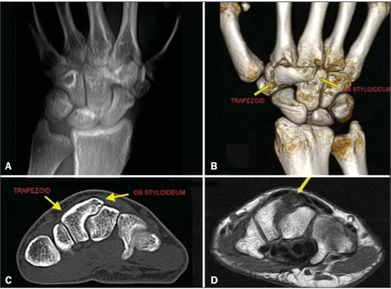
A 29-year-old White female presented with chronic pain on dorsiflexion of the right hand and a hard prominence, which was painful on palpation, at the base of the second and third metacar-pal muscles. An X-ray of the hand (Figure 1A) revealed a bony prominence in the region identified as palpable in the physical examination, as well as showing that there was lack of definition of the joint space between the trapezoid and the capitate. In multiplanar and three-dimensional computed tomography recon-structions, which provided greater detail (Figures 1B and 1C), an os styloideum was seen to be fused to the trapezoid bone and in neoarticulation with the base of the third metacarpal. Magnetic
resonance imaging showed a hypointense signal on a T1-weighted image (Figure 1D) and increased intensity in a T2-weighted short-tau inversion-recovery sequence, with bone edema adjacent to the neoarticulation, which is indicative of apophysitis.
Os styloideum is an anatomical variation characterized by an accessory ossicle on the dorsum of the wrist, between the trap-ezoid and capitate, at the base of the second and third metacarpal bones(1). When it produces symptoms, mainly local pain, it is known as a carpal boss(2,3). The true incidence of carpal boss syndrome is unknown; it is probably underestimated and often confused, clini-cally, with other causes of tumor in the dorsum of the carpus(4).
Although a carpal boss can be classified as acquired (osteo-phytic), congenital (secondary to os styloideum), or of mixed etiol-ogy, the clinical presentations appear to be similar across the
Figure 1. A: Digital X-ray showing a lack of definition of the joint space between the trapezoid and the capitate. B,C: Three-di-mensional computed tomography recon-struction and axial computed tomography slice showing an os styloideum fused to the trapezoid and in neoarticulation with the capitate. D: Magnetic resonance imaging in a T1-weighted sequence, showing os styloideum with bone edema adjacent to the neoarticulation (apophysitis).
A
B
Letters to the Editor
Radiol Bras. 2017 Mai/Jun;50(3):199–208
201
groups(3). Os styloideum is also known as the ninth carpal bone(5). The main difficulty in recognizing a carpal boss lies in the nonspecificity of the symptoms, which are often attributed to dor-sal cysts, given that the two conditions are quite similar in terms of their location(4).
The case reported here represents the rarest form of congeni-tal carpal boss, in which the os styloideum is fused to the trap-ezoid, which occurs in only 0.5% of cases. More commonly (in 94.0% of cases), it is fused to the base of the second and third metacarpal, merged with the capitate (in 3.5%) or (in 2.0%) iso-lated(2,6). The clinical presentation of carpal boss is highly vari-able(2): the condition can be asymptomatic or can produce spon-taneous pain, precipitated by excessive use of the joint or by pal-mar flexion of the wrist.
Knowledge of the disease and imaging studies are fundamen-tal for the diagnosis of carpal boss and for distinguishing it from its main differential diagnoses, which include synovial cysts, frac-tures, osteoarthrosis, exostoses, bone neoplasms, and soft-tissue neoplasms(7). Tomography studies allow the relationship between the accessory ossicle and the adjacent bones to be analyzed, and magnetic resonance imaging is important for the evaluation of the integrity of bones, entheses, and ligaments(5). The proximity of the carpal boss to the short and long radial extensor tendons of the carpus can cause insertional tenosynovitis, aggravating the symptoms, especially in athletes who perform repetitive move-ments, specifically those involving forced flexion of the wrist (5,8,9). The treatment for carpal boss is usually conservative, typi-cally involving the use of anti-inflammatory drugs and, in some cases, immobilization of the wrist(6,7). However, surgical excision
can be required in cases that are refractory to the standard treat-ment(6,7).
REFERENCES
1. Karmazyn B, Siddiqui AR. Painful os styloideum in a child. Pediatr Radiol. 2002;32:370–2.
2. Gomes AF, Paganella VC, Paganella MC, et al. Computed tomography and magnetic resonance imaging findings of os styloideum in a symp-tomatic athlete. Radiol Bras. 2010;43:207–9.
3. Apple JS, Martinez S, Nunley JA. Painful os styloideum: bone scintigra-phy in carpe bossu disease. AJR Am J Roentgenol. 1984;142:181–2.
4. Castro AA, Skare TL, Nassif PAN, et al. Sonographic diagnosis of carpal tunnel syndrome: a study in 200 hospital workers. Radiol Bras. 2015;48: 287–91.
5. Poh F. Carpal boss in chronic wrist pain and its association with partial osseous coalition and osteoarthritis – a case report with focus on MRI findings. Indian J Radiol Imaging. 2015;25:276–9.
6. Conway WF, Destouet JM, Gilula LA, et al. The carpal boss: an overview of radiographic evaluation. Radiology. 1985;156:29–31.
7. Park MJ, Namdari S, Weiss AP. The carpal boss: review of diagnosis and treatment. J Hand Surg Am. 2008;33:446–9.
8. Kissel P. Conservative management of symptomatic carpal bossing in an elite hockey player: a case report. J Can Chiropr Assoc. 2009;53:282–9. 9. Linscheid RL, Dobyns JH. Athletic injuries of the wrist. Clin Orthop Relat
Res. 1985;(198):141–51.
Gabriel Clève Nicolodi1, Cesar Rodrigo Trippia1, Maria
Fernanda F. S. Caboclo1, Raphael Rodrigues de Lima1,
Wagner Peitl Miller1
1. Hospital São Vicente – Funef, Curitiba, PR, Brazil. Mailing address: Dr. Gabriel Clève Nicolodi. Rua Francisco Rocha, 1435, ap. 31, Bigor-rilho. Curitiba, PR, Brazil, 80730-390. E-mail: [email protected].
Prenatal diagnosis of sirenomelia in the second trimester of pregnancy using two-dimensional ultrasound, three-dimensional ultrasound and magnetic resonance imaging
Dear Editor,
A 30-year-old woman was referred at 23 weeks of gestation due to olygohydramnios, together with short fetal femur length and cystic hygroma. It was the first pregnancy for a non-consanguin-eous couple with a family history of neural tube defects. The pa-tient reported chronic arterial hypertension during her pregnancy. The previous ultrasound findings were confirmed at our facility. Two-dimensional (2D) ultrasound showed fusion of the lower limbs, and color Doppler ultrasound revealed no vascularization of the lower limbs (Figure 1A). Three-dimensional (3D) ultrasound in the rendering mode confirmed the findings of the 2D
ultra-sound (Figure 1B). For a better understanding of the fetal mor-phology due to the olygohydramnios, magnetic resonance imag-ing (MRI) was performed. The MRI scan showed myelomenin-gocele and bilateral renal agenesis, as well as showing no identi-fiable characteristics of the lower limbs (Figure 1C). Termina-tion of the pregnancy was authorized at 29 weeks of gestaTermina-tion. The stillborn infant weighed 1120 g. Pathologic investigation showed sirenomelia (sympus apus), lumbar myelomeningocele, and interventricular communication (Figure 2). Radiographic studies showed only one femur (sirenomelia type VII according to the Stocker and Heifetz classification).
Sirenomelia is a rare congenital anomaly with an estimated incidence of 1:60,000 live births(1). It is defined by fused lower limbs, a single umbilical artery, and genitourinary anomalies(2). In ap-proximately 25–30% of cases, sirenomelia is accompanied by other
Figure 1. Prenatal findings of sirenomelia at 26 weeks and 5 days of gestation: 2D ultrasound with color Doppler in the axial plane shows myelomeningocele. Note that the mass is very close to the neck (arrow, A); same view at 3D ultrasound in the rendering mode (B), and at T2-weighted MRI sequence in the sagittal plane (C). Note that the mass of lumbar origin (myelomeningocele) is very close to the cervical region of the fetus (arrow, C).
A B C