Submitted 23 December 2014
Accepted 8 June 2015
Published25 June 2015
Corresponding author
David G. Fernig, [email protected]
Academic editor Sandhya Visweswariah
Additional Information and Declarations can be found on page 21
DOI10.7717/peerj.1060
Copyright 2015 Sun et al.
Distributed under
Creative Commons CC-BY 4.0
OPEN ACCESS
HaloTag is an e
ff
ective expression and
solubilisation fusion partner for a range
of fibroblast growth factors
Changye Sun1,3, Yong Li1,3, Sarah E. Taylor1, Xianqing Mao2, Mark C. Wilkinson1and David G. Fernig1
1Department of Biochemistry, Institute of Integrative Biology, University of Liverpool, Liverpool,
UK
2Department of Oncology, Laboratory of Cellular and Molecular Oncology, Luxembourg
Institute of Health, Luxembourg
3These authors contributed equally to this work.
ABSTRACT
The production of recombinant proteins such as the fibroblast growth factors (FGFs) is the key to establishing their function in cell communication. The production of recombinant FGFs inE. coliis limited, however, due to expression and solubility problems. HaloTag has been used as a fusion protein to introduce a genetically-encoded means for chemical conjugation of probes. We have expressed 11 FGF proteins with an N-terminal HaloTag, followed by a tobacco etch virus (TEV) protease cleavage site to allow release of the FGF protein. These were purified by heparin-affinity chromatography, and in some instances by further ion-exchange chromatography. It was found that HaloTag did not adversely affect the expression of FGF1 and FGF10, both of which expressed well as soluble proteins. The N-terminal HaloTag fusion was found to enhance the expression and yield of FGF2, FGF3 and FGF7. Moreover, whereas FGF6, FGF8, FGF16, FGF17, FGF20 and FGF22 were only expressed as insoluble proteins, their N-terminal HaloTag fusion counterparts (Halo-FGFs) were soluble, and could be successfully purified. However, cleavage of Halo-FGF6, -FGF8 and -FGF22 with TEV resulted in aggregation of the FGF protein. Measurement of phosphorylation of p42/44 mitogen-activated protein kinase and of cell growth demonstrated that the HaloTag fusion proteins were biologically active. Thus, HaloTag provides a means to enhance the expression of soluble recombinant proteins, in addition to providing a chemical genetics route for covalent tagging of proteins.
Subjects Biochemistry
Keywords Fibroblast growth factor, Recombinant protein expression, HaloTag, Fusion protein
INTRODUCTION
has been important in probing the mechanisms of, for example, their transport (Duchesne et al., 2012;Lin, 2004;Yu et al., 2009). Chemical labelling has disadvantages compared to genetically encoded labelling, since with the latter it is easier to predict the structural and hence functional consequences of labelling, which can be achieved bothin vitro
andin vivo. While fluorescent proteins remain a mainstay of genetic labelling, they have
limitations. These have been overcome, for example, by non-covalent tagging of proteins on hexahistidine sequences with Tris-Ni2+
nitriloacetic acid (Huang et al., 2009;Lata et al., 2005;Tinazli et al., 2005), which has allowed diverse labelling strategies, ranging from fluorescent dyes (Uchinomiya et al., 2009) and quantum dots (Roullier et al., 2009;
Susumu et al., 2010) to gold nanoparticles (Duchesne et al., 2008). However, non-covalent coupling is reversible and exchange may occur in this instance with histidine-rich patches on endogenous proteins.
HaloTag is a mutant of a bacterial haloalkane dehalogenase, which reacts with chloroalkane ligands to form a covalent bond that represents the covalent intermediate of the enzyme’s normal catalytic cycle (Los et al., 2008). Fluorescent dyes (Los et al., 2008) and quantum dots (Zhang et al., 2006b) carrying a chloroalkane group have been used to label HaloTag fusion proteins for fluorescence imaging. This approach is particularly versatile, since it combines the power of a genetically encoded tag (the HaloTag protein) with covalent labelling.
Consequently, we set out to produce N-terminal HaloTag fusions of different FGFs. In the course of this work, we observed that the N-terminal HaloTag fusion had a substantial effect on the expression of the more recalcitrant FGFs, consistent with the observation that HaloTag is a potential solubilisation tag for recombinant proteins (Ohana et al., 2009). Thus, whereas expression of FGF1 and FGF10 was somewhat reduced and that of FGF2 increased, expression of FGF7, which can be toxic (Ron et al., 1993), was no longer so, while expression of soluble FGF3, FGF6, FGF7, FGF8, FGF16, FGF17, FGF20 and FGF22 was markedly enhanced. This is in contrast to previous reports where FGFs such as FGF6 (Pizette et al., 1991), FGF8 (Loo & Salmivirta, 2002;Macarthur et al., 1995;Vogel, Rodriguez & IzpisuaBelmonte, 1996), FGF16 (Danilenko et al., 1999) and FGF20 (Jeffers et al., 2002;
Kalinina et al., 2009) have been found to be mainly expressed in inclusion bodies, even as truncated proteins, and so require refolding. Thus, HaloTag provides not just a means to label proteins covalently and specifically, but is also a useful solublisation partner for the production of recombinant proteins.
MATERIALS AND METHODS
Materials
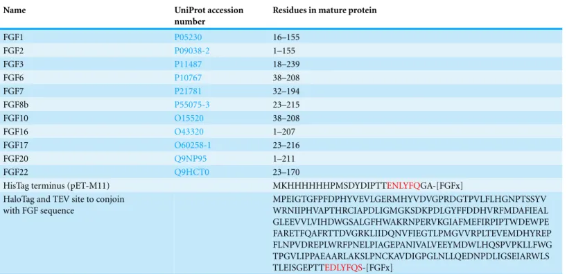
Table 1 Peptide sequences of FGFs, the N-terminal HisTag constructs and the N-terminal HaloTag constructs.FGF names, sequences and amino acid numbering are according to the UniProt entry. FGF1 is an N-terminal truncated protein (Ke et al., 1990). FGF2 does not possess a secretory signal sequence, whereas there is no signal peptide recognised in Uniprot for FGF16 and FGF20; consequently full length protein sequence was expressed. For all other FGFs, the protein expressed was without the Uniprot determined secretory signal sequence. FGFx refers any one of the FGFs. TEV cleavage sites are in red.
Name UniProt accession
number
Residues in mature protein
FGF1 P05230 16–155
FGF2 P09038-2 1–155
FGF3 P11487 18–239
FGF6 P10767 38–208
FGF7 P21781 32–194
FGF8b P55075-3 23–215
FGF10 O15520 38–208
FGF16 O43320 1–207
FGF17 O60258-1 23–216
FGF20 Q9NP95 1–211
FGF22 Q9HCT0 23–170
HisTag terminus (pET-M11) MKHHHHHHPMSDYDIPTTENLYFQGA-[FGFx] HaloTag and TEV site to conjoin
with FGF sequence
MPEIGTGFPFDPHYVEVLGERMHYVDVGPRDGTPVLFLHGNPTSSYV WRNIIPHVAPTHRCIAPDLIGMGKSDKPDLGYFFDDHVRFMDAFIEAL GLEEVVLVIHDWGSALGFHWAKRNPERVKGIAFMEFIRPIPTWDEWPE FARETFQAFRTTDVGRKLIIDQNVFIEGTLPMGVVRPLTEVEMDHYREP FLNPVDREPLWRFPNELPIAGEPANIVALVEEYMDWLHQSPVPKLLFWG TPGVLIPPAEAARLAKSLPNCKAVDIGPGLNLLQEDNPDLIGSEIARWLS TLEISGEPTTEDLYFQS-[FGFx]
of the protein sequences corresponding to the above cDNAs are listed inTable 1. Enzymes for cloning were from: NcoI, BamHI and T4 ligase (NEB, Hitchin, UK); KOD Hot Start DNA polymerase (Merck, Hertfordshire, UK); In-Fusion®HD Cloning Kit (Clontech, Takara Bio Europe SAS, Saint-Germain-en-Laye, France). Bacterial cells: DH5α, BL21 (DE3) pLysS and SoluBL21 were a gift from Olga Mayans, University of Liverpool. The sources of other materials were as follows: LB broth and LB agar (Merck, Hertfordshire, Germany); Soniprep 150 Plus (MSE, London, UK); Affi-Gel®Heparin Gel (Bio-Rad, Hertfordshire, UK), CM Sepharose Fast Flow, DEAE Sepharose Fast Flow, HiTrap Q HP column; empty disposable PD-10 Columns; ¨AKTApurifier 100 plus (GE Healthcare, Buckinghamshire, UK).
Figure 1 Cloning strategy for plasmids encoding Halo-FGFs.DNA encoding HaloTag was inserted 5′
of the FGF2 coding sequence with the In-Fusion HD enzyme. Subsequently, a NotI cleavage site was added 5′to the BamHI site and other FGFs were exchanged into the plasmid using the digestion-ligation
cloning method. A cartoon structure of Halo-FGF is presented in the middle of this figure.
(Sigma-Aldrich, Dorset, UK), anti-mouse IgG, horseradish peroxidase-linked antibody (Cell Signalling, NEB), polyvinylidene fluoride (PVDF) transfer membrane (Millipore UK, Hertfordshire, UK), enhanced chemiluminescence (ECL) Western blotting reagents (GE Healthcare, Little Chalfont, UK), Hyperfilm (GE Healthcare, Little Chalfont, UK).
DNA cloning of hexahistidine tagged FGFs (His-FGFs) and HaloTag tagged FGFs (Halo-FGFs)
DNA encoding FGF1, FGF3, FGF6, FGF8, FGF10, FGF16, FGF17, FGF20 and FGF22 was cloned into pET-M11 such that the resulting protein would have a N-terminal 6xhis tag followed by a tobacco etch virus (TEV) cleavage site (ENLYFQ). FGF2 and FGF7 DNA se-quences were previously cloned into pET-14b and pET-M11, respectively (Xu et al., 2012).
A plasmid encoding Halo-FGF2 was produced by adding a HaloTag encoding DNA sequence in-frame 5′
to a DNA sequence encoding full-length FGF2. This construct was then used to produce the other DNAs encoding Halo-FGFs (Fig. 1). The plasmid pET-14b-fgf2 contains NcoI and BamHI cleavage sites 5′
and 3′
of
Table 2 Concentrations of NaCl in 50 mM Tris-Cl buffer (pH 7.4) used for heparin affinity chromatog-raphy of FGFs.[NaCl] for lysate is the concentration of NaCl in the sample applied to the column.
Name [NaCl] for lysate (M) [NaCl] for wash (M) [NaCl] for elution (M)
FGF1 0.6 0.6 1.0
FGF2 0.6 0.6 1.5
FGF3 0.3 0.6 1.0
FGF6 0.3 0.4 1.0
FGF7 0.3 0.3 1.0
FGF8 0.6 0.6 1.5
FGF10 0.6 0.6 1.0
FGF16 0.3 0.4 1.0
FGF17 0.6 0.6 1.0
FGF20 0.3 0.4 1.0
FGF22 0.6 0.8 1.5
TCCCGGCTGCCATGGAGCTCTGAAAGTACAGATC, primers (NcoI cleavage site underlined), and inserted into the linearized vector using In-Fusion enzyme. A TEV cleavage site (Fig. 1: green ellipsoid) was also included at the C-terminus of HaloTag to allow release of the FGF. A NotI cleavage site was also inserted 5′
of the BamHI to provide an additional 3′
cleavage sites for cloning. The other cDNAs (FGF1, FGF3, FGF6, FGF7, FGF8, FGF10, FGF16, FGF17, FGF20 and FGF22) were exchanged into the established pET-14b-Halo-fgf2plasmid by double-digestion with NcoI and BamHI/NotI enzymes and ligation using T4 ligase (Fig. 1).
Protein expression and purification of His-FGFs and Halo-FGFs His-FGF7, because it is toxic like native FGF7 (Ron et al., 1993), was transformed into BL21 (DE3) pLysS (F– ompT hsdSB(rB–, mB–) gal dcm (DE3) pLysS (CamR)) for subsequent protein expression and purification. FGF2, the other His-FGFs and Halo-FGFs were transformed into SoluBL21(F–ompThsdSB(rB–, mB–)gal dcm (DE3)). The bacteria containing FGF encoding plasmids were cultured at 37◦
C until the OD600 values were between 0.4 and 0.6, and then protein expression at 16◦
C was induced by adding 1 mM isopropylβ-D-1-thiogalactopyranoside (IPTG). The bacteria were harvested by centrifugation at 4◦
C, 14,000 g for 10 min and the pellets frozen at−80◦C.
The bacterial pellets were resuspended with the corresponding 50 mM Tris-Cl lysate buffers (pH 7.4) (Table 2), and the cells were disrupted by 5–6 cycles of sonication (30 s sonication, 60 s pause) on ice. Cell debris and insoluble proteins were removed by centrifugation at 4◦
C, 30,000 g for 30 min. Then, the presence of soluble FGFs was tested by analysis of whole cells, the supernatant and pellet by separation of polypeptides on 12% (w/v) SDS-PAGE and coomassie staining.
agarose. For each FGF, different concentrations of NaCl (in 50 mM Tris-Cl pH 7.4) were used for washing and elution (Table 2) by following the previous measurements on the electrolyte sensitivity of their heparin binding assessed by Western blot (Asada et al., 2009). The yields of His-FGFs and Halo-FGFs were quantified by measuring the absorbance at 280 nm and the level of impurities were estimated by analysis of coomassie stained SDS-PAGE gels with ImageJ-Analyze-Gels (Ferreira & Rasband, 2012). The soluble His-FGFs eluted from heparin affinity chromatography were further purified by Ni2+
affinity chromatography. Due to the negative charge on the surface of HaloTag and positive charge on the surface of FGFs, Halo-FGFs could bind to both cation- and anion-exchange stationary phases. Thus, Halo-FGF1, Halo-FGF2, Halo-FGF3, Halo-FGF7 and Halo-FGF10 were purified by chromatography on a 5 mL HiTrap Q HP column. Samples were applied in 0.15 M NaCl in PB buffer (2.7 mM KCl, 10 mM Na2HPO4,
1.8 mM KH2PO4, pH 7.4) and eluted with a gradient running to 0.8 M NaCl in the
same buffer. Halo-FGF6 and Halo-FGF20 were purified by chromatography on a 3 mL column of CM Sepharose Fast Flow followed by a 3 mL column of DEAE Sepharose Fast Flow. Samples were again applied in 0.15 M NaCl in PB buffer and eluted with 0.4 M NaCl in the same buffer. The purified His-FGFs and Halo-FGFs were analysed by 12% (w/v) SDS-PAGE followed by coomassie staining.
Purification of FGFs by removing HaloTag from Halo-FGFs
To test the accessibility of the TEV cleavage site, some Halo-FGFs, including Halo-FGF2, Halo-FGF17, Halo-FGF6, Halo-FGF8 and Halo-FGF22 eluted with high concentration of NaCl in 50 mM Tris buffer from heparin agarose chromatography and Halo-FGF20 purified with heparin, DEAE and CM chromatography, were incubated with 2.5% (mol/mol) TEV protease at 4◦
C overnight. In cases where the digestion products were cloudy, they were clarified by centrifugation for 30 min at 13,000 g, 4◦
C. Samples were then analysed on a 12% (w/v) SDS-PAGE. The supernatants of the TEV digestions of Halo-FGF6 and of Halo-FGF20 were applied onto a 2 mL heparin agarose column, and washed as before (Table 2). FGF6 and FGF20 were eluted with PB buffer containing 1 M NaCl or 0.1 M arginine and 1 M NaCl, respectively. After TEV digestion, FGF17 was further purified on a 1 mL HiTrap SP HP cation-exchange column by washing with 0.3 M NaCl and eluting with 1 M NaCl, both in 50 mM Tris-Cl, pH 7.4. All of the fractions from the purification steps were analysed by 12% (w/v) SDS-PAGE.
Cell culture
Measurement of p44/42MAPKphosphorylation
Cells were cultured in 3 cm dishes until near confluence. Then, the dishes were washed twice with phosphate-buffered saline (PBS) and 2.5 mL step-down medium (SDM: DMEM with 250 ng BSA, 0.75% (w/v) sodium bicarbonate and 4 mM L-glutamine) was added for 24 h (Rama 27 cells) or 48 h (HaCaT cells). Rama 27 and HaCaT cells were then incubated with different FGFs for 15 min, as described in the figure legends. After the incubation, the cells were washed twice with ice-cold PBS and collected by scraping in 2X SDS-PAGE lysis buffer (4% (w/v) SDS, 20% (v/v) glycerol, 12% (v/v) Tris-Cl (pH 6.8), 2.5% (v/v)β-mercaptoethanol, 0.02% (w/v) bromophenol blue, 1 tablet of protease inhibitor and 6.8 mL distilled water). The cell lysates were heated for 10 min at 98◦
C prior to SDS-PAGE.
Western blot
After separation by 10% (w/v) SDS-PAGE, polypeptides were transferred onto a PVDF membrane. The membrane was blocked with 5% (w/v) skimmed milk in 1X TBST (50 mM Tris-Cl, 150 mM NaCl and 0.05% Tween-20 (v/v), pH 7.5) for 2 h. After two washes with TBST, the membrane was incubated with phospho-p44/42MAPKantibody (1:1,000 dilution in TBST) on a shaker overnight at 4◦
C. Secondary anti-mouse antibody (1:1,000 dilution) was added to the membrane after three washes with TBST, 5 min each, for 1 h at room temperature. Following three washes with TBST to remove the excess secondary antibody, the membrane was covered with 1 mL ECL solution and signal was detected with Hyperfilm. The same membrane was stripped with 2.5% (w/v) SDS in TBST at 50◦
C for 1 h and reblocked as above, before probing withβ-actin antibody (1:10,000 dilution). The Western blot band intensity was quantified in the same way as SDS-PAGE bands and the signal intensities of phospho-p44/42MAPKwere normalised by dividing by the intensity of the band corresponding toβ-actin and then by that of the BSA control samples.
Cell growth assay
Cell growth in Rama 27 fibroblasts was measured as before (Smith, Winslow & Rudland, 1984). Rama 27 cells were dispensed into a 24 well cell culture plates at 2,000 cells/well. After 24 h the cells were washed twice with PBS and cultured in SDM, as described for the p44/42MAPKphosphorylation assay for 24 h. The SDM was then replaced and the appropriate proteins added, as described in the figure legend. After 68 h incubation, cells were trypsinised and the number of cells counted.
RESULTS AND DISCUSSION
Expression of soluble FGFs
Group 1: soluble FGFs
After induction, bands corresponding to the expected molecular size of His-FGF1, FGF2 and His-FGF10 were apparent in the whole cell lysates (Figs. 2A,2Cand2E, lane L, green arrow). His-FGF1 and His-FGF10 were expressed at a higher level than FGF2 inE. coliSoluBL21. After centrifugation of the cell lysates, bands corresponding to the molecular size of all three FGFs were mainly recovered in the soluble fraction (supernatant), rather than in the insoluble fraction (pellet;Figs. 2A,2Cand2E, lanes S and P). Chromatography of the supernatants on heparin demonstrated that little of the expressed protein was present in the flow-through fraction (Figs. 2A,2Cand2E, lane T). Weak bands corresponding to His-FGF1 and His-FGF10, but not FGF2, were observed in the wash fraction (Figs 2Aand2E, lane Wa), which may represent aggregated or less well-folded protein. The majority of the three FGFs was recovered in the high NaCl eluate (Figs. 2A,2Cand2E, lane Hep), which demonstrated that these soluble FGFs bound heparin strongly. This indicated that they were likely to be properly folded, because the canonical, highest affinity heparin binding site of FGFs depends on the tertiary structure of the proteins (Xu et al., 2012).
The bands corresponding to Halo-FGF1, Halo-FGF2 and Halo-FGF10 were clearly observed in the whole cell lysates and these proteins were all highly expressed in SoluBL21 cells (Figs. 2B,2Dand2F, lane L, red arrow). Similarly to the His-FGF1, FGF2 and His-FGF10, after centrifugation of the whole cell lysates, the bands corresponding to the three Halo-FGFs were observed in the soluble fractions (Figs. 2B,2Dand2F, lanes S and P). Chromatography of the soluble fractions on heparin indicated that most of Halo-FGF2 and Halo-FGF10 had bound to the column, but there was a substantial amount of Halo-FGF1 in the flow-through (Figs. 2B,2Dand2F, lane T). This may be due to the capacity of the column for Halo-FGF1 being lower than for His-FGF1. All three Halo-FGFs were eluted from the heparin affinity column at the expected NaCl concentration (Figs. 2B, 2Dand2F, lane Hep).
The yield of Halo-FGF1 and Halo-FGF10 was similar to that of the corresponding his-tagged proteins (Table 3). However, since the Halo-FGF proteins are considerably larger than the corresponding His-tagged FGF1 and FGF10, this represents a decrease in the molar amounts of FGF produced. In contrast, the yield of Halo-FGF2 was 4-fold higher (Table 3), which is only partly accounted for by the increased size of the fusion protein. The low yield of full-length FGF2 has been ascribed to the presence of secondary structure at the 5′
end of the FGF2 mRNA (Knoerzer et al., 1989), and the presence of the upstream HaloTag sequence may mitigate this effect.
Group 2: low expression proteins
Figure 2 Expression and heparin affinity purification of His-FGF1, FGF2, His-FGF10, Halo-FGF1,
Halo-FGF2 and Halo-FGF10.Following induction of expression with IPTG, cells were lysed by
Table 3 Summary of the molecular sizes and yields of His-FGFs and Halo-FGFs.The molecular weight of the proteins was calculated from their amino acid sequence. The concentrations and volumes of His-FGFs and Halo-His-FGFs recovered from heparin affinity chromatography were measured. The impurities identified by SDS-PAGE were quantified using ImageJ relative to the band corresponding to His-FGF and to Halo-FGF and the amount of protein in the eluate from heparin chromatography adjusted accordingly, to provide an estimate of the yield.
FGFs Molecular weight (kDa) Yield (mg/L)
HisTag HaloTag HisTag HaloTag
FGF1 19.1 50.9 14 16
FGF2 17.3
No Tag
52.2 2.5
No Tag
11
FGF3 28.2 60.0 0.5 11
FGF6 22.3 54.1 n.d.a 27
FGF7 22.2 54.0 0.6 5.6
FGF8 25.7 57.5 n.d.a 1.7
FGF10 22.7 54.5 7.7 9.3
FGF16 26.9 58.7 n.d.a 1.0
FGF17 25.8 57.6 n.d.a 1.5
FGF20 26.9 58.6 n.d.a 10
FGF22 20.5 52.3 n.d.a 2.0
Notes.
aNot detected. Insufficient soluble protein for reliable quantification.
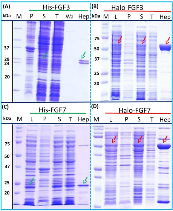
Transformation of SoluBL21with the plasmid encoding Halo-FGF7 yielded the expected number of colonies, indicating that the fusion protein was not toxic. Bands corresponding to the molecular size of Halo-FGF3 and Halo-FGF7 were observed in the cell lysates (Figs. 3Band3D, lane L, red arrow) and in the soluble fraction obtained after centrifu-gation, whereas the pellet has relatively weaker bands (Figs. 3Band3D, lanes P and S), indicating that Halo-FGF3 and Halo-FGF7 were soluble. Heparin chromatography of the soluble factions demonstrated that large amounts of Halo-FGF3 and Halo-FGF7 retained their heparin binding interaction with the polysaccharide (Figs. 3Band3D, lane Hep).
The yields of Halo-FGF3 and of Halo-FGF7 were 21-fold and 9-fold greater than of the corresponding His-tagged FGF (Table 3). Thus, the presence of the HaloTag N-terminal fusion increased the amounts of FGF3 and FGF7 substantially, even after taking into account the larger size of these fusion proteins (Table 3).
Group 3: insoluble proteins
been reported that FGF20 could also be solubilised by high concentrations of arginine (Maity, Karkaria & Davagnino, 2009), which suggests that FGF20 in the lysis buffer has a tendency to aggregate. However, arginine would compete for binding of FGFs to heparin, which reduces the utility of this approach to solubilisation.
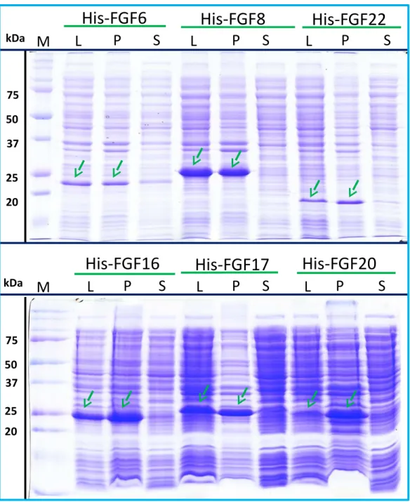
As illustrated by SDS-PAGE, all of the bands corresponding to the molecular size of Halo-FGF6, Halo-FGF8, Halo-FGF22, Halo-FGF17, Halo-FGF16 and Halo-FGF20 were clearly observed in the whole lysates, which suggested that all six proteins expressed well in
E. coli(Fig. 5, lanes L, red arrow), particularly Halo-FGF6, Halo-FGF17, Halo-FGF16
and Halo-FGF20. Although some material corresponding to the expected molecular size of these Halo-FGFs was observed in the pellet after centrifugation of the cell lysates (Fig. 5, lanes P), there were strong bands corresponding to Halo-FGF6, Halo-FGF16 and Halo-FGF20 and weak bands corresponding to Halo-FGF8, Halo-FGF17 and Halo-FGF22 present in the soluble fractions (Fig. 5, lanes S). Following application to a heparin affinity column, most of Halo-FGF6 in the supernatant bound to heparin and was eluted by 1 M NaCl in Tris-Cl (Fig. 5A, lanes S, T and Hep). Halo-FGF8 Halo-FGF17 and Halo-FGF22 also bound to the heparin-affinity column reasonably efficiently, whereas a considerable amount of Halo-FGF16 and Halo-FGF20 did not bind (Figs. 5B–5E, lanes S and T). All four proteins could be recovered from heparin chromatography with high concentration NaCl-containing elution buffers (Table 2) (Figs. 5B–5E, lane Hep). When the Halo-FGF20 in the flow-through fraction (Fig. 5F, lane T) was applied to a second heparin-affinity chromatography column, a large amount of Halo-FGF20 was found to bind and could be eluted (Fig. 5F, lane Hep2). A considerable amount of Halo-FGF16 also failed to bind to the heparin affinity column (Fig. 5E, lane T), though the bound protein was eluted with high NaCl (Fig. 5E, lane Hep). This suggests that the capacity of the heparin affinity column for Halo-FGF20 was exceeded. The same explanation may underlie the presence of Halo-FGF16 in the flow-through fraction, though this protein was present at a slightly lower level. However, nothing is known about the preference of either FGF16 or FGF20 for binding structures in the polysaccharide, if so these were relatively rare in heparin, the column capacity might easily be exceeded. Alternatively, the Halo-FGF16 in the flow through fraction may represent protein that is in small aggregates and/or not properly folded.
Further purification of some Halo-FGFs
Four Halo-FGFs, Halo-FGF1, Halo-FGF7, Halo-FGF6 and Halo-FGF20 were chosen to determine whether the Halo-FGFs could be easily subjected to further purification, since there was clear evidence for impurities following heparin-affinity chromatography. Halo-FGF1 and Halo-FGF7 were successfully purified by Q anion-exchange chromatography (Figs. 6Aand6B, lane Q), which depends on the acidic isoelectric point of the HaloTag (pI: 4.77). For Halo-FGF6 and Halo-FGF20, advantage was taken of the acidic HaloTag and positive surfaces of FGFs, to enable a two-step ion-exchange purification of the eluate from heparin-affinity chromatography, using both DEAE anion and CM cation ion-exchange chromatography (Figs. 6Cand6D, lane DEAE and CM). The isolated Halo-FGFs are relatively pure, as is shown on the gels (Fig. 6).
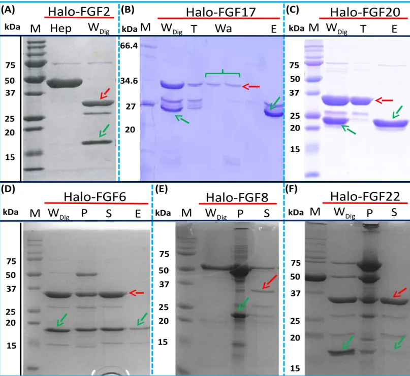
Purification of FGFs by removing the HaloTag with TEV protease The inclusion of a TEV site between the sequence of the HaloTag and FGF proteins provides a means to remove the HaloTag fusion partner in those instances where the HaloTag is not required for analysis (or when it may interfere with such analyses). Halo-FGF2 was first incubated with TEV protease to test whether the fusion protein could be cleaved by TEV. SDS-PAGE of the TEV digestion product of Halo-FGF2 shows that almost all of the protein was cleaved into the 35 kDa HaloTag (Fig. 7A, red arrow) and the 18 kDa FGF2 (Fig. 7A, green arrow). Thus, the cleavage site is fully accessible to TEV protease. Both Halo-FGF17 and Halo-FGF20 were also well digested by TEV protease and subsequently soluble FGF17 (Fig. 7B, green arrow) and FGF20 (Fig. 7C, green arrow) were purified by cation-exchange and heparin chromatography, respectively.
Most of FGF6 (Fig. 7D, lane WDig, green arrow) and FGF22 (Fig. 7F, lane WDig, green
arrow) and a small proportion of FGF8 were also released from HaloTag (Figs. 7D–7F, lane WDigand S, red arrow), but these proteins were observed to aggregate upon cleavage. This
suggested that these proteins were not very stable, at least in the buffer conditions used here, and required the HaloTag N-terminal fusion to remain soluble. The soluble FGF6 released by cleavage (Fig. 7D, lane S, green arrow) was applied to a heparin affinity column, but was observed to be concentrated at the top of the column where it formed a white aggregate. Very little protein was eluted with 1 M NaCl in PB buffer (Fig. 7D, lane E, green arrow). The disappearance of FGF8 and FGF22 in the soluble fractions after TEV digestion (Figs. 7Eand7F, lane S) showed that these two proteins were also not very soluble in the present buffer conditions without the HaloTag fusion partner.
Biological activities of FGFs and Halo-FGFs on Rama 27 fibrob-lasts and HaCaT keratinocytes
Figure 6 Further purification of the heparin affinity eluate of Halo-FGF1, Halo-FGF6, Halo-FGF7 and
Halo-FGF20 by ion-exchange chromatography.The soluble Halo-FGF1 and Halo-FGF7 eluted from
Figure 7 Cleavage of Halo-FGFs by TEV and purification.The eluates of Halo-FGF2, Halo-FGF17, Halo-FGF6, Halo-FGF8 and Halo-FGF22 from heparin-affinity chromatography and the Halo-FGF20 purified by heparin and ion-exchange chromatography were digested by TEV protease to separate the HaloTag and the FGF. Halo-FGF6, Halo-FGF8 and Halo-FGF22 became turbid after digestion and these samples were clarified by centrifugation. Then, the samples containing FGF6 and FGF20 were subjected to heparin chromatography and that of FGF17 to SP HP cation-exchange chromatography. Lanes M, markers; Hep, eluate from heparin chromatography;WDig, whole digestion product of Halo-FGFs
Figure 9 Quantification of p44/42MAPKphosphorylation.The band intensities from two experiments were quantified with imageJ and normalised to the BSA control to compare the similarities and diff er-ences of stimulation of phosphorylation p44/42MAPKby different FGFs. Results are the mean with the actual values from two independent experiments.
stimulation of phosphorylation of p44/42MAPK(Fig. 8A). In contrast, the 25 pM or 50 pM HaloTag protein alone did not appreciably stimulate p44/42MAPKphosphorylation. Therefore, the activity of Halo-FGF2 in this assay is equivalent to that of FGF2 (Fig. 9). In the case of FGF1, the N-terminal HaloTag also did not affect the ability of the growth factor to stimulate the phosphorylation of p44/42MAPK(Fig. 8A). FGF6 is not soluble without the HaloTag, so only the activity of the fusion protein could be tested, and it was found to stimulate the phosphorylation of p44/42MAPKto an extent similar to that observed with FGF1 and FGF2 (Fig. 8A). Since FGF6 has the same receptor preference as FGF1 and FGF2 (Zhang et al., 2006a), this suggests Halo-FGF6 was fully active.
activity (Zhang et al., 2006a). When 500 pM HaloTag was added to the cells, there was no detectable increase in phosphorylation of p44/42MAPK, whereas Halo-FGF8, Halo-FGF20 and FGF20 at concentrations comparable to those used in previous work (Zhang et al., 2006a) were all found to stimulate the phosphorylation of p44/42MAPK(Fig. 8B). In contrast, Halo-FGF16, Halo-FGF17 and FGF17 did not cause a detectable increase in phosphorylation of p44/42MAPK(Fig. 9). These data indicate that Halo-FGF8, FGF20 and Halo-FGF20 have biological activities on Rama 27 fibroblasts. The absence of stimulation of phosphorylation of p44/42MAPK by Halo-FGF16 may reflect the fact that the ability of this FGF to activate FGFR1c is considerably lower than that of FGF8, FGF17 and FGF20 (Zhang et al., 2006a). However, the absence of stimulation of phosphorylation of p44/42MAPK by FGF17 and Halo-FGF17 is more puzzling. One explanation may be that FGF16, and perhaps FGF17, do not cause the FGFR to activate strongly early biochemical signals that converge on p44/42MAPK. To test this, the capacity of Halo-FGF16, Halo-FGF17 and FGF17 to stimulate cell growth was measured in Rama 27 fibroblasts. The results show that 10 nM HaloTag only weakly stimulated the growth of Rama27 fibroblasts. Halo-FGF16 caused the number of cells to double compared to the negative control, and this level was significantly (p=0.015, Tukey test, OriginPro 9) above that observed in the presence of HaloTag alone (Fig. 8C). Halo-FGF17 and FGF17 were even more effective, as they caused a 3- to 4-fold increase in the number of cells (Fig. 8C). These results demonstrated that Halo-FGF16, FGF17 and Halo-FGF17 possess biological activities of similar potency as observed by others in growth assays (Zhang et al., 2006a).
The activity of members of the FGF7 subfamily were tested on HaCaT keratinocytes, as this cell type expresses the cognate receptor for these FGFs, FGFR2b (Ron et al., 1993). HaCaT cells have previously been shown to express more p42MAPkthan p44MAPk (Delehedde et al., 2002). The data show clearly that HaloTag alone did not stimulate the phosphorylation of p44/42MAPK(Fig. 8D). In contrast, FGF3, FGF7 and FGF10, and the corresponding HaloTag fusion proteins stimulated p44/42MAPK phosphorylation (Fig. 8C). FGF22, which is only soluble as a HaloTag fusion protein, also stimulated p44/42MAPKphosphorylation to an extent similar to that seen with the other members of the subfamily (Fig. 8D). Thus, these Halo-FGFs retain full biological activity in this assay.
CONCLUSION
the HaloTag can enhance expression of soluble protein and provide a means to label FGF protein with different fluorescent dyes and quantum dots, e.g.,Los et al. (2008);Zhang et al. (2006b)it is clearly a versatile and useful tool for these two purposes and, therefore, worthwhile exploring as a part of experimental strategy with these aims.
ACKNOWLEDGEMENT
Xianqing Mao would like to thank Monika Dieterle for help with cloning Halo-FGF3.
ADDITIONAL INFORMATION AND DECLARATIONS
Funding
The Cancer and Polio Research Fund and North West Cancer Research provided financial support. The funders had no role in study design, data collection and analysis, decision to publish, or preparation of the manuscript.
Grant Disclosures
The following grant information was disclosed by the authors: Cancer and Polio Research Fund and North West Cancer Research.
Competing Interests
The authors declare there are no competing interests.
Author Contributions
• Changye Sun and Yong Li conceived and designed the experiments, performed the experiments, analyzed the data, contributed reagents/materials/analysis tools, wrote the paper, prepared figures and/or tables, reviewed drafts of the paper.
• Sarah E. Taylor and Xianqing Mao performed the experiments, contributed reagents/materials/analysis tools, reviewed drafts of the paper.
• Mark C. Wilkinson performed the experiments, reviewed drafts of the paper.
• David G. Fernig conceived and designed the experiments, analyzed the data, wrote the paper, reviewed drafts of the paper.
REFERENCES
Asada M, Shinomiya M, Suzuki M, Honda E, Sugimoto R, Ikekita M, Imamura T. 2009. Glycosaminoglycan affinity of the complete fibroblast growth factor family.Biochimica et Biophysica ACTA/General Subjects1790:40–48DOI 10.1016/j.bbagen.2008.09.001.
Beenken A, Mohammadi M. 2009.The FGF family: biology, pathophysiology and therapy.Nature Reviews Drug Discovery8:235–253DOI 10.1038/nrd2792.
Boukamp P, Petrussevska RT, Breitkreutz D, Hornung J, Markham A, Fusenig NE. 1988. Normal keratinization in a spontaneously immortalized aneuploid human keratinocyte cell line.Journal of Cell Biology106:761–771DOI 10.1083/jcb.106.3.761.
Costa S, Almeida A, Castro A, Domingues L. 2014.Fusion tags for protein solubility, purification, and immunogenicity inEscherichia coli: the novel Fh8 system.Frontiers in Microbiology5:63
Danilenko DM, Montestruque S, Philo JS, Li TS, Hill D, Speakman J, Bahru M, Zhang MS, Kon-ishi O, Itoh N, Chirica M, Delaney J, Hernday N, Martin F, Hara S, Talvenheimo J, Narhi LO, Arakawa T. 1999.Recombinant rat fibroblast growth factor-16: structure and biological activity.
Archives of Biochemistry and Biophysics361:34–46DOI 10.1006/abbi.1998.0967.
Delehedde M, Lyon M, Vidyasagar R, McDonnell TJ, Fernig DG. 2002.Hepatocyte growth factor/scatter factor binds to small heparin-derived oligosaccharides and stimulates the proliferation of human HaCaT keratinocytes.Journal of Biological Chemistry277:12456–12462
DOI 10.1074/jbc.M111345200.
Delehedde M, Seve M, Sergeant N, Wartelle I, Lyon M, Rudland PS, Fernig DG. 2000.Fibroblast growth factor-2 stimulation of p42/44(MAPK) phosphorylation and I kappa B degradation is regulated by heparan sulfate/heparin in rat mammary fibroblasts.Journal of Biological Chemistry275:33905–33910DOI 10.1074/jbc.M005949200.
Duchesne L, Gentili D, Comes-Franchini M, Fernig DG. 2008.Robust ligand shells for biological applications of gold nanoparticles.Langmuir24:13572–13580DOI 10.1021/la802876u. Duchesne L, Octeau V, Bearon RN, Beckett A, Prior IA, Lounis B, Fernig DG. 2012.Transport of
fibroblast growth factor 2 in the pericellular matrix is controlled by the spatial distribution of its binding sites in heparan sulfate.PLoS Biology10:e1001976DOI 10.1371/journal.pbio.1001361. Ferreira T, Rasband W. 2012.ImageJ user guide—Analyze: Gels.Available athttp://rsbweb.nih.
gov/ij/docs/guide/146-30.html#toc-Subsection-30.13(accessed 09 December 2014). Huang ZH, Hwang P, Watson DS, Cao LM, Szoka FC. 2009.Tris-nitrilotriacetic acids of
subnanomolar affinity toward hexahistidine tagged molecules. Bioconjugate Chemistry 20:1667–1672DOI 10.1021/bc900309n.
Itoh N. 2007.The FGF families in humans, mice, and zebrafish: their evolutional processes and roles in development, metabolism, and disease.Biological & Pharmaceutical Bulletin 30:1819–1825DOI 10.1248/bpb.30.1819.
Jeffers M, McDonald WF, Chillakuru RA, Yang MJ, Nakase H, Deegler LL, Sylander ED, Rittman B, Bendele A, Sartor RB, Lichenstein HS. 2002.A novel human fibroblast growth factor treats experimental intestinal inflammation.Gastroenterology 123:1151–1162
DOI 10.1053/gast.2002.36041.
Kalinina J, Byron SA, Makarenkova HP, Olsen SK, Eliseenkova AV, Larochelle WJ,
Dhanabal M, Blais S, Ornitz DM, Day LA, Neubert TA, Pollock PM, Mohammadi M. 2009. Homodimerization controls the fibroblast growth factor 9 subfamily’s receptor binding and heparan sulfate-dependent diffusion in the extracellular matrix.Molecular and Cellular Biology 29:4663–4678DOI 10.1128/MCB.01780-08.
Ke YQ, Fernig DG, Smith JA, Wilkinson MC, Anandappa SY, Rudland PS, Barraclough R. 1990.High level production of human acidic fibroblast growth factor inEscherichia coli
cells inhibition of DNA synthesis in Rat mammary fibroblasts at high concentrations of growth factor.Biochemical and Biophysical Research Communications 171:963–971
DOI 10.1016/0006-291X(90)90778-L.
Knoerzer W, Binder HP, Schneider K, Gruss P, Mccarthy JEG, Risau W. 1989.Expression of synthetic genes encoding bovine and human basic fibroblast growth-factors (bFGFs) in
escherichia-coli.Gene75:21–30DOI 10.1016/0378-1119(89)90379-X.
Lin XH. 2004.Functions of heparan sulfate proteoglycans in cell signaling during development.
Development131:6009–6021DOI 10.1242/dev.01522.
Loo BM, Salmivirta M. 2002.Heparin/heparan sulfate domains in binding and signaling of fibroblast growth factor 8b. Journal of Biological Chemistry 277:32616–32623
DOI 10.1074/jbc.M204961200.
Los GV, Encell LP, McDougall MG, Hartzell DD, Karassina N, Zimprich C, Wood MG, Learish R, Ohane RF, Urh M, Simpson D, Mendez J, Zimmerman K, Otto P, Vidugiris G, Zhu J, Darzins A, Klaubert DH, Bulleit RF, Wood KV. 2008.HatoTag: a novel protein labeling technology for cell imaging and protein analysis.ACS Chemical Biology3:373–382
DOI 10.1021/cb800025k.
Macarthur CA, Lawshe A, Xu JS, Santosocampo S, Heikinheimo M, Chellaiah AT, Ornitz DM. 1995.Fgf-8 isoforms activate receptor splice forms that are expressed in mesenchymal regions of mouse development.Development121:3603–3613.
Maity H, Karkaria C, Davagnino J. 2009.Effects of pH and arginine on the solubility and stability of a therapeutic protein (fibroblast growth factor 20): relationship between solubility and stability.Current Pharmaceutical Biotechnology10:609–625DOI 10.2174/138920109789069297. Ohana RF, Encell LP, Zhao K, Simpson D, Slater MR, Urh M, Wood KV. 2009.HaloTag7: a
genetically engineered tag that enhances bacterial expression of soluble proteins and improves protein purification.Protein Expression and Purification68:110–120
DOI 10.1016/j.pep.2009.05.010.
Ornitz DM. 2000.FGFs, heparan sulfate and FGFRs: complex interactions essential for development.Bioessays22:108–112
DOI 10.1002/(SICI)1521-1878(200002)22:2<108::AID-BIES2>3.0.CO;2-M. Pizette S, Batoz M, Prats H, Birnbaum D, Coulier F. 1991.Production and
functional-characterization of human recombinant FGF-6 protein.Cell Growth and Differentiation 2:561–566.
Ron D, Bottaro DP, Finch PW, Morris D, Rubin JS, Aaronson SA. 1993. Expression of biologically-active recombinant keratinocyte growth-factor—structure-function analysis of amino-terminal truncation mutants.Journal of Biological Chemistry268:2984–2988. Roullier V, Clarke S, You C, Pinaud F, Gouzer G, Schaible D, Marchi-Artzner V, Piehler J,
Dahan M. 2009.High-affinity labeling and tracking of individual histidine-tagged proteins in live cells using Ni2+
Tris-nitrilotriacetic acid quantum dot conjugates.Nano Letters9:1228–1234
DOI 10.1021/nl9001298.
Rudland PS, Twiston Davies AC, Tsao SW. 1984.Rat mammary preadipocytes in culture produce a trophic agent for mammary epithelia-prostaglandin E2.Journal of Cellular Physiology 120:364–376DOI 10.1002/jcp.1041200315.
Smith JA, Winslow DP, Rudland PS. 1984.Different growth factors stimulate cell division of rat mammary epithelial, myoepithelial and stromal cell lines in culture.Journal of Cellular Physiology119:120–126DOI 10.1002/jcp.1041190310.
Susumu K, Medintz IL, Delehanty JB, Boeneman K, Mattoussi H. 2010. Modification of poly(ethylene glycol)-capped quantum dots with nickel nitrilotriacetic acid and
self-assembly with histidine-tagged proteins.Journal of Physical Chemistry C114:13526–13531
DOI 10.1021/jp103872j.
Tinazli A, Tang JL, Valiokas R, Picuric S, Lata S, Piehler J, Liedberg B, Tampe R. 2005.
Turner N, Grose R. 2010.Fibroblast growth factor signalling: from development to cancer.Nature Reviews Cancer10:116–129DOI 10.1038/nrc2780.
Uchinomiya S, Nonaka H, Fujishima S, Tsukiji S, Ojida A, Hamachi I. 2009.Site-specific covalent labeling of His-tag fused proteins with a reactive Ni(II)-NTA probe.Chemical Communications 39:5880–5882DOI 10.1039/b912025d.
Vogel A, Rodriguez C, IzpisuaBelmonte JC. 1996.Involvement of FGF-8 in initiation, outgrowth and patterning of the vertebrate limb.Development122:1737–1750.
Xu RY, Ori A, Rudd TR, Uniewicz KA, Ahmed YA, Guimond SE, Skidmore MA, Siligardi G, Yates EA, Fernig DG. 2012.Diversification of the structural determinants of fibroblast growth factor-heparin interactions implications for binding specificity.Journal of Biological Chemistry 287:40061–40073DOI 10.1074/jbc.M112.398826.
Yu S, Burkhardt M, Nowak M, Ries J, Petr´asek Z, Scholpp S, Schwille P, Brand M. 2009.FGF8 morphogen gradient is formed by a source–sink mechanism with freely diffusing molecules.
Nature461:533–536DOI 10.1038/nature08391.
Zhang XQ, Ibrahimi OA, Olsen SK, Umemori H, Mohammadi M, Ornitz DM. 2006a.Receptor specificity of the fibroblast growth factor family— the complete mammalian FGF family.Journal of Biological Chemistry281:15694–15700DOI 10.1074/jbc.M601252200.
Zhang Y, So MK, Loening AM, Yao HQ, Gambhir SS, Rao JH. 2006b.HaloTag protein-mediated site-specific conjugation of bioluminescent proteins to quantum dots.Angewandte
Chemie-International Edition45:4936–4940DOI 10.1002/anie.200601197.