ISOLATION AND CHARACTERIZATION OF ENDOPHYTIC BACTERIA ISOLATED FROM THE LEAVES OF
THE COMMON BEAN (
PHASEOLUS VULGARIS
)
Leonardo Emanuel de Oliveira Costa; Marisa Vieira de Queiroz*; Arnaldo Chaer Borges; Celia Alencar de Moraes; Elza
Fernandes de Araújo
Universidade Federal de Viçosa, Viçosa, MG, Brasil.
Submitted: February 21, 2011; Returned to authors for corrections: April 06, 2011; Approved: June 07, 2012.
ABSTRACT
The common bean is one of the most important legumes in the human diet, but little is known about the
endophytic bacteria associated with the leaves of this plant. The objective of this study was to characterize
the culturable endophytic bacteria of common bean (
Phaseolus vulgaris
) leaves from three different
cultivars (Vermelhinho, Talismã, and Ouro Negro) grown under the same field conditions. The density of
endophytic populations varied from 4.5 x 10
2to 2.8 x 10
3CFU g
-1of fresh weight. Of the 158 total isolates,
36.7% belonged to the Proteobacteria, 32.9% to Firmicutes, 29.7% to Actinobacteria, and 0.6% to
Bacteroidetes. The three
P. vulgaris
cultivars showed class distribution differences among Actinobacteria,
Alphaproteobacteria and Bacilli. Based on 16S rDNA sequences, 23 different genera were isolated
comprising bacteria commonly associated with soil and plants. The genera
Bacillus
,
Delftia
,
Methylobacterium
,
Microbacterium
,
Paenibacillus
,
Staphylococcus
and
Stenotrophomonas
were isolated
from all three cultivars. To access and compare the community structure, diversity indices were calculated.
The isolates from the Talismã cultivar were less diverse than the isolates derived from the other two
cultivars. The results of this work indicate that the cultivar of the plant may contribute to the structure of the
endophytic community associated with the common bean. This is the first report of endophytic bacteria
from the leaves of
P. vulgaris
cultivars. Future studies will determine the potential application of these
isolates in biological control, growth promotion and enzyme production for biotechnology.
Key words:
Endophytic bacteria, common bean,
Phaseolus vulgaris
, 16S rDNA, diversity indices.
INTRODUCTION
The phyllosphere is the habitat for a large diversity of
microorganisms. Although bacteria are the predominant
microorganisms present in phyllospheres, others such as
filamentous fungi are also important members. Phyllosphere
bacteria may be found on the surface of plants (epiphytes) as
well as in the interior of plant tissues (endophytes) (3, 32, 40).
Endophytic bacteria can be defined as those that can be isolated
from healthy, superficially disinfected plant tissues and do not
cause any damage to the host plant (15, 17).
The population density of endophytic bacteria can vary
from 10
2to 10
9(6, 12, 25, 39, 44) and depends on many
factors, including the plant being studied, the part under
analysis (31, 46), the developmental stage of the plant (17, 44),
the plant cultivar (genotype) (15, 44) and the interaction with
other organisms, as well as other environmental-related factors
(17).
The interaction between endophytic bacteria and their host
plants is not completely understood. However, many isolates
seem to have beneficial effects on their hosts (58). These
beneficial effects include promoting host growth and biological
control of phytopathogens (17, 21).
The common bean (
Phaseolus vulgaris
) is one of the most
important legumes in the human diet and serves as a significant
source of proteins (10). The relationship between
Rhizobium
and other nitrogen-fixing bacteria in the root nodules of beans
has been extensively studied (13, 37, 38). Recently,
López-López
et al
. (34) reported the isolation of endophytic bacteria
from the seeds and roots of the common bean. However, little
is known about endophytic bacteria inhabiting the aerial tissues
of the common bean. Therefore, the objective of this study was
to isolate the culturable, endophytic bacteria from the leaves of
three different common bean cultivars growing in field
conditions and characterize the community of culturable
bacteria. To our knowledge, this is the first report on
endophytic bacteria from the leaves of different cultivars of the
common bean.
MATERIALS AND METHODS
Plant materials
Samples were collected from three common bean cultivars
during the winter of 2007: Talismã (TAL), Ouro Negro (ONG),
and Vermelhinho (VER). The cultivars were planted in an
experimental field in the town of Coimbra – MG (altitude: 690
m; latitude: 20º 45’ S; longitude 42º 51’ W). During sowing,
350 kg ha
-1of the 8-28-16 NPK (percentage of nitrogen,
phosphorus and potassium) formula was applied, and 25 days
after their emergence, the plants were covered in 150 kg ha
-1of
ammonium sulfate. The leaves of the superior portion of the
plant (20 cm above the soil) were collected in the vegetative
phase 45 days after sowing.
Sample preparation and bacterial isolation
The collected leaves were washed in running water and
those with superficial injury that was visible to the naked eye
were excluded. Each isolation procedure was done in triplicate
for each cultivar. Each triplicate was composed of
approximately 2 g of leaves belonging to two different plants
being evaluated, totaling six plants per cultivar. The
disinfection and isolation were performed according to Araujo
et al.
(1) with minor modifications. Briefly, the leaves were
disinfected superficially through the following protocol: 70%
alcohol for 1 min, sodium hypochlorite (2.5% Cl
-) for 4 min,
ethanol for 30 s, and finally 3 rinses in sterile, distilled water.
To confirm the disinfection protocol, aliquots of the sterile
water used in the final rinse were plated in 10% TSA (1.5 g/L
of triptone, 0.5 g/L of soy peptone, 1.5 g/L of NaCl, 15 g/L of
agar, pH 7.3) at 28 ºC for 15 days and the plates are examined
for the presence or absence of microorganismal growth colony.
Initially, the leaves were ground with 6 mL of aqueous
solution (0.9 % NaCl) using a sterile mortar and pestle. The
tissue extract was subsequently incubated at 28 ºC for 3 hours
to allow the complete release of endophytic microorganisms
from the host tissue. For the isolation of endophytic bacteria,
the tissue extract was diluted in an aqueous solution (0.9 %
NaCl) and plated on five 10% TSA plates for each dilution
(10
-1and 10
-2). The plates were incubated for up to 15 days at
28ºC. Colonies were selected on days 2, 5, 10, and 15 of
incubation and purified in 10% TSA. For each petri dish
evaluated, the colonies were selected according to their time of
growth and morphology (color, size, shape). After 15 days of
incubation, all of the colonies were counted and expressed as
CFU per gram of fresh tissue.
Identification and phylogenetic analysis of endophytic
bacteria
for 5 minutes at 14000
g
and resuspended in 1 ml of TE buffer
(mM Tris-HCl, 1 mM EDTA, pH 8.0), centrifuged,
resuspended in 500 µl of TE buffer and finally adding 0.5 g of
glass pearls (0.1 mm in diameter) (Sigma-Aldrich, USA) and
15 µl of 20% SDS. The cells were then homogenized for 30 s
in a vortex mixer (AP56 – Phoenix), 500 µl of buffered phenol
was added, and the solution was mixed and centrifuged for 5
min at 14000
g
. The aqueous phase was extracted once with
phenol-chloroform (1:1) and once more with chloroform.
Following the extraction of the aqueous phase, 20 µl of 5M
NaCl was added, the DNA was precipitated with isopropanol
(5 min at room temperature) and collected by centrifugation for
10 min at 14000
g
. The DNA pellet was washed with 70%
ethanol, air dried and resuspended in 30 µl of autoclaved,
ultrapure water.
The amplification of 16S rDNA was carried out in a
reaction with a final volume of 25 µl containing 1 µl (0.5-10
ng) of total DNA, 2.5 µl (0.2 µM) of the P027F primer
(5’-GAGAGTTTGATCCTGGCTAG-3’), 2.5 µl (0.2 µM) of the
1378R primer (5’-CGGTGTGTACSSGGCCCGGGAACG-3’),
1.6 µl (200 µM) of each dNTP, 2.5 µl of 5x IB buffer
(Phoneutria; Belo Horizonte, Brazil); 1µl (1U) of Taq DNA
polymerase (Phoneutria; Belo Horizonte, Brazil), and 2.5 µl
(25 µg) of BSA (Promega). A negative control (PCR mix
without DNA) was included in all PCR experiments. The PCR
reaction conditions were as follows: 94ºC for 4 min, followed
by 30 cycles of denaturation at 94ºC for 30 s, annealing at 63ºC
for 1 min and extension at 72ºC for 1 min, before a final
extension at 72ºC for 7 min. The PCR products were purified
and sequenced by Macrogen Inc. (Seoul, South Korea) using
an ABI3730 XL automatic DNA sequencer and the primers
P027F and 1378R.
The identification of the isolates was performed using the
Ribosomal Database Project (14, 61) and BLAST
(
http://blast.ncbi.nlm.nih.gov/blast/Blast.cgi
) in NCBI. We
used the Sequence Match application and BLAST to verify the
similarity of experimental sequences with the reference
sequences in the databases (14) and classified them at the
genus level.
The DNA sequences of 34 reference strains (“type
strain”), 2 strains obtained from the Ribosomal Database
Project, and 34 representative strains from experimental
isolates were aligned using the Ribosomal Database Project.
Phylogenetic trees were constructed using the
Neighbor-Joining (NJ) algorithm in
MEGA
version 4 (56), the Maximum
Parsimony (MP) and Maximum Likelihood (ML) algorithm in
Paup* (52), and the Bayesian Analysis (BA) algorithm in
MrBayes 3.1 (23). The Neighbor Joining method was corrected
by the Tamura-Nei multiple base substitution model (55) and
by the GAMA distribution (0.4899) established by Modeltest
3.7. The parameters for Maximum Likelihood (GTR+I+G)
were selected by AIC in Modeltest 3.7 (45). The Bayesian
parameters (GTR+I+G) were selected by AIC in MrModeltest
2.3 (42). A total of 1000 replications were used for the
bootstrap tests of the NJ and MP methods, while the ML test
had 100 replications. The MB was performed in two
independent runs with four Markov chain Monte Carlo
(MCMC). A total of 10,000,000 generations were run, with
trees being sampled every 1000 generations and the first
1,000,000 trees being discarded. Non-rooted trees were
calculated using the 16S rDNA sequence of
Methanocaldococcus jannaschii
DSM 2661 as an outgroup.
The 16S rDNA sequences of each isolate were deposited in the
NCBI GENBANK database under the accession numbers
HM355592 to HM355749.
Diversity indices
The diversity indices were calculated in the PAST
program version 2.01(20), and the expected number of
genotypes in the R program version 2.11.1 (47) using the
Vegan library (43).
RESULTS
Endophytic bacteria isolation and identification
TSA medium varied from 4.5 x 10
2to 2.8 x 10
3CFU g
-1per
fresh weight. A total of 158 (about 40 % of the total counted)
isolates was obtained, of which 31.01% (49) were isolated
from the Talismã cultivar, 37.34% (59) from the Ouro Negro
cultivar and 31.65% (50) from the Vermelhinho cultivar (Table
1).
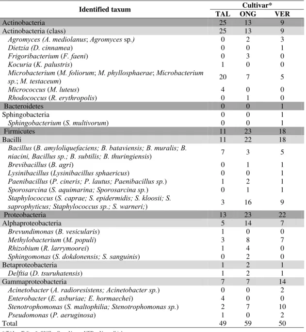
Table 1.
Endophytic isolates obtained from three
Phaseolus vulgaris
cultivars.
Cultivar*
Identified taxum
TAL ONG
VER
Actinobacteria
25
13
9
Actinobacteria (class)
25
13
9
Agromyces (A. mediolanus
;
Agromyces
sp
.)
0 2
3
Dietzia
(D. cinnamea
)
0 0
1
Frigoribacterium
(
F. faeni
)
0 3
0
Kocuria
(
K. palustris
)
1 0
0
Microbacterium
(
M. foliorum
;
M. phyllosphaerae
;
Microbacterium
sp.
;
M. testaceum
)
20 7
5
Micrococcus
(
M. luteus
) 4
0
0
Rhodococcus
(
R. erythropolis
)
0 1
0
Bacteroidetes
0
0
1
Sphingobacteria 0
0
1
Sphingobacterium
(
S. multivorum
)
0 0
1
Firmicutes
11
23
18
Bacilli
11
22
18
Bacillus
(
B. amyloliquefaciens; B. bataviensis; B. muralis; B.
niacini, Bacillus sp.; B. subtilis; B. thuringiensis
)
7 3
5
Brevibacillus
(
B. agri
)
0 1
1
Lysinibacillus
(
Lysinibacillus sphaericus
)
0 0
1
Paenibacillus
(
P. cineris; P. lautus; Paenibacillus sp.
)
1 2
1
Sporosarcina
(
S. aquimarina; Sporosarcina sp.
)
0 1
1
Staphylococcus
(
S. caprae; S. epidermidis; S. kloosii; S.
saprophyticus; Staphylococcus sp.; S. warneri;
)
3 16
9
Proteobacteria
13
23
22
Alphaproteobacteria
5
14
7
Brevundimonas
(
B. vesicularis
)
1 0
0
Methylobacterium
(
M. populi
)
3 8
7
Rhizobium
(
R. larrymoorei
)
1 4
0
Sphingomonas
(
S. dokdonensis; S. sanguinis
)
0 2
0
Betaproteobacteria
1
2
1
Delftia
(
D. tsuruhatensis
)
1 2
1
Gammaproteobacteria
7
7
14
Acinetobacter
(
A. radioresistens; Acinetobacter sp.
)
0 0
2
Enterobacter
(
E. asburiae; E. hormaechei
)
4 0
0
Stenotrophomonas
(
S. maltophilia; Stenotrophomonas sp.
)
2 7
10
Pseudomonas
(
P. aeruginosa
)
1 0
2
Total 49
59
50
* TAL = Talismã; ONG = Ouro Negro; VER = Vermelhinho.
Identification and phylogenetic analyses of endophytic
bacteria
Sequencing of 16S rDNA was performed in all 158
isolates. Based on the nucleotide sequences each of the isolates
was assigned to 23 different genera (Table 1). In terms of
(Sphingobacteriaceae) of the genus
Sphingobacterium
. The
highest number of isolates belonged to the Bacilli class
(32.9%), comprised of bacteria from the families
Staphylococcaceae (17.7%), Bacillaceae (10.1%),
Paenibacillaceae (3.8%) and Planococcaceae (1.3%). The
second most prevalent class in isolates was Actinobacteria
(29.7%), which includes Microbacteriaceae (24.7%),
Micrococcaceae (3.1%), Nocardiaceae (0.6%) and Dietziaceae
(0.6%). Among the isolates identified as Proteobacteria, the
dominant class in the isolate collection was
Gammaproteobacteria (17.71%), with isolates belonging to the
families Xanthomonadaceae (12.0%), Enterobacteriaceae
(2.5%), Pseudomonadaceae (1.9%) and two (1.3%) isolates
from the family Moraxellaceae. Isolates from the
Alphaproteobacteria (16.5%) comprised representatives from
the families Methylobacteriaceae (11.4%), Rhizobiaceae
(3.2%), Sphingomonadaceae (1.3%) and one isolate from the
family Caulobacteraceae. Betaproteobacteria (2.5%) contains
only members from the family Comamonadaceae (2.5%).
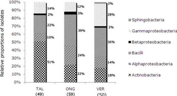
The relative composition of the bacterial isolates by
cultivars is shown in Figure 1 according to class. Differences in
the proportions of the classes Actinobacteria,
Alphaproteobacteria and Bacilli were observed between the
three
P. vulgaris
cultivars. Sphingobacteria were isolated only
from the Vermelhinho cultivar, which also exhibited
differences in the proportion of isolates belonging to
Gammaproteobacteria compared to isolates from the other two
cultivars. The proportion of Betaproteobacteria was the same in
all cultivars and all isolates of this class belonged to the genus
Delftia
.
Figure 1.
Bacterial class distribution of the culturable endophytic isolates obtained from three
Phaseolus vulgaris
cultivars:
Talismã cultivar (TAL); Ouro Negro cultivar (ONG); Vermelhinho cultivar (VER).
Partial 16S rDNA gene sequences (approximately 1200
bp) from the isolates were used together with sequences taken
from the Ribosomal Database Project for construction of
phylogenetic trees using four different methods
(Neighbor-Joining, Maximum Parsimony, Maximum Likelihood and
Bayesian). The tree obtained by the Bayesian method is shown
in Figure 2.
method; the terminal node that contained the isolate BAC3114
had bootstrap values below 90 for both the ML and NJ
methods. The terminal node that contained the isolate
BAC2073 had bootstrap values below 90 for the methods MP,
ML and NJ. The phylum Bacteroidetes aligned with bacteria
from the phylum Proteobacteria.
Figure 2. Phylogenetic tree showing the relationship between the 16S rDNA gene sequences from representative isolates of endophytic
bacteria from three
P. vulgaris
cultivars. Terminal nodes in bold have bootstrap values greater than or equal to 94 in the three methods
used (NJ, MP, ML) and presented
a posteriori
probabilities greater than or equal to 0.99. Terminal nodes with
a posteriori
probabilities
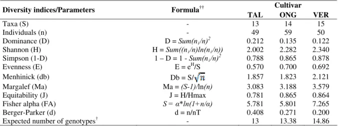
Diversity indices
The diversity index that was calculated in the PAST program
and the expected number of genotypes for each cultivar estimated
in the R program can be visualized in Table 2.
Table 2.
Number of taxa, individuals, diversity index and expected number of genotypes for each cultivar.
Cultivar
Diversity indices/Parameters
Formula
††TAL ONG VER
Taxa (S)
-
13
14
15
Individuals (n)
-
49
59
50
Dominance (D)
D =
Sum(n
i/n)
20.212
0.135
0.122
Shannon (H)
H =
Sum((n
i/n)ln(n
i/n))
2.002
2.282
2.340
Simpson (1-D)
1 – D = 1 -
Sum(n
i/n)
20.788
0.865
0.878
Evenness (E)
E = e
H/S 0.570
0.700
0.692
Menhinick (db)
Db = S/
1.857 1.823 2.121
Margalef (Ma)
Ma =
(S-1)/
ln
(n)
3.083
3.188
3.579
Equitability (J)
J = H/Hmax
0.781
0.865
0.864
Fisher alpha (FA)
S =
α
*ln(1+n/
α
)
5.781
5.801
7.265
Berger-Parker (d)
d = n/nT
0.408
0.271
0.200
Expected number of genotypes
†-
13
13.38
14.86
† Calculated in R program version 2.11.1
†† n = number of individuals; ni = number of individuals of taxon i; S = number of taxa; Nt = number of individuals in the dominant taxon; Hmax = log S.
* Fisher´s alpha.
DISCUSSION
Isolation and identification of endophytic bacteria was
performed from the leaves of three common bean (
P. vulgaris
)
cultivars grown under the same field conditions in Minas
Gerais during the winter season. The population densities of
culturable bacteria in this study were similar to the population
density of isolates obtained from soybean leaves growing in
herbicide-free soil by Kuklinsky-Sobral
et al.
(30).
All identified isolates corresponded to genera commonly
isolated from either the rhizosphere or bacteria associated with
plants. Species from the genera
Agromyces
,
Bacillus
,
Brevibacillus
,
Delftia
,
Dietzia
,
Enterobacter
,
Methylobacterium
,
Microbacterium
,
Micrococcus
,
Paenibacillus
,
Pseudomonas
,
Rhizobium
,
Rhodococcus
,
Sphingobacterium
and
Stenotrophomonas
have already been
isolated from rhizospheric soil and as endophytic bacteria in
many previous studies (4, 5, 8, 18, 19, 24, 27–30, 36, 48–51,
54, 57–60). Additionally, species from the genera
Acinetobacter
,
Brevundimonas
,
Frigoribacterium
,
Kocuria
,
Sphingomonas
,
Sporosarcina
and
Staphylococcus
have been
isolated or reported in studies of culturable and non-culturable
endophytic bacteria (5, 8, 27, 29, 30, 48, 50, 51, 58).
Many of the bacterial genera encountered in this work
were previously reported by Lopez-Lopez
et al.
, (34), and
many species of genera
Bacillus
were found by Walker
et al.
(63) in bean seeds. However, some of the species are not the
same. The presence of certain genera in different bean cultivars
suggest that they are better adapted to live as endophytic
bacteria in
P. vulgaris
than other genera. The genera isolated in
this work that have not been previously reported for
P. vulgaris
are as follows:
Agromyces
,
Brevibacillus
,
Brevundinomonas
,
Delftia
,
Dietzia
,
Frigoribacterium
,
Lysinibacillus
,
Sphingobacterium
,
Sporosarcina
and
Stenotrophomonas
.
isolate analyses also indicated that the cultivar of the plant may
contribute to the determination of associated bacteria. Some of
the genera had been isolated with greater frequency from a
particular cultivar, for example, the genus
Microbacterium
from TAL, the genus
Staphylococcus
from ONG and
Stenotrophomonas
from VER. The differences between the
number and type of isolates in each cultivar may suggest
distinct endophytic communities in each cultivar. The
differences in diversity of the endophytic communities of the
cultivars may also be observed by the comparison of the
relative class percentages presented in Figure 1.
To better visualize the community structure of the three
common bean cultivars studied, diversity indices (Table 2)
were calculated. The diversity indices obtained show that the
diversity of bacterial isolates from cultivar Talismã was lower
than the diversity of isolates obtained from the other two
cultivars while the diversity of bacterial isolates from the
cultivar Vermelhinho was the highest. Moreover, the indices
Dominance_D and Berger-Parker clearly show that a single
taxa
of the cultivar Talismã is more abundant in the
community, and the number of isolates shown in Table 1 reveal
that this is the genus
Microbacterium
.
Bacteria usually associated with common bean leaf
diseases belong to the genera
Curtobacterium
(22),
Pseudomonas
(33) and
Xanthomonas
(62). None of the isolates
belong to
Curtobacterium
or
Xanthomonas
, while all the
isolates belonging to
Pseudomonas
aligned with different
strains of
Pseudomonas aeruginosa
with scores of 0.999.
The levels of NPK and ammonium sulfate applied to the
plants were in accordance with the recommendations for
producers in Brazil. However, this high level of nitrogen
probably inhibited the nodulation of the bean roots and the
association with other nitrogen-fixing bacteria. A few
Rhizobium
,
Pseudomonas
,
Methylobacterium
and
Enterobacter
species have already been described in the literature as
nitrogen-fixing and nodule-forming organisms in the roots of
many Leguminosae (7, 26, 37, 53). The five
Rhizobium
isolates
aligned with sequences of
Rhizobium larrymoorei
, which was
originally isolated from tumors affecting aerial parts of
Ficus
benjamina
(9). Some bacterial species considered pathogenic
for certain plant species have been isolated as endophytic in
other species; from the polar tree, Ulrich
et al.
(58) isolated
endophytes with high similarity to known plant pathogens,
such as
Clavibacter michiganensis
,
Pseudomonas syringae
and
Xanthomonas populi.
Maes
et. al.
(35) also showed that
Brenneria salicis
could be isolated as an endophyte from
poplar (
Populus
) and alder (
Alnus
). It is unclear whether these
endophytic bacterial species confer some benefit to the host
plant or if they merely use the host as a survival strategy in the
environment to reach plants on which they can develop disease.
The study of endophytic microorganisms is important to
comprehend their interaction with their host plants.
Additionally, endophytic microorganisms may have
biotechnological applications. The potential of the isolated
endophytic bacteria to promote bean plant growth and their
biocontrol potential in diseases that affect the aerial parts of
this important legume for the human diet will be addressed in
future studies.
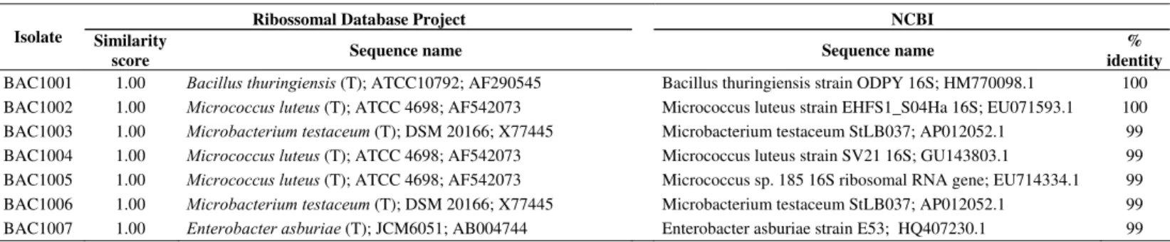
Table S1.
Identity of the 16S rDNA gene sequences of the isolates with the sequences deposited in the database.
Ribossomal Database Project NCBI
Isolate Similarity
score Sequence name Sequence name
Table S1.
Continuation
Ribossomal Database Project NCBI
Isolate Similarity
score Sequence name Sequence name
% identity BAC1008 1.00 Microbacterium testaceum (T); DSM 20166; X77445 Microbacterium testaceum strain ESS21; EF602568.1 99 BAC1009 0.98 Microbacterium testaceum (T); DSM 20166; X77445 Microbacterium testaceum strain PCSB7 16S; HM449703.1 98 BAC1010 1.00 Enterobacter hormaechei (T); CIP 103441; AJ508302 Enterobacter hormaechei strain Ni-1 16S; HM446004.1 99 BAC1011 1.00 Rhizobium larrymoorei (T); 3-10; Z30542 Agrobacterium larrymoorei strain 13638E 16S; EU741094.1 100 BAC1012 1.00 Stenotrophomonas maltophiliaAB008509 (T); ATCC 13637T; Stenotrophomonas maltophilia strain AhsB4 16S; HM143858.1 100 BAC1013 1.00 Microbacterium testaceum (T); DSM 20166; X77445 Microbacterium testaceum StLB037; AP012052.1 99 BAC1014 1.00 Microbacterium testaceum (T); DSM 20166; X77445 Stenotrophomonas maltophilia strain AhsB4 16S; HM143858.1 100 BAC1015 0.98 Bacillus niacini (T); IFO15566; AB021194 Bacillus sp. DL006 16S; GQ355276.1 98 BAC1016 1.00 Staphylococcus epidermidis (T); ATCC 14990; D83363 Staphylococcus epidermidis strain LCR40 16S; FJ976549.1 100 BAC1017 1.00 Bacillus thuringiensis (T); ATCC10792; AF290545 Bacillus thuringiensis strain ODPY 16S; HM770098.1 100 BAC1018 1.00 Microbacterium testaceum (T); DSM 20166; X77445 Microbacterium testaceum StLB037; AP012052.1 99 BAC1019 1.00 Microbacterium testaceum (T); DSM 20166; X77445 Microbacterium testaceum strain ESS21 16S; EF602568.1 99 BAC1020 1.00 Microbacterium testaceum (T); DSM 20166; X77445 Microbacterium testaceum StLB037; AP012052.1 99 BAC2021 1.00 Rhizobium larrymoorei (T); 3-10; Z30542 Agrobacterium larrymoorei strain 13638E 16S; EU741094.1 99 BAC2022 1.00 Microbacterium testaceum (T); DSM 20166; X77445 Microbacterium testaceum StLB037; AP012052.1 99 BAC2023 0.99 Methylobacterium populi (T); BJ001; ATCC BAA-705;
NCIMB 13946; AY251818 Methylobacterium sp. SuP10 16S; EU912450.1 100
BAC2024 1.00 Stenotrophomonas maltophilia (T); ATCC 13637T;
AB008509 Stenotrophomonas maltophilia strain AhsB4 16S; HM143858.1 100
BAC2025 1.00 Rhizobium larrymoorei (T); 3-10; Z30542 Agrobacterium larrymoorei strain 13638E 16S; EU741094.1 100 BAC2026 1.00 Staphylococcus epidermidis (T); ATCC 14990; D83363 Staphylococcus epidermidis strain F71028 16S; HQ908659.1 100 BAC2027 1.00 Brevibacillus agri (T); NRRL NRS-1219; D78454 Brevibacillus agri partial 16S strain R-20121; AJ586388.1 99 BAC2028 0.99 Methylobacterium populi (T); BJ001; ATCC BAA-705;
NCIMB 13946; AY251818 Methylobacterium populi BJ001 16S; CP001029.1 99
BAC2029 1.00 Microbacterium testaceum (T); DSM 20166; X77445 Microbacterium testaceum StLB037; AP012052.1 99 BAC2030 1.00 Staphylococcus warneri (T); L37603 Staphylococcus warneri strain FUA2075 16 S; HQ694734.1 99 BAC2031 1.00 Stenotrophomonas maltophilia (T); ATCC 13637T;
AB008509 Stenotrophomonas maltophilia strain AhsB4 16S; HM143858.1 99
BAC2032 1.00 Staphylococcus warneri (T); L37603 Staphylococcus warneri strain FUA2075 16 S; HQ694734.1 99 BAC2033 0.99 Methylobacterium populi (T); BJ001; ATCC BAA-705;
NCIMB 13946; AY251818 Methylobacterium populi BJ001; CP001029.1 99
BAC2034 1.00 Staphylococcus epidermidis (T); ATCC 14990; D83363 Staphylococcus epidermidis gene for 16S; AB617573.1 100 BAC2035 1.00 Staphylococcus epidermidis (T); ATCC 14990; D83363 Staphylococcus epidermidis strain NM62-4 16S; HM218280.1 100 BAC2036 0.98 Microbacterium testaceum (T); DSM 20166; X77445 Microbacterium hominis strain 1P10AE; EU977655.1 99 BAC2037 0.98 Sphingomonas dokdonensis (T); DS-4; DQ178975 Sphingomonas dokdonensis strain 2P01AE; EU977661.1 99 BAC2038 0.98 Sporosarcina aquimarina (T); SW28(T); AF202056 Sporosarcina luteola gene for 16S; AB473560.1 99 BAC2039 1.00 Staphylococcus caprae (T); ATCC 35538T; AB009935 Staphylococcus capitis strain EHFS2_AU1Hc 16S; EU071603.1 100 BAC2040 0.99 Methylobacterium populi (T); BJ001; ATCC BAA-705;
NCIMB 13946; AY251818 Methylobacterium extorquens AM1; CP001510.1 99
BAC3041 1.00 Staphylococcus epidermidis (T); ATCC 14990; D83363 Staphylococcus epidermidis strain NM62-4 16S; HM218280.1 100 BAC3042 1.00 Bacillus thuringiensis (T); ATCC10792; AF290545 Bacillus thuringiensis serovar colmeri 16S; EU429660.1 100 BAC3043 1.00 Microbacterium foliorum (T); DSM 12966; P 333/02;
AJ249780 Microbacterium foliorum strain 720 16S; EU714376.1 99
BAC3044 0.97 Bacillus bataviensis (T); type strain: LMG 21832;
AJ542507 Bacillus sp. R-30632 partial 16S; AM910246.1 99
BAC3045 0.98 Sphingobacterium multivorum (T); IFO 14947; D14025 Sphingobacterium sp. G-2-27-2 16S; EF102865.1 99 BAC3046 1.00 Staphylococcus epidermidis (T); ATCC 14990; D83363 Staphylococcus epidermidis strain F71028 16S; HQ908659.1 100 BAC3047 1.00 Microbacterium testaceum (T); DSM 20166; X77445 Microbacterium sp. Fek04 16S; EU741023.1 99 BAC3048 1.00 Bacillus amyloliquefaciens (T); CR-502; AY603658 Bacillus amyloliquefaciens LL3; CP002634.1 100 BAC3049 1.00 Staphylococcus kloosii (T); ATCC 43959T; AB009940 Staphylococcus kloosii strain FR2_36con 16S; EU934080.1 100 BAC3050 0.99 Microbacterium phyllosphaerae (T); DSM 13468; P
369/06; AJ277840 Microbacterium foliorum strain 720 16S; EU714376.1 99
BAC3051 0.99 Methylobacterium populi (T); BJ001; ATCC BAA-705;
NCIMB 13946; AY251818 Methylobacterium sp. SuP10 16S; EU912450.1 99
BAC3052 1.00 Stenotrophomonas maltophilia (T); ATCC 13637T;
Table S1.
Continuation
Ribossomal Database Project NCBI
Isolate Similarity
score Sequence name Sequence name
% identity
BAC3053 0.97 Bacillus bataviensisAJ542507 (T); type strain: LMG 21832; Bacillus sp. R-30632 partial 16S; AM910246.1 98 BAC3054 0.99 Methylobacterium populi (T); BJ001; ATCC BAA-705;
NCIMB 13946; AY251818 Methylobacterium sp. SuP10 16S; EU912450.1 100
BAC3055 0.99 Staphylococcus warneri (T); L37603 Staphylococcus warneri strain FUA2075 16S; HQ694734.1 98 BAC3056 0.92 Sporosarcina koreensis F73; DQ073393 Sporosarcina ginsengisoli strain CR5 16S; HQ331532.1 90 BAC3057 0.82 Acinetobacter radioresistens INBS1; AM495259 Acinetobacter radioresistens strain TY37SsD 16S; HQ406757.1 81 BAC3058 1.00 Acinetobacter radioresistens (T); DSM 6976; X81666 Acinetobacter radioresistens strain S13 16S; GU145275.1 99 BAC3059 0.98 Lysinibacillus sphaericus; KNUC228; EF166045 Lysinibacillus sphaericus strain IMAU80223 16S; GU125639.1 97 BAC1061 0.92 Microbacterium testaceum (T); DSM 20166; X77445 Microbacterium sp. CSBd gene for 16S; AB552874.1 91 BAC1062 1.00 Staphylococcus epidermidis (T); ATCC 14990; D83363 Staphylococcus epidermidis strain NM62-4 16S; HM218280.1 100 BAC1063 1.00 Bacillus thuringiensis (T); ATCC10792; AF290545 Bacillus thuringiensis strain ODPY 16S; HM770098.1 100 BAC1064 0.99 Methylobacterium populi (T); BJ001; ATCC BAA-705;
NCIMB 13946; AY251818 Methylobacterium extorquens DM4 str. DM4; FP103042.2 100 BAC1065 1.00 Microbacterium testaceum (T); DSM 20166; X77445 Microbacterium testaceum strain DSM 20166 16S; NR_026163.1 99 BAC1066 1.00 Microbacterium testaceum (T); DSM 20166; X77445 Microbacterium testaceum StLB037; AP012052.1 99 BAC1067 1.00 Microbacterium foliorumAJ249780 (T); DSM 12966; P 333/02; Microbacterium foliorum strain 720 16S; EU714376.1 99 BAC1068 0.99 Methylobacterium populi (T); BJ001; ATCC BAA-705;
NCIMB 13946; AY251818 Methylobacterium extorquens AM1; CP001510.1 99
BAC1069 0.99 Methylobacterium populi (T); BJ001; ATCC BAA-705;
NCIMB 13946; AY251818 Methylobacterium extorquens AM1; CP001510.1 100
BAC1070 0.99 Brevundimonas vesicularis (T); ATCC 11426 (T); AJ007801
Brevundimonas vesicularis DNA for 16S strain LMG 11141;
AJ227781.1 99
BAC2071 1.00 Staphylococcus epidermidis (T); ATCC 14990; D83363 Staphylococcus epidermidis strain LCR40 16S; FJ976549.1 100 BAC2072 1.00 Frigoribacterium faeni (T); 801; Y18807 Frigoribacterium sp. PDD-24b-20 16S; HQ256793.1 99 BAC2073 0.99 Sphingomonas sanguinis (T); IFO 13937; D13726 Sphingomonas pseudosanguinis partial 16S; AM412238.1 99 BAC2074 0.98 Staphylococcus warneri (T); L37603 Staphylococcus pasteuri partial 16S strain PSM NO.15; FR846535.1 98 BAC2075 0.99 Frigoribacterium faeni (T); 801; Y18807 Frigoribacterium sp. 301 16S; AF157479.1 99 BAC2076 1.00 Rhizobium larrymoorei (T); 3-10; Z30542 Agrobacterium larrymoorei strain 2R46 16S; EF178437.1 100 BAC2077 1.00 Staphylococcus epidermidis (T); ATCC 14990; D83363 Staphylococcus epidermidis strain F71028 16S; HQ908659.1 100 BAC2078 1.00 Bacillus subtilis subsp. subtilis (T); DSM10; AJ276351 Bacillus subtilis strain M-15 16S; HQ401271.1 100 BAC2079 1.00 Microbacterium testaceum (T); DSM 20166; X77445 Microbacterium sp. CSBd gene for 16S; AB552874.1 100 BAC2080 1.00 Microbacterium testaceum (T); DSM 20166; X77445 Microbacterium sp. Fek04 16S; EU741023.1 99 BAC3081 1.00 Brevibacillus agri (T); NRRL NRS-1219; D78454 Brevibacillus agri strain PLIV 16S; HQ166189.1 100 BAC3082 0.99 Methylobacterium populi (T); BJ001; ATCC BAA-705;
NCIMB 13946; AY251818 Methylobacterium sp. SuP10 16S; EU912450.1 99
BAC3083 0.99 Methylobacterium populi (T); BJ001; ATCC BAA-705;
NCIMB 13946; AY251818 Methylobacterium sp. SuP10 16S; EU912450.1 99
BAC3084 1.00 Paenibacillus cineris (T); type strain:LMG 18439;
AJ575658 Paenibacillus sp. 3492BRRJ 16S; JF309261.1 100
BAC3085 1.00 Staphylococcus epidermidis (T); ATCC 14990; D83363 Staphylococcus epidermidis strain NM62-4 16S; HM218280.1 100 BAC3087 1.00 Microbacterium foliorum (T); DSM 12966; P 333/02;
AJ249780 Microbacterium foliorum strain 720 16S; EU714376.1 99
BAC3088 0.99 Methylobacterium populiNCIMB 13946; AY251818 (T); BJ001; ATCC BAA-705; Methylobacterium sp. DC2c-19 gene for 16S; AB552870.1 99 BAC3089 1.00 Staphylococcus warneri (T); L37603 Staphylococcus warneri strain FUA2075 16S; HQ694734.1 99 BAC3090 0.99 Methylobacterium populi (T); BJ001; ATCC BAA-705;
NCIMB 13946; AY251818 Methylobacterium sp. SuP10 16S; EU912450.1 99
BAC1091 1.00 Paenibacillus cineris (T); type strain:LMG 18439;
AJ575658 Paenibacillus cineris partial 16S; AJ575658.1 99
Table S1.
Continuation
Ribossomal Database Project NCBI
Isolate Similarity
score Sequence name Sequence name
% identity
BAC1098 1.00 Kocuria palustrisY16263 (T); TAGA27 (DSM 11925, type strain); Kocuria palustris strain cT220 16S; JF303036.1 99 BAC1099 1.00 Microbacterium testaceum (T); DSM 20166; X77445 Staphylococcus warneri strain FUA2075 16S; HQ694734.1 100 BAC1100 1.00 Microbacterium testaceum (T); DSM 20166; X77445 Staphylococcus saprophyticus strain OTUC3 16S; FJ210844.1 100 BAC2101 1.00 Staphylococcus saprophyticus subsp. saprophyticus (T);
ATCC 15305 (= MAFF 911473); D83371 Staphylococcus saprophyticus strain OTUC3 16S; FJ210844.1 100 BAC2102 1.00 Staphylococcus warneri (T); L37603 Staphylococcus warneri strain FUA2075 16S; HQ694734.1 100 BAC2103 0.99 Methylobacterium populi (T); BJ001; ATCC BAA-705;
NCIMB 13946; AY251818 Methylobacterium extorquens gene for 16S rRNA; AB298401.1 99 BAC2104 1.00 Delftia tsuruhatensis (T); T7; AB075017 Delftia tsuruhatensis strain IPPBC R15 16S; HQ436355.1 100 BAC2105 0.99 Methylobacterium populi (T); BJ001; ATCC BAA-705;
NCIMB 13946; AY251818 Methylobacterium extorquens gene for 16S; AB298401.1 99
BAC2106 0.86 Bacillus cereus me-5; EU652058 Bacillus cereus partial 16S; FR749846.1 85
BAC2107 1.00 Staphylococcus warneri (T); L37603 Staphylococcus warneri strain FUA2075 16S; HQ694734.1 99 BAC2108 0.99 Methylobacterium populi (T); BJ001; ATCC BAA-705;
NCIMB 13946; AY251818 Methylobacterium extorquens gene for 16S rRNA; AB298401.1 99 BAC2109 1.00 Staphylococcus warneri (T); L37603 Staphylococcus warneri strain FUA2075 16S; HQ694734.1 99 BAC2110 1.00 Frigoribacterium faeni (T); 801; Y18807 Frigoribacterium faeni partial 16S; AM410686.1 99 BAC3111 0.99 Pseudomonas aeruginosa (T); DSM50071; X06684 Pseudomonas aeruginosa strain CRC5 16S; HQ995502.1 100 BAC3112 1.00 Staphylococcus warneri (T); L37603 Staphylococcus warneri strain FUA2075 16S; HQ694734.1 100 BAC3113 0.99 Methylobacterium populi (T); BJ001; ATCC BAA-705;
NCIMB 13946; AY251818 Methylobacterium chloromethanicum gene for 16S; AB175630.1 99 BAC3114 0.97 Dietzia cinnamea (T); type strain:IMMIB RIV-399;
AJ920289 Dietzia timorensis gene for 16S; AB377289.1 100
BAC3115 0.99 Pseudomonas aeruginosa (T); DSM50071; X06684 Pseudomonas aeruginosa strain MTH8 16S; HQ202541.1 100 BAC3116 0.95 Staphylococcus warneri (T); L37603 Staphylococcus warneri strain FUA2075 16S; HQ694734.1 94 BAC3117 1.00 Agromyces mediolanus (T); DSM 20152; X77449 Agromyces mediolanus gene for 16S; D45054.1 99 BAC3118 1.00 Agromyces mediolanus (T); DSM 20152; X77449 Agromyces mediolanus strain c18 16S; FJ950540.1 100 BAC3119 1.00 Staphylococcus warneri (T); L37603 Staphylococcus warneri strain FUA 3088 16S; GQ222399.1 99 BAC3120 0.88 Stenotrophomonas maltophilia; AY484506 Stenotrophomonas maltophilia strain AhsB4 16S; HM143858.1 87 BAC3121 1.00 Stenotrophomonas maltophilia (T); ATCC 13637T;
AB008509 Stenotrophomonas maltophilia strain AhsB4 16S; HM143858.1 100
BAC3122 1.00 Stenotrophomonas maltophilia (T); ATCC 13637T;
AB008509 Stenotrophomonas maltophilia strain AhsB4; HM143858.1 100
BAC3123 0.99 Stenotrophomonas maltophilia (T); ATCC 13637T;
AB008509 Stenotrophomonas maltophilia strain AhsB4 16S; HM143858.1 99
BAC3124 0.98 Agromyces mediolanus (T); DSM 20152; X77449 Agromyces mediolanus strain c18 16S; FJ950540.1 97 BAC3125 1.00 Stenotrophomonas maltophilia (T); ATCC 13637T;
AB008509 Stenotrophomonas maltophilia strain JKR32b 16S; HQ671069.1 100
BAC2126 0.99 Microbacterium testaceum (T); DSM 20166; X77445 Microbacterium trichotecenolyticum strain 3370 16S; EU714362.1 99 BAC2127 0.99 Stenotrophomonas maltophiliaAB008509 (T); ATCC 13637T; Stenotrophomonas maltophilia strain AhsB4 16S; HM143858.1 99 BAC2128 1.00 Stenotrophomonas maltophilia (T); ATCC 13637T;
AB008509 Stenotrophomonas maltophilia strain AhsB4 16S; HM143858.1 100
BAC2129 1.00 Agromyces mediolanus (T); DSM 20152; X77449 Agromyces mediolanus strain c18 16S; FJ950540.1 100 BAC2130 0.92 Agromyces mediolanus DSM 20152; X77449 Agromyces mediolanus strain c70 16S; FJ950561.1 91 BAC2131 1.00 Staphylococcus epidermidis (T); ATCC 14990; D83363 Staphylococcus epidermidis gene for 16S; AB617573.1 100 BAC2132 0.99 Methylobacterium populi (T); BJ001; ATCC BAA-705;
NCIMB 13946; AY251818 Methylobacterium populi strain TNAU10 16S; EF116588.1 98 BAC2133 0.99 Paenibacillus lautus (T); NRRL NRS-666T; D78473 Paenibacillus lautus strain DS19 16S; EU834247.1 99 BAC2134 1.00 Stenotrophomonas maltophilia (T); ATCC 13637T;
AB008509 Stenotrophomonas maltophilia strain JKR32b 16S; HQ671069.1 99
BAC2135 1.00 Stenotrophomonas maltophilia (T); ATCC 13637T;
AB008509 Stenotrophomonas maltophilia strain AhsB4 16S; HM143858.1 100
BAC1136 0.99 Stenotrophomonas maltophilia (T); ATCC 13637T;
AB008509 Stenotrophomonas maltophilia strain AhsB4 16S; HM143858.1 98
Table S1.
Continuation
Ribossomal Database Project NCBI
Isolate Similarity
score Sequence name Sequence name
% identity BAC1140 1.00 Bacillus thuringiensis (T); ATCC10792; AF290545 Bacillus thuringiensis strain ODPY 16S; HM770098.1 100 BAC1141 1.00 Bacillus thuringiensis (T); ATCC10792; AF290545 Bacillus thuringiensis strain ODPY 16S; HM770098.1 100 BAC2142 1.00 Rhizobium larrymoorei (T); 3-10; Z30542 Agrobacterium larrymoorei strain 13638E 16S; EU741094.1 100 BAC2143 0.95 Staphylococcus warneri (T); L37603 Staphylococcus warneri strain FUA2075 16S; HQ694734.1 95 BAC2144 0.88 Staphylococcus saprophyticus ATCC 15305; AP008934 Staphylococcus saprophyticus strain T86 16S; HQ407261.1 88 BAC2145 0.81 Paenibacillus lautus JCM 9073; AB073188 Paenibacillus lactis strain ZYb1 16S; FJ445392.1 80 BAC2147 0.98 Bacillus bataviensis (T); type strain: LMG 21832;
AJ542507 Bacillus circulans strain RIGLD BC1 16S; HQ315829.1 98
BAC3148 1.00 Stenotrophomonas maltophilia (T); ATCC 13637T;
AB008509 Stenotrophomonas sp. 2A9S2 16S; HQ246220.1 100
BAC3149 1.00 Stenotrophomonas maltophilia (T); ATCC 13637T;
AB008509 Stenotrophomonas sp. 2A9N6 16S; HQ246302.1 100
BAC3150 1.00 Stenotrophomonas maltophilia (T); ATCC 13637T;
AB008509 Stenotrophomonas maltophilia strain AhsB4 16S; HM143858.1 100
BAC3151 1.00 Bacillus thuringiensis (T); ATCC10792; AF290545 Bacillus thuringiensis strain NBB6 16S; HQ256544.1 100 BAC1152 1.00 Staphylococcus warneri (T); L37603 Staphylococcus warneri strain FUA2075 16S; HQ694734.1 100 BAC2153 1.00 Microbacterium testaceum (T); DSM 20166; X77445 Microbacterium testaceum strain BAC2153 16S; HM355741.1 100 BAC3154 1.00 Microbacterium testaceum (T); DSM 20166; X77445 Microbacterium testaceum strain BAC3154 16S; HM355742.1 100 BAC3155 1.00 Stenotrophomonas maltophilia (T); ATCC 13637T;
AB008509 Stenotrophomonas sp. 2A9N6 16S; HQ246302.1 100
BAC1156 0.92 Delftia tsuruhatensis (T); T7; AB075017 Delftia tsuruhatensis strain BN-HKY6 16S; HQ731453.1 92 BAC1157 1.00 Microbacterium foliorum (T); DSM 12966; P 333/02;
AJ249780 Microbacterium foliorum strain DS42 16S; EU834263.1 99
BAC2158 1.00 Delftia tsuruhatensis (T); T7; AB075017 Delftia tsuruhatensis strain BN-HKY6 16S; HQ731453.1 100 BAC3159 1.00 Delftia tsuruhatensis (T); T7; AB075017 Delftia tsuruhatensis strain BN-HKY6 16S; HQ731453.1 100 BAC2160 1.00 Stenotrophomonas maltophilia (T); ATCC 13637T;
AB008509 Stenotrophomonas maltophilia strain BAC3148 16S; HM355736.1 100
BAC2162 1.00 Rhodococcus erythropolis (T); ATCC 4277T; X81929 Rhodococcus erythropolis strain BAC2162 16S; HM355749.1 100
ACKNOWLEDGEMENTS
We would like to thank the Brazilian institutions CAPES
(Coordenação de Aperfeiçoamento de Pessoal de Nível
Superior), CNPq (Conselho Nacional de Desenvolvimento
Científico e Tecnológico) and FAPEMIG (Fundação de
Amparo à Pesquisa do Estado de Minas Gerais) for financial
support.
REFERENCES
1. Araújo, W. L.; Lima, A.O.S.; Azevedo, J.L.; Marcon, J.; Kuklinsky-Sobral, J.; Lacava, P.T. (2002) Manual: Isolamento de microrganismos endofíticos. Departamento de Genética Escola Superior de Agricultura “Luiz de Queiroz” – Universidade de São Paulo, Piracicaba, SP. 2. Araujo, W.L.; Marcon, J.; Maccheroni, W.Jr.; van Elsas, J.D.; van
Vuurde, J.W.L.; Azevedo, J.L. (2002). Diversity of endophytic bacterial populations and their interaction with Xylella fastidiosa in citrus plants.
Appl. Environ. Microbiol. 68(10): 4906-4914.
3. Arnold, A.E.; Maynard, Z.; Gilbert, G.S.; Coley, P.D.; Kursar, T.A. (2000). Are tropical fungal endophytes hyperdiverse? Ecol. Lett. 3(4): 267-274.
4. Azanza, M.; Azanza, R.; Vargas, V.; Hedreyda, C. (2006). Bacterial endosymbionts of Pyrodinium bahamense var. compressum. Microb. Ecol. 52(4): 756-764.
5. Barzanti, R.; Ozino, F.; Bazzicalupo, M.; Gabbrielli, R.; Galardi, F.; Gonnelli, C.; Mengoni, A. (2007). Isolation and characterization of endophytic bacteria from the nickel hyperaccumulator plant Alyssum bertolonii. Microb. Ecol. 53(2): 306-316.
6. Bell, C.R.; Dickie, G.A.; Harvey, W.L.G.; Chan, J. (1995). Endophytic bacteria in grapevine. Can. J. Microbiol. 41(1): 46-53.
7. Benhizia, Y.; Benhizia, H.; Benguedouar, A.; Muresu, R.; Giacomini, A.; Squartini, A. (2004). gamma proteobacteria can nodulate legumes of the genus Hedysarum. Syst. Appl. Microbiol. 27(4): 462-468.
8. Berg, G.; Krechel, A.; Ditz, M.; Sikora, R.A.; Ulrich, A.; Hallmann, J. (2005). Endophytic and ectophytic potato-associated bacterial communities differ in structure and antagonistic function against plant pathogenic fungi. FEMS Microbiol. Ecol. 51(2): 215-229.
9. Bouzar, H.; Jones, J. (2001). Agrobacterium larrymoorei sp. nov., a pathogen isolated from aerial tumours of Ficus benjamina. Int. J. Syst.
10. Broughton, W.J.; Hernández, G.; Blair, M.; Beebe, S.; Gepts, P.; Vanderleyden, J. (2003). Beans (Phaseolus spp.) – model food legumes. Plant Soil 252(1): 55-128.
11. Cambours, M.A.; Nejad, P.; Granhall, U.; Ramstedt, M. (2005). Frost-related dieback of willows. Comparison of epiphytically and endophytically isolated bacteria from different Salix clones, with emphasis on ice nucleation activity, pathogenic properties and seasonal variation. Biomass Bioenergy 28(1): 15-27.
12. Chi, F.; Shen, S.-H.; Cheng, H.P.; Jing, Y.X.; Yanni, Y.G.; Dazzo, F.B. (2005). Ascending migration of endophytic rhizobia, from roots to leaves, inside Rice plants and assessment of benefits to rice growth physiology. Appl. Environ. Microbiol. 71(11): 7271-7278.
13. Cocking, E.C. (2003). Endophytic colonization of plant roots by nitrogen-fixing bacteria. Plant Soil 252(1): 169-175.
14. Cole, J.R.; Wang, Q.; Cardenas, E.; Fish, J.; Chai, B.; Farris, R.J.; Kulam-Syed-Mohideen, A.S.; McGarrell, D.M.; Marsh, T.; Garrity, G.M.; Tiedje, J.M. (2009). The Ribosomal Database Project: improved alignments and new tools for rRNA analysis. Nucleic Acids Res. 37(S1): D141-145.
15. Compant, S., Duffy, B., Nowak, J., Clement, C., Barka, E.A. (2005). Use of Plant Growth-Promoting Bacteria for Biocontrol of Plant Diseases: Principles, Mechanisms of Action, and Future Prospects. Appl. Environ.
Microbiol. 71 (9): 4951-4959.
16. Fromin, N.; Achouak, W.; Thiéry, J.M.; Heulin, T. (2001). The genotypic diversity of Pseudomonas brassicacearum populations isolated from roots of Arabidopsis thaliana: influence of plant genotype.
FEMS Microbiol. Ecol. 37(1): 21-29.
17. Hallmann, J.; QuadtHallmann, A.; Mahaffee, W.F.; Kloepper, J.W. (1997). Bacterial endophytes in agricultural crops. Can. J. Microbiol. 43(10): 895-914.
18. Hallmann, J.; Rodríguez-Kábana, R.; Kloepper, J.W. (1999). Chitin-mediated changes in bacterial communities of the soil, rhizosphere and within roots of cotton in relation to nematode control. Soil Biol.
Biochem. 31(4): 551-560.
19. Han, J.; Xia, D.; Li, L.; Sun, L.; Yang, K.; Zhang, L. (2009). Diversity of culturable bacteria isolated from root domains of Moso Bamboo (Phyllostachys edulis). Microb Ecol 58, 363-373.
20. Hammer, Ø.; Harper, D.A.T.; Ryan, P.D. (2001). PAST: Paleontological statistics software package for education and data analysis. Palaeontol.
Electronica 4(1), 1-9. http://palaeo-electronica.org/2001_1/past/
issue1_01.htm.
21. Hardoim, P.R.; van Overbeek, L.S.; and Elsas, J.D.v. (2008). Properties of bacterial endophytes and their proposed role in plant growth. Trends
Microbiol. 16: 463-471.
22. Hedges, F. (1922). A bacterial wilt of the bean caused by Bacterium flaccumfaciens nov. sp. Science 55(1425): 433-434.
23. Huelsenbeck, J.P.; Ronquist, F. (2001). MRBAYES: Bayesian inference
of phylogenetic trees. Bioinformatics 17(8): 754-755.
24. Idris, R.; Trifonova, R.; Puschenreiter, M.; Wenzel, W.; Sessitsch, A. (2004). Bacterial communities associated with flowering plants of the Ni hyperaccumulator Thlaspi goesingense. Appl. and Environ. Microbiol. 70(5): 2667-2677.
25. Jacobs, M.J.; Bugbee, W.M.; Gabrielson, D.A. (1985). Enumeration, location, and characterization of endophytic bacteria within sugar-beet roots. Can. J. Bot. 63(7): 1262-1265.
26. Jourand, P.; Giraud, E.; Bena, G.; Sy, A.; Willems, A.; Gillis, M. Dreyfus, B.; Lajudie, P. (2004). Methylobacterium nodulans sp. nov., for a group of aerobic, facultatively methylotrophic, legume root-nodule-forming and nitrogen-fixing bacteria. Int. J. Syst. Evol. Microbiol. 54(6): 2269-2273.
27. Kang, S.; Cho, H.; Cheong, H.; Ryu, C.; Kim, J.; Park, S. (2007). Two bacterial entophytes eliciting both plant growth promotion and plant defense on pepper (Capsicum annuum L.). J. Microbiol. Biotechnol. 17(1): 96-103.
28. Kuffner, M.; Puschenreiter, M.; Wieshammer, G.; Gorfer, M.; Sessitsch, A. (2008). Rhizosphere bacteria affect growth and metal uptake of heavy metal accumulating willows. Plant Soil 304(1): 35-44.
29. Kuklinsky-Sobral, J.; Araújo, W.L.; Mendes, R.; Geraldi, I.O.; Pizzirani-Kleiner, A.A.; Azevedo, J.L. (2004). Isolation and characterization of soybean-associated bacteria and their potential for plant growth promotion. Environ. Microbiol. 6(11): 1244-1251.
30. Kuklinsky-Sobral, J.; Araújo, W.L.; Mendes, R.; Pizzirani-Kleiner, A.A.; Azevedo, J.L. 2005. Isolation and characterization of endophytic bacteria from soybean (Glycine max) grown in soil treated with glyphosate herbicide. Plant Soil 273(1): 91-99.
31. Lamb, T.G.; Tonkyn, D.W.; Kluepfel, D.A. (1996). Movement of
Pseudomonasaureofaciens from the rhizosphere to aerial plant tissue.
Can. J. Microbiol. 42(11): 1112-1120.
32. Lindow, S.E.; Brandl, M.T. (2003). Microbiology of the phyllosphere.
Appl. Environ. Microbiol. 69(4): 1875-1883.
33. Lindow, S.E.; Arny, D.C.; Upper, C.D. (1978). Distribution of ice nucleation-active bacteria on plants in nature. Appl. Environ. Microbiol. 36(6): 831-838.
34. López-López A., Rogel, M.A.; Ormeño-Orrillo, E.; Martínez-Romero, J.; Martínez-Romero, E. (2010). Phaseolus vulgaris seed-borne endophytic community with novel bacterial species such as Rhizobium
endophyticum sp. nov. Syst. Appl. Microbiol. 33, 322-327
35. Maes, M.; Huvenne, H.; Messens, E. (2009). Brenneria salicis, the bacterium causing watermark disease in willow, resides as an endophyte in wood. Environ Microbiol 11, 1453-1462.
37. Martínez-Romero, E. (2003). Diversity of Rhizobium-Phaseolus vulgaris symbiosis: overview and perspectives. Plant Soil 252(1): 11-23.
38. Martínez-Romero, E. (2009). Coevolution in Rhizobium-legume symbiosis? DNA Cell Biol. 28(8): 361-370.
39. Misaghi, I.J.; Donndelinger, C.R. (1990). Endophytic bacteria in symptom-free cotton plants. Phytopathology 80(9): 808-811.
40. Monier, J.-M.; Lindow, S.E. (2004). Frequency, size, and localization of bacterial aggregates on bean leaf surfaces. Appl. Environ. Microbiol. 70(1): 346-355.
41. Moore, F.P.; Barac, T.; Borremans, B.; Oeyen, L.; Vangronsveld, J.; van der Lelie, D.; Campbell, C.D.; Moore, E.R.B. (2006). Endophytic bacterial diversity in poplar trees growing on a BTEX-contaminated site: the characterization of isolates with potential to enhance phytoremediation. Syst. Appl. Microbiol. 29(7): 539-556.
42. Nylander, J.A.A. (2004). MrModeltest 2.3. Program distributed by the author. In: Evolutionary Biology Centre, Uppsala University.
43. Oksanen, J.; Blanchet, F.G.; Kindt, R.; Legendre, P.; O'Hara, R.B.; Simpson, G.L.; Solymos, P.; Stevens, M.H.H.; Wagner. H. (2010). vegan: Community Ecology Package. R package version 1.17-3. http://CRAN.R-project.org/package=vegan
44. Overbeek, L.v.; Elsas, J.D.v. (2008). Effects of plant genotype and growth stage on the structure of bacterial communities associated with potato (Solanum tuberosum L.). FEMS Microbiol. Ecol. 64(2): 283-296. 45. Posada, D.; Crandall, K. (1998). MODELTEST: testing the model of
DNA substitution. Bioinformatics 14(9): 817-818.
46. QuadtHallmann, A.; Kloepper, J.W. (1996). Immunological detection and localization of the cotton endophyte Enterobacter asburiae JM22 in different plant species. Can. J. Microbiol. 42(11): 1144-1154.
47. 47. R Development Core Team 2010. R: A language and environment for statistical computing. R Foundation for Statistical Computing, Vienna, Austria. ISBN 3-900051-07-0, Available at: http://www.R-project.org. Accesed 08 February 2011.
48. Rijavec, T.; Lapanje, A.; Dermastia, M.; Rupnik, M. (2007). Isolation of bacterial endophytes from germinated maize kernels. Can. J. Microbiol. 53(6): 802-808.
49. Rivas, R.; Trujillo, M.; Mateos, P.; Martinez-Molina, E.; Velaquez, E. (2004). Agromyces ulmi sp nov., a xylanolytic bacterium isolated from Ulmus nigra in Spain. Int. J. Syst. Evol. Microbiol. 54(6): 1987-1990. 50. Sun, L.; Qiu, F.; Zhang, X.; Dai, X.; Dong, X.; Song, W. (2008).
Endophytic bacterial diversity in rice (Oryza sativa L.) roots estimated by 16S rDNA Sequence Analysis. Microb. Ecol. 55(3): 415-424. 51. Surette, M.; Sturz, A.; Lada, R.; Nowak, J. (2003). Bacterial endophytes
in processing carrots (Daucus carota L. var. sativus): their localization,
population density, biodiversity and their effects on plant growth. Plant Soil 253(2): 381-390.
52. Swofford, D.L. (2003). PAUP*. Phylogenetic analysis using parsimony (*and other methods). In. Sunderland, Massachusetts.: Sinauer Associates.
53. Sy, A.; Giraud, E.; Jourand, P.; Garcia, N.; Willems, A.; de Lajudie, P.; Prin, Y.; Neyra, M.; Gillis, M.; Boivin-Masson, C.; Dreyfus, B. (2001). Methylotrophic Methylobacterium bacteria nodulate and fix nitrogen in symbiosis with legumes. J. Bacteriol. 183(1): 214-220.
54. Takeuchi, M.; Hatano, K. (2001). Agromyces luteolus sp nov.,
Agromyces rhizospherae sp nov and Agromyces bracchium sp nov., from
the mangrove rhizosphere Int. J. Syst. Evol. Microbiol. 51(4): 1529-1537.
55. Tamura, K.; Nei, M. (1993). Estimation of the number of nucleotide substitutions in the control region of mitochondrial DNA in humans and chimpanzees. Mol. Biol. Evol. 10(3): 512-526.
56. Tamura, K.; Dudley, J.; Nei, M.; Kumar, S. (2007). MEGA4: Molecular evolutionary genetics analysis (MEGA) Software Version 4.0. Mol. Biol.
Evol. 24(8): 1596-1599.
57. Tian, F.; Ding, Y.; Zhu, H.; Yao, L.; Du, B. (2009). Genetic diversity of siderophore-producing bacteria of tobacco rhizosphere. Braz. J.
Microbiol. 40(2): 276-284.
58. Ulrich, K.; Ulrich, A.; Ewald, D. (2008). Diversity of endophytic bacterial communities in poplar grown under field conditions. FEMS
Microbiol. Ecol. 63(2): 169-180.
59. Van Aken, B.; Peres, C.M.; Doty, S.L.; Yoon, J.M.; Schnoor, J.L. (2004). Methylobacterium populi sp. nov., a novel aerobic, pink-pigmented, facultatively methylotrophic, methane-utilizing bacterium isolated from poplar trees (Populus deltoidesxnigra DN34). Int. J. Syst.
Evol. Microbiol. 54(4): 1191-1196.
60. Vivas, A.; Biró, B.; Ruíz-Lozano, J.M.; Barea, J.M.; Azcón, R. (2006). Two bacterial strains isolated from a Zn-polluted soil enhance plant growth and mycorrhizal efficiency under Zn-toxicity. Chemosphere 62(9): 1523-1533.
61. Wang, Q.; Garrity, G.M.; Tiedje, J.M.; Cole, J.R. (2007). Naive bayesian classifier for rapid assignment of rRNA sequences into the new bacterial taxonomy. Appl. and Environ. Microbiol. 73(16): 5261-5267.
62. Webster, D.M.; Atkin, J.D.; Cross, J.E. (1983). Bacterial blights of snap beans and their control. Plant Disease 67(10): 935-940.
63. Walker, R.; Powell, A.A; Seddon, B. (1998). Bacillus isolates from the spermosphere of peas and dwarf French beans with antifungal activity against Botrytis cinerea and Pythium species. J. Appl. Microbiol. 84, 791-801.