http://dx.doi.org/10.1590/0037-8682-0234-2014
Major Article
INTRODUCTION
Address to: Dr. Renato Andreotti. Lab. Biologia Molecular/Sanidade Animal/ EMBRAPA Gado de Corte. Av. Rádio Maia, Vila Popular, 79106-550 Campo Grande, MS, Brasil.
Phone: 55 67 3368-2173
e-mail: [email protected] Receveid 29 September 2014 Accepted 24 November 2014
Geographical distribution of
Trypanosoma cruzi
in
triatomine vectors in the State of Mato Grosso do Sul, Brazil
Marlon Cezar Cominetti
[1], Bárbara Guimarães Csordas
[1],
Rodrigo Casquero Cunha
[2]and Renato Andreotti
[3][1].Programa de Pós-graduação em Doenças Infecciosas e Parasitárias, Faculdade de Medicina, Universidade Federal de Mato Grosso do Sul, Campo Grande, MS. [2]. Programa de Pós-Graduação em Biotecnologia, Universidade Federal de Pelotas, Pelotas, RS. [3]. Laboratório de Biologia Molecular, Sanidade Animal, EMBRAPA Gado de Corte, Campo Grande, MS.
ABSTRACT
Introduction: This work presents the initial fi ndings of a molecular epidemiological investigation of Trypanosoma cruzi in triatomine insects in State of Mato Grosso do Sul. Methods: A total of 511 triatomines from different regions of the state were examined. Deoxyribonucleic acid (DNA) was extracted from the intestinal contents of the insects using phenol-chloroform-isoamyl alcohol (25:24:1). Polymerase chain reaction (PCR) using primers 121/122 targeting DNA kinetoplast (kDNA) was then performed to identify T. cruzi, and positive samples were subjected to PCR using the primer pair TcSC5D-F/R followed by restriction fragment length polymorphism (RFLP) with the restriction enzymes SphI and HpaI (1 U/reaction), cloning and sequencing. Results: One hundred samples were positive for T. cruzi, and three discrete typing units (DTUs) were identifi ed (TcI, TcII, and TcBat). Triatoma sordida had the highest T. cruzi occurrence (83.3%), and DTUs were found in three samples: 58.3% of the samples were TcI, 33.3% were TcII and 8.3% were TcBat. There was a clear geographical distribution of the DTUs throughout the state, with TcI, TcII and TcBat located in the center, TcI located in the east, and TcII located in the west.
Conclusions: This study showed the occurrence of overlapping DTUs in State of Mato Grosso do Sul. The distributions of the DTUs were different, with TcI, TcII and TcBat in the center of the state, TcI predominantly in the east, and TcII in the west.
Further studies may reveal a more defi ned mosaic distribution of DTUs in MS.
Keywords: Trypanosoma cruzi DTUs. PCR. Restriction fragment length polymorphism. Microscopy examination.
Trypanosoma cruzi, a protozoan flagellate belonging to the order Kinetoplastida and family Trypanosomatidae, is the etiologic agent of Chagas disease, which is one of the most important parasitic infections in Latin America, surpassed only by malaria. Over 10 million people are infected with this parasite. The disease is a complex zoonosis, with mammals serving as reservoirs and hosts1, and is endemic to South and Central America2 as well as some states in the United States of America3.
Transmission is primarily vectorial and occurs through parasite penetration of the host through injured skin or mucosa. The invertebrate vectors of T. cruzi are the triatomine insects Hemiptera, subfamily Reduviidae. Among the 138 described
species of triatomines, only four play a direct role in the epidemiology of the parasite: Triatoma brasiliensis (Neiva, 1911), Panstrongylus megistus (Burmeister, 1835), Triatoma pseudomaculata (Corrêa e Espínola, 1964) and Triatoma sordida (Stal, 1859). In a study of triatomines in the State of Mato Grosso do Sul (MS), T. sordida was the most frequently observed species parasitized by flagellate protozoa4. The
presence of three major of triatomine species was confi rmed
in MS: Triatoma brasiliensis (Neiva, 1911), P. megistus
(Burmeister, 1835) and T. sordida (Stal, 1859), with signifi cant infestation rates in domestic and peridomestic areas only for
T. sordida (9.3% and 86.6%, respectively). T. brasiliensis and
P. megistus exhibited less than 0.2% infestation4.
Infection by T. cruzi is maintained in cycles of wild transmission over a broad range of mammalian reservoir hosts5. Human infection occurs due to the natural distribution of T. cruzi
in triatomines that are adapted to the domestic environment or are peridomestic. However, infection also occurs orally through the ingestion of triatomines or food contaminated with the parasite, which is the most common form of transmission among wild animals, as well as through blood transfusion, organ transplantation and congenital infection6.
METHODS
in nature7, although there are complex sexual processes in
T. cruzi8. Nonetheless, the parasite presents considerable genetic diversity9-11.
Although intraspecifi c polymorphisms occur, an analysis of
the isoenzyme patterns of T. cruzi isolates revealed three groups
that are classifi ed as zymodemes: Z1, Z2 and Z312,13. Subsequent studies using markers based on ribosomal gene and mini-exon (spliced leader) sequences revealed two major lineages, T. cruzi I (TcI) and T. cruzi II (TcII), and indicated the existence of hybrid lines (TcI/II)7,9,13-16. DNA sequencing analysis revealed that the TcI group is a relatively homogeneous clade, whereas TcII is
divided into fi ve subgroups (a-e), with two or three distinct
phylogenetic clades (IIa-c) and two hybrid strains (IId and IIe) that are derived from the clades of the IIb and IIc subtypes13,17-20.
A new classifi cation of the types and subtypes divides T. cruzi
into six strains called discrete typing units (DTUs), i.e., TcI, TcII, TcIII, TcIV, TcV, and TcVI21, wherein TcI corresponds to the group TcI, TcII to subgroup TcIIb, TcIII to TcIIc, TcIV to TcIIa, TcV to TcIId, and TcVI to TcIIe. More recently, a new DTU has been described, TcBat22.
The objective of this study was to investigate the distribution of T. cruzi DTUs from samples of triatomines collected from entomology cores of the State Secretariat of Health of MS using molecular methods.
Study area
The State of Mato Grosso do Sul is located in the Midwest region of Brazil and has an area of 357,145,532km2 with 79 municipalities, an estimated population of 2,619,657 inhabitants, and a population density23 of 6.86 inhabitants/km2. Approximately two-thirds of the state is a part of the Cerrado, a heterogeneous, floristic savannah that covers more than 2 million km2 and extends from Central Brazil to parts of Bolivia and Paraguay24. In the western area of the state lies the Pantanal, one of the richest
fl oodplains in the world, with an abundance and diversity of wildlife,
in addition to habitats with a complex mosaic of resources25. All insects were collected from the municipalities of Jaraguari (May to August 2009 and September 2011), Rochedo, Caarapó, Douradina, Antônio João, Dourados, Terenos, São Gabriel do Oeste, Aparecida do Taboado, Paranaíba, Rio Verde de Mato Grosso, Corumbá, Miranda and Aquidauana
(August 2011 to November 2012) in the State of Mato Grosso do Sul using the method described by Cominetti et al.26. The selection of municipalities was based on the collection of material by the regional units of the Coordenadoria de Controle de Vetores do Estado de Mato Grosso do Sul (CCV-MS).
Survey of triatomine fauna
Insects were collected by teams from regional units of the Coordenadoria de Controle de Vetores do Estado de Mato Grosso do Sul (CCV-MS). When found, the insects
were collected and identifi ed using the methodology described
by Cominettiet al.26
The state is currently divided into fi ve Vector Technician
cores, as established by the Coordenadoria de Controle de Vetores (CCV) of the Secretaria Estadual de Saúde (SES). These
cores were responsible for the collection and identifi cation of
insects as well as for parasitological examinations, i.e., both fresh and thin-layer smear preparations.
After the tests, the collected triatomines were placed in tubes containing 70% alcohol and sent to the Animal Health unit of Embrapa Beef Cattle for molecular tests, as described by Cominettiet al.27
Identifi cation of triatomines and
microscopic examination
Triatomines were identifi ed using the dichotomous keys
proposed by Carcavallo et al.28 Flagellated protozoa were detected using the method described by Souza29.
DNA extraction and PCR
DNA was extracted from insects as described by Westenberger et al.30. The integrity of the DNA samples was determined via electrophoresis on a 0.8% agarose gel that was subsequently stained with ethidium bromide (0.5µg.mL-1) and examined under ultraviolet light.
The DNA was quantified using a spectrophotometer (GeneQuant™ pro; Biochrom). An A260nm/A280nm ratio above 1.8 was established as ideal, and the concentration of each sample was adjusted to 20ng.µL-1.
The following primers, described by Wincker et al.31,
were used for the molecular identifi cation of T. cruzi: 121 (5'-AAATAATGTACGGG(T/L)GAGATGCATGA-3') and 122 (5'-GGTTCGATTGGGGTTGGTGTAATATA-3'). This primer
pair permitted the amplifi cation of a 330-bp fragment of the T. cruzi kinetoplast (kDNA). The amplifi cation scheme that
was used was previously described by Schijmanet al.32. Under natural conditions, triatomines are often co-infected with T. cruzi and Trypanosoma rangeli. Therefore, the samples were also subjected to PCR for T. rangeli using the primers TRF3 (5'-CCCCATACAAAACACCCTT-3') and TrR8 (5'-TGGAATGACGGTGCGGCGAC-3'), which target the conserved subtelomeric region of this species (SubTr, GenBank
accession number AF426020). The amplifi cation protocol was
previously described by Chiurillo et al.33
Trypanosoma cruzi-positive samples were submitted to a second PCR targeting the TcSC5D gene (genome CL-Brener loci: TcCLB.473111.10, TcCLB.507853.10). To this end, the primers TcSC5D-fwd (5'-GGACGTGGCGTTTGATTTAT-3') and TcSC5D-rev (5'-TCCCATCTTCTTCGTTGACT-3') were used,
which amplify an 832-bp fragment. The amplifi cation protocol that
RESULTS The reactions were performed in a fi nal volume of 25µL
containing 1X PCR buffer (Tris-HCl 10mM, pH 8.3; 50mM KCl), 1.5mM MgCl2, 0.2mM dNTPs, 0.2pmol each primer, 1U of Taq DNA polymerase (Platinum®, Invitrogen) and 20ng of genomic DNA.
After electrophoresis in an agarose gel (2%) and staining with ethidium bromide, the amplification products were visualized under UV light.
Identifi cation of DTUs using restriction enzymes All restriction enzymes were purchased from Promega
(Southampton, UK). A 20-µL aliquot of the amplifi cation
product of the TcSC5D gene was digested in a single reaction with 1U of HpaI (R6305) and 1U of SphI (R6265) and heated to 37ºC for 1h. The resulting restriction fragments were visualized under ultraviolet(UV) light after electrophoresis on an agarose gel (2%) and staining with ethidium bromide.
The process for identifying the DTUs followed the protocol described by Cosentino and Agüero34 and is shown in a
simplifi ed form in Figure 1.
Sequencing
The amplifi ed gene product was purifi ed using a TcSC5D
PurelinkTM Kit (Invitrogen), followed by cloning into the pGEM-Teasy plasmid (Promega) according to the recommendations of the manufacturer. Sequencing was then performed using the method described by Sanger35 and an ABI 3730 DNA Analyzer (Applied Biosystems). The sequencing reactions were performed using the universal T7 primer for sequencing (5’-AATACGACTCACTATAG-3’) and the BigDye® Terminator
655bp 177bp
Restriction sites Strains ofT. cruzi
231bp
337bp
337bp
231bp
177bp 160bp
DTU Tcl
DTU Tcll
DTU Tclll
Sphl Hpal
601bp
495bp
832bp
495bp
601bp
495bp
DTU Tcbat DTU TcV / Tcvl DTU TclV
FIGURE 1 - Marker lineage using the TcSC5D gene. Simplifi ed schematic representation of the TcSC5D amplicon and the polymorphic restriction sites HpaI/SphI, showing the presence/absence of T. cruzi in each strain (modifi ed from Cosentino and Agüero34).
DTU: discrete typing units; T.: Trypanosoma.
v3.1 Cycle Sequencing Kit. The races were performed in 36-cm capillary tubes using POP7 polymer. The obtained sequences were analyzed, and the plasmid sequences were
identifi ed and removed using BioEdit software36. The obtained sequences were compared with the sequences in the GenBank database, and a BLAST search (http://blast.ncbi.nlm.nih.gov/ Blast.cgi) was performed to determine the sequence identity37. Phylogenetic analyses were performed using the Geneious v.4.8.5 (Biomatters) software package38.
DNA samples from 511 triatomines from 14 different regions of State of Mato Grosso do Sul were examined. Of
these, 100 (19.6%) from eight municipalities were confi rmed
positive using PCR with primers 121/122, which target the kinetoplast deoxyribonucleic acid (kDNA) of the parasite. Of
the 100 positive samples, 12 (12%) were amplifi ed with primers
TcSC5D-fwd/TcSC5D-rev, which target the TcSC5D gene. It was not possible to amplify the samples from the municipalities of Corumbá and Dourados using the TcSC5D-fwd/TcSC5D-rev primers (Table 1).
Trypanosoma cruzi-positive triatomines were uncovered in samples from six municipalities. It was possible to identify
T. cruzi DTUs in 12 of the samples. Three DTUs were found in T. sordida, which was the most commonly infected species (83.3%), followed by T. matogrossensis and P. megistus
TABLE 1 - Molecular identifi cation of Trypanosoma cruzi using primers 121/122 and the DTUs identifi ed using primers TcSC5D-fwd/ TcSC5D-rev in triatomines found in municipalities of the State of Mato Grosso do Sul, Brazil.
Positive
Municipality Triatomine Number* 121/122 TcSC5D-fwd/rev DTU
Jaraguari Triatoma sordida 260 76 5 TcI
Terenos Triatoma sordida 48 1 1 TcII
Triatoma matogrossensis 9 5 1 TcII
Aquidauana Triatoma sordida 12 2 1 TcII
Rhodnius sp. 1 - -
-Aparecida do Taboado Triatoma sordida 54 10 2 TcI
Rochedo Triatoma sordida 64 3 1 TcBat
Caarapó Panstrongylus megistus 1 1 1 TcII
Dourados Panstrongylus megistus 1 1 -
-Corumbá Triatoma sordida 5 1 -
-Miranda Triatoma sordida 7 - -
-Rio Verde de Mato Grosso Triatoma sordida 17 - -
-São Gabriel do Oeste Triatoma matogrossensis 8 - -
-Paranaíba Triatoma sordida 22 - -
-Antônio João Triatoma sordida 1 - -
-Douradina Triatoma sordida 1 - -
-Total 511 100 12
*number of triatomines captured. DTU: discrete typing units.
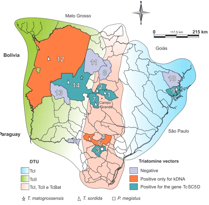
TcI, TcII and TcBat located in the center, TcI in the east, and TcII in the west (Figure 2).
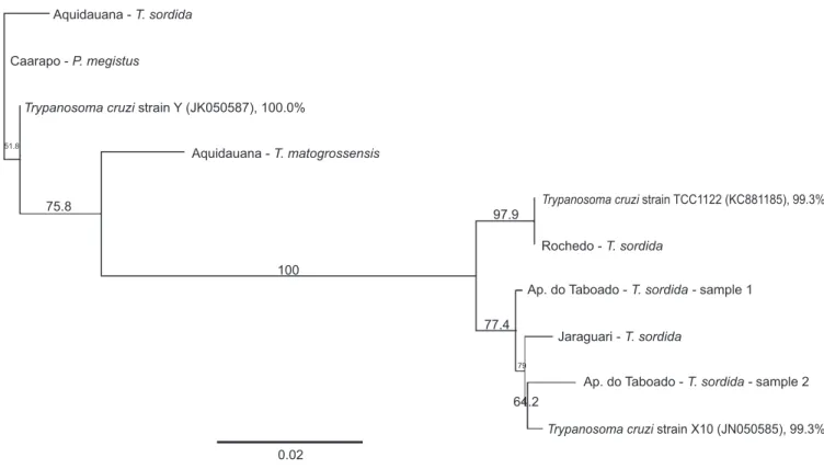
The PCR product generated from the TcSC5D gene was cloned and sequenced. After alignment and analysis of the nucleotide sequence, TcI, TcII and TcBat were found in 98-100% of the samples (Figure 3). It was not possible to sequence some
of the samples; however, identifi cation via RFLP revealed that
DTUs were present in triatomines in those samples.
The reactions using primers for T. rangeli (data not shown)
did not produce overlapping data, confi rming that the amplicons were specifi c for T. cruzi.
DISCUSSION
The most commonly captured triatomine was T. sordida, which was similar to what was observed in previous studies, showing that this species, which is usually considered secondary, is the most frequently encountered species in MS4,26,27. Triatoma sordida is native to the Cerrado, the biome in which State of Mato Grosso do Sul as well as the transition areas of Maranhão, Piauí, Bahia, the Pantanal and the eastern Chaco are located39.
This was also the species that harbored the greatest variety of DTUs (TcI, TcII and TcBat). As this species was the most frequently encountered, the likelihood of fi nding the largest
number of infected insect species as well as a greater variety of DTUs was higher.
Insect vectors usually occupy wild environments. However, if their habitat is degraded, they can relocate near human habitations, such as corrals, sties or chicken coops as well as other peridomiciliary constructions. Although all captured triatomines were found in peridomiciles, their mere coexistence with an insect vector increases the chances of infection40. One notable factor is that TcII was associated with the severe forms of Chagas disease that were found south of the Amazon region41,42; however, other factors are also associated with a higher or lower pathogenicity of the parasite43, and its presence near residences is therefore reason to focus attention on the control of triatomines, even though this DTU was found less frequently in this study.
Trypanosoma cruzi is extremely successful, as observed by its continental distribution and its broad host range, which was replicated in our State of Mato Grosso do Sul data. The DTUs found in this work are common in MS, and other studies have
identifi ed their presence in the Pantanal region22,44. Figure 2 shows that the central region of State of Mato Grosso do Sul had the largest variety of DTUs, presenting TcI, TcII and TcBat
(fi rst report of natural infection in triatomines).
FIGURE 2 - Map of the State of Mato Grosso do Sul, Brazil, highlighting the cities where the triatomines were captured and the distribution of the DTUs found in infected insects. 1: Jaraguari; 2: Rochedo; 3: Caarapó; 4: Douradina; 5: Antônio João; 6: Dourados; 7: Terenos; 8: São Gabriel do Oeste; 9: Aparecida do Taboado; 10: Paranaíba; 11: Rio Verde de Mato Grosso; 12: Corumbá; 13: Miranda; 14: Aquidauana. DTU: discrete typing units; kDNA: kinetoplast deoxyribonucleic acid. T.: Triatoma; P.: Panstrongylus.
the central and eastern areas of the state. Other studies have demonstrated the presence of TcI in São Paulo (SP)45 and Minas Gerais (MG)46, neighboring states to the East of MS, and in the City of Aparecida do Taboado. However, the occurrence of more than one DTU was observed in SP and MG. One of the factors
that may be associated with fi nding only one TcI in the eastern area
of MS may be that it is the most anthropized portion of the state47, with large agropastoral formations that reduce the availability of meat for triatomines. As T. sordida was the most abundant species, is predominantly ornithophilous and was obtained from
henhouse peridomiciles, fi nding other DTUs in the studied sites might be diffi cult. The incidence of TcII was higher in the west,
which is similar to the results of previous work with vectors and other wild animals, but this DTU was found mainly in the Pantanal region44,48. One possible explanation for this localization is that the region is preserved (in relation to the eastern state), leading to an increased supply of hosts as food sources for the insect vectors, thus facilitating the movement of the parasite. Studies indicate the occurrence of at least approximately 124 species of mammals in the Pantanal region alone49-52, but this is only an
estimate, and further studies may confi rm or refute this hypothesis.
However, as the dispersion and isolation of T. cruzi is limited by the dispersion of their hosts, which include several orders of mammals and triatomines53, this idea deserves more attention.
Bolivia
Paraguay São Paulo
Mato Grosso
Goiás
1
2
3
5
6
7
8
4
10
9
0 117,5 km 215 km
11
12
13
4
5
9
8
6
7
14
Negative
Positive only for kDNA
Positive for the gene Tc SC5D
Triatomine vectors
TcI TcII
TcI, TcII e TcBat DTU
T. matogrossensis T. sordida P. megistus Campo
Other studies have demonstrated that TcIII is present in central MS45, which is the same area where TcI is prevalent, more
specifi cally, in the community of Furnas do Dionísio, Jaraguari.
This is a rural Quilombola community located 45km from Campo Grande, the capital of MS. The community comprises approximately 1,031ha, with modest brick houses that are often near wood sties, chicken coops and pens. However, although Marcili et al.45 found TcIII circulating in dogs, the present study showed that only TcI circulated in triatomines and TcII circulated in bats (data not shown) in this community. TcI is a DTU that is widely distributed from South to North America, with most of the isolates from the Amazon region54-57. Although TcI is known to be predominantly wild and the source of more frequent, serious infections in humans in Central America, Venezuela58 and even in the Amazon region, studies have shown that TcI can manifest severe forms of Chagas disease, especially when co-infection occurs in humans with different DTUs42,59. In this case, the epidemiological situation of the studied community has drawn attention because it is located between hills, resulting in a high frequency of infected insects and a large number of
natural and artifi cial ecotopes, as described by Cominetti et
al.26; it is therefore an ideal environment for triatomine colony infestations. Moreover, the observed TcII infection of bats leads to the question of whether there is overlap between peridomestic and sylvatic cycles that leads to an exchange of the T. cruzi
types found in the area. Eco-epidemiological and molecular
Aquidauana -T. sordida
Aquidauana -T. matogrossensis Caarapo -P. megistus
Trypanosoma cruzistrain Y (JK050587), 100.0%
Trypanosoma cruzistrain TCC1122 (KC881185), 99.3%
Trypanosoma cruzistrain X10 (JN050585), 99.3% Rochedo -T. sordida
Ap. do Taboado -T. sordida -sample 1
Ap. do Taboado -T. sordida -sample 2 Jaraguari -T. sordida
51.8
79 75.8
100
0.02
97.9
64.2 77.4
FIGURE 3 - Partial sequence of the TcSC5d gene. Phylogenetic position of the samples of different genotypes in relation to other DTUs of Trypanosoma cruzi. The analysis was performed with 1,000 bootstrap replicates. The number in parentheses next to the accession number is the GenBank. The scale bar indicates the nucleotide substitutions per site. DTU: discrete typing units. T.: Triatoma; P.: Panstrongylus.
studies are needed in this region and are now being performed to elucidate this question.
The same types found in the central areas of MS, except for TcBat, were also found in the State of Paraná60, which is a route of passage for DTUs (Figure 2). However, it can be inferred from our data, in contrast to other studies44,45,48, that it is not
possible to delimit DTUs to a specifi c region. The overlap of
DTUs can result from the variety of intricate types of dispersion of the parasite due to the peculiar mechanisms of vector reinfestation61 the wide variety of hosts, which are abundant and conspicuous in the Cerrado19, and from environmental heterogeneity62.
Despite the fact that were did not demonstrate mixed infection by TcI and TcII in insect vectors, as reported by Abolis et al.60, this phenomenon may also occur in MS because the conditions for these mixed infections in triatomines are present
in this state. Three DTUs were identifi ed in the center of the state
and in nearby municipalities. In addition, the Cerrado biome,
which covers a large portion of MS, has signifi cant horizontal variation, with open fi elds, capons, woods, forests and wetlands
that potentially coexist in the same region19. These data add more strength to the idea that the distribution of the parasite is very wide and dynamic.
fi nding extends the distribution of this DTU in the state, as it
was previously described only in the Pantanal region of MS22,48.
Additionally, this is the fi rst report of this DTU in a naturally
infected triatomine, which had not been previously described, even after attempts at laboratory infection22. However, it is expected that the incidence of this DTU is more frequent because it is most closely related to TcI, the most widely distributed DTU
in MS, and has fi nally been identifi ed to infect a triatomine. In
the same region, TcII was found in a sample of DNA extracted from the blood of an armadillo (Euphractus sexcinctus) (data not shown).
This study demonstrates the occurrence of overlapping DTUs in the State of Mato Grosso do Sul. The geographical distributions of the DTUs follow different patterns, with TcI, TcII and TcBat in the center, TcI predominantly in the east, and TcII in the west. Furthermore, TcI was the most frequent DTU in triatomines, followed by TcII and TcBat. This study provides greater insight into the distribution of T. cruzi in Brazil, but
further studies may reveal a more defi ned mosaic distribution
of DTUs in MS over the long term.
Support for this work was provided by Embrapa Gado de Corte, Conselho Nacional de Desenvolvimento
Científi co e Tecnológico (CNPQ) and Fundação de Apoio ao
Desenvolvimento do Ensino, Ciência e Tecnologia do Estado de Mato Grosso do Sul (Fundect-MS). A PhD scholarship was provided by Coordenação de Aperfeiçoamento de Pessoal de Nível Superior (CAPES).
ACKNOWLEDGMENTS
The authors wish to thank the Laboratório Regional de Entomologia, Núcleo Regional Saúde, and Secretaria de Estado de Saúde for assistance and Dr. Marta M.G. Teixeira (Universidade de São Paulo, Brazil) for the generous gift of
T. cruzi and T. rangeli genomic DNA.
The authors declare that there is no confl ict of interest. CONFLICT OF INTEREST
FINANCIAL SUPPORT
REFERENCES
1. Miles MA, Feliciangeli MD, Arias AR. American trypanosomiasis (Chagas disease) and the role of molecular epidemiology in guiding control strategies. BMJ 2003; 7404:1444-1448.
2. Guhl F, Schofi eld CJ. Population genetics and control of Triatominae.
Parasitol Today 1996; 5:169-170.
3. Roellig DM, Brown EL, Barnabé C, Tibayrenc M, Steurer FJ, Yabsley MJ. Molecular typing of Trypanosoma cruzi isolates, United States. Emerg Infect Dis 2008; 7:1123-1125.
4. Almeida PS, Ceretti Júnior W, Obara MT, Santos HR, Barata JM. Survey of Triatominae (Hemiptera: Reduviidae) fauna in domestic
environments and natural infection by Trypanosomatidae in the State of Mato Grosso do Sul. Rev Soc Bras Med Trop 2008; 4:374-380.
5. Yeo M, Acosta N, Llewellyn M, Sanchez H, Adamson S, Miles GA, et al. Origins of Chagas disease: Didelphis species are natural hosts
of Trypanosoma cruzi I and armadillos hosts of Trypanosoma cruzi
II, including hybrids. Int J Parasitol 2004; 35:225-233.
6. Rassi Jr A, Rassi A, Marin-Neto JA. Chagas disease. Lancet 2010; 375:1388-1402.
7. Tibayrenc M, Neubauer K, Barnabé C, Guerrini F, Skarecky D, Ayala F. Genetic characterization of six parasitic protozoa: parity betweem ramdom primer DNA typing and multilocus enzyme electrophoresis. Proc Natl Acad Sci USA 1993; 4:1335-1339. 8. Gaunt MW, Yeo M, Frame IA, Stothard JR, Carrasco HJ, Taylor MC,
et al. Mechanism of genetic exchange in American trypanosomes. Nature 2003; 6926:936-939.
9. Miles MA, Toye PJ, Oswald SC, Godfrey DG. The identifi cation by
isoenzyme patterns of two distinct strain-groups of Trypanosoma
cruzi circulating independently in a rural area of Brazil. Trans R
Soc Trop Med Hyg 1997; 3:217-225.
10. Miles MA, Cedillos RA, Povoa MM, Souza AA, Prata A, Macedo V. Do radically dissimilar Trypanosoma cruzi strains (zymodemes) cause Venezuelan and Brazilian forms of Chagas’ disease? Lancet 1981; 1:1338-1340.
11. Miles MA, Arias JR, Souza AA. Chagas’ disease in the Amazon basin: V. Periurban palms as habitats of Rhodnius robustus and
Rhodnius pictipes–triatomine vectors of Chagas’ disease. Mem Inst
Oswaldo Cruz 1983; 78:391-398
12. Miles MA, Souza A, Povoa M, Shaw JJ, Lainson R, Toye PJ. Isozymic heterogeneity of Trypanosoma cruzi in the fi rst autochthonous
patients with Chagas' disease in Amazonian Brazil. Nature 1978; 5656:819-821.
13. Devera R, Fernandes O, Coura JR. Should Trypanosoma cruzi be called "cruzi" complex? A review of the parasite diversity and the potential of selecting population after in Vitro culturing and mice infection. Mem Inst Oswaldo Cruz 2003; 1:1-12.
14. Fernandes O, Souto RP, Castro JA, Pereira JB, Fernandes NC, Junqueira AC, et al. Brazilian isolates of Trypanosoma cruzi from
human and triatomines classifi cated into two lineages using
mini-exon and ribosomal RNA sequence. Am J Trop Med Hyg 1998; 6:807-811.
15. Souto RP, Fernandes O, Macedo AM, Campbell DA, Zingales B. DNA markers defi ne two major phylogenetic lineages of
Trypanosoma cruzi. Mol Biochem Parasitol 1996; 2:141-152.
16. Zingales B, Souto RP, Mangia RH, Lisboa CV, Campbell DA, Coura
JR, et al. Molecular epidemiology of American trypanosomiasis in Brazil based on dimorphisms of rRNA and mini-exon gene sequences. Int J Parasitol 1998; 1:105-112.
17. Brisse S, Verhoef J, Tibayrenc M. Characterisation of large and small subunit rRNA and miniexon genes further supports the distinction of six Trypanosoma cruzi lineages. Int J Parasitol 2001; 11:1218-1226.
18. Coura JR. Trypanosomiasis, chagas disease. Ciênc Cult 2003; 1: 30-33.
19. Machado CA, Ayala FJ. Nucleotide sequences provide evidence of genetic exchange among distantly related lineages of Trypanosoma
cruzi. Proc Natl Acad Sci USA 2001; 13:7396-7401.
20. Silva AV, Bosco SMG, Langoni H, Bagagli E. Study of Toxoplasma infection in Brazilian wild mammals: Serological evidence in
Dasypus novemcinctus Linnaeus, 1758 and Euphractus sexcinctus
Wagler, 1830. Vet Parasitol 2006; 1:81-83.
21. Zingales B, Andrade SG, Briones MR, Campbell DA, Chiari
intraspecifi c nomenclature: second revision meeting recommends
TCI to TCVI. Mem Inst Oswaldo Cruz 2009; 7:1051-1054.
22. Marcili A, Lima L, Cavazzana M, Junqueira AC, Veludo HH, Maia Da Silva F, et al. A new genotype of Trypanosoma cruzi associated with bats evidenced by phylogenetic analyses using SSU rDNA, cytochrome b and Histone H2B genes and genotyping based on ITS1 rDNA. Parasitology 2009; 6:641-655.
23. Instituto Brasileiro de Geografi a e Estatística (IBGE) [Internet].
Brasilia: IBGE. Available at: http://www.ibge.gov.br/estadosat/
perfi l.php?sigla=ms.
24. Simon MF, Grether R, Queiroz LP, Skema C, Pennington RT, Hughes CE. Recent assembly of the Cerrado, a neotropical plant
diversity hotspot, by in situ evolution of adaptations to fi re.
Proc Natl Acad Sci USA 2009; 48:20359-20364.
25. Harris MB, Tomas WM, Mourão G, Silva CJ, Guimarães E, Sonoda F.
Desafi os para proteger o Pantanal brasileiro: ameaças e iniciativas
em conservação. Megadiversidade 2005; 1:156-164.
26. Cominetti MC, Andreotti R, Oshiro ET, Dorval MEMC. Epidemiological factors related to the transmission risk of
Trypanosoma cruzi in a Quilombola community, State of
Mato Grosso do Sul, Brazil. Rev Soc Bras Med Trop 2011; 5:575-581. 27. Cominetti MC, Almeida RF, Gonçalves GM, Andreotti R. Monitoring
Trypanosoma cruzi infection in triatomines using PCR in Mato
Grosso do Sul, Brazil. Rev Soc Bras Med Trop 2013; 3:277-280. 28. Carcavallo RU, Rodrigues MEF, Galvão C, Rocha DS, Girón IG,
Arocha MAO, et al. Habitats e fauna relacionada. In: Carcavallo RU, Girón IG, Jurberg J, Lent H, editors. Atlas dos vetores da doença de Chagas nas Américas. Rio de Janeiro: Fundação Oswaldo Cruz; 1997. p. 107-244.
29. Souza MA. Morphobiological characterization of Trypanosoma
cruzi Chagas, 1909 and its distinction from other trypanosomes.
Mem Inst Oswaldo Cruz 1999; 94:205-210.
30. Westenberger SJ, Sturm NR, Yanega D, Podlipaev SA, Zeledón R,
Campbell DA, et al. Trypanosomatid biodiversity in Costa Rica: genotyping of parasites from Heteroptera using the spliced leader RNA gene. Parasitology 2004; 129:537-547
31. Wincker P, Britto C, Pereira JB, Cardoso MA, Oelemann W, Morel CM.
Use of a simplifi ed polymerase chain reaction procedure to detect
Trypanosoma cruzi in blood samples patients in a rural endemic
area. Am J Trop Med Hyg 1994; 51:771-777.
32. Schijman AG, Bisio M, Orellana L, Sued M, Duffy T, Jaramillo AMM, et al. International study to evaluate PCR methods for detection of Trypanosoma cruzi DNA in blood samples from Chagas disease patients. PLoS Negl Trop Dis 2011; 5:e931.
33. Chiurillo MA, Crisante G, Rojas A, Peralta A, Dias M, Guevara P, et al. Detection of Trypanosoma cruzi and Trypanosoma rangeli
infection by duplex PCR assay based on telomeric sequences. Clin Diagn Lab Immunol 2003; 10:775-779
34. Cosentino RO, Agüero F. A simple strain typing assay for
Trypanosoma cruzi: discrimination of major evolutionary lineages
from a single amplifi cation product. PLoS Negl Trop Dis 2012;
7:e1777.
35. Sanger F, Nicklen S, Coulson AR. DNA sequencing with chain-terminating inhibitors. Proc Natl Acad Sci 1977; 74:5463-5467 36. Hall TA. BioEdit: a user-friendly biological sequence alignment
editor and analysis program for Windows 95/98/NT. Nucl Acids Symp Ser 1999; 41:95-98.
37. Altschul SF, Gish W, Miller W, Myers EW, Lipman DJ. Basic local alignment search tool. J Mol Biol 1990; 215:403-410.
38. Drummond AJ, Ashton BMC, Heled J, Kearse M, Moir R, Stones-Havas S, et al. Geneious v 4.8. [Software]. 2009. Available at: http:// www.geneious.com/
39. Forattini OP. Biogeography, origin, and distribution of triatominae domiciliarity in Brazil. Rev Saude Publica 1980; 6:964-968. 40. Toledo MJO, Kühl JB, Silva SV, Gasperi V, Araújo SM.
Biogeography, origin, and distribution of triatominae domiciliarity in Brazil. Rev Saude Publica 1997; 3:197-203.
41. Burgos JM, Altcheh J, Bisio M, Duffy T, Valadares HM, Seidenstein
ME, et al. Direct molecular profi ling of minicircle signatures and
lineages of Trypanosoma cruzi bloodstream populations causing congenital Chagas disease. Int J Parasitol 2007; 37:1319-1327. 42. del Puerto R, Nishizawa JE, Kikuchi M, Iihoshi N, Roca Y,
Avilas C, et al. Lineage analysis of circulating Trypanosoma cruzi
parasites and their association with clinical forms of Chagas disease in Bolivia. PLoS Negl Trop Dis 2010; 4:e687.
43. Macedo AM, Machado CR, Oliveira RP, Pena SD. Trypanosoma
cruzi: genetic structure of populations and relevance of genetic
variability to the pathogenesis of chagas disease. Mem Inst Oswaldo Cruz 2004; 99:1-12.
44. Herrera HM, Lisboa CV, Pinho AP, Olifi ers N, Bianchi RC, Rocha FL,
et al. The coati (Nasua nasua, Carnivora, Procyonidae) as a reservoir host for the main lineages of Trypanosoma cruzi in the Pantanal region, Brazil. Trans R Soc Trop Med Hyg 2008; 11:1133-1339. 45. Marcili A, Lima L, Valente VC, Valente SA, Batista JS, Junqueira
AC, et al. Comparative phylogeography of Trypanosoma cruzi
TCIIc: new hosts, association with terrestrial ecotopes, and spatial clustering. Infect Genet Evol 2009; 6:1265-1274.
46. Viana EN. Dinâmica de reinfestações por triatomíneos e alterações ambientais na ecoepidemiologia da doença de Chagas em área de
Triatoma sordida Stål 1859 (Hemiptera, Reduviidae, Triatominae)
no norte de Minas Gerais, Brasil [PhD thesis]. [Belo Horizonte]: Universidade Federal de Minas Gerais, Instituto de Ciências Biológicas; 2001. 280 p.
47. Machado RB, Ramos Neto MB, Pereira P, Caldas E, Gonçalves D, Santos N, et al. Estimativas de perda da área do Cerrado brasileiro. Brasilia: Conservation International do Brasil; 2004.
48. Cavazzana Jr M, Marcili A, Lima L, Silva FM, Junqueira AC, Veludo HH, et al. Phylogeographical, ecological and biological patterns shown by nuclear (ssrRNA and gGAPDH) and mitochondrial (Cyt b) genes of trypanosomes of the subgenus Schizotrypanum parasitic in Brazilian bats. Int J Parasitol 2010; 3:345-55.
49. Alho CJR, Lacher TE. Mammalian conservation in the Pantanal of Brazil. In: Mares MA, Schmidly DJ, editors. Latin American mammalogy: history, biodiversity and conservation. University of Oklahoma Press; 1991. p. 280-294.
50. Mourão G, Coutinho M, Mauro R, Campos Z, Tomás W, Magnusson
W. Aerial surveys of caiman, marsh deer and pampas deer in the Pantanal wetland of Brazil. Biol Cons 2000; 92:175-183.
51. Tomas WM, Lima Borges PA, Rocha HJF, Sá Filho R, Kutchenski Jr F, Udry TV. The potential of the Aquidauana and Miranda rivers, in the Pantanal Wetland, for the conservation of the giant river otter (Pteronura brasilienis). In: Dantas M, Resende EK, Comastri Filho JA, editors. Anais do III Simpósio sobre Recursos Naturais e Socioeconômicos do Pantanal. Corumbá: Empresa Brasileira de Pesquisa Agropecuária - Embrapa-Pantanal; 2002. p. 1-12.
52. Sanderson EW, Chetkiewicz CLB, Medellin RA, Rabinowitz A,
Redford KH, Robinson JG, et al. Un análisis geográfi co del estado
de conservación y distribución de los jaguars através de su área de distribución. In: Medellín RA, Equihua C, Chetkiewicz CLB, Crawshaw Jr PG, Rabinowitz A, Redford KH, et al, editors. El jaguar en el Nuevo milenio. Mexico: Universidad Nacional Autônoma de México, Nova York: Wildlife Conservation Society; 2002. p. 551-560.
54. Miles MA, Povoa M, Souza AA, Lainson R, Shaw JJ, Ketteridge DS. Chagas disease in the Amazon Basin. II. The distribution of
Trypanosoma cruzi zymodemes 1 and 3 in Pará State, north Brazil.
Trans R Soc Trop Med Hyg 1981; 75:667-674.
55. Fernandes O, Santos SS, Cupolillo E, Mendonça B, Derre R, Junqueira ACV, et al. A mini-exon multiplex polymerase chain reaction to distinguish the major groups of Trypanosoma cruzi and
T. rangeli in the Brazilian Amazon. Trans R Soc Trop Med Hyg
2001; 95:97-99.
56. Maia da Silva F, Naiff RD, Marcili A, Gordo M, D’Affonseca Neto JA, Naiff MF, et al. Infection rates and genotypes of Trypanosoma
rangeli and T. cruzi infecting free-ranging Saguinus bicolor
(Callitrichidae), a critically endangered primate of the Amazon Rainforest. Acta Trop 2008; 107:168-173.
57. Marcili A, Valente V, Valente A, Junqueira ACV, Maia da Silva F, Naiff R, et al. Trypanosoma cruzi in Brazilian Amazonia: lineages TCI and TCIIa in wild primates, Rhodnius spp. and in humans with Chagas disease associated with oral transmission. Int J Parasitol 2009; 39:615-623.
58. Miles MA, Cedillos RA, Póvoa MM, Souza AA, Prata A, Macedo V. Do radically dissimilar Trypanosoma cruzi strains (zymodemes)
cause Venezuelan and Brazilian forms of Chagas' disease? Lancet 1981; 8234:1338-1340.
59. Burgos JM, Diez M, Vigliano C, Bisio M, Risso M, Duffy T,
et al. Molecular identifi cation of Trypanosoma cruzi Discrete Typing Units in end-stage chronic Chagas heart disease and reactivation after heart transplantation. Clin Infect Dis 2010; 51:485-495. 60. Abolis NG, Araújo SM, Toledo MJ, Fernandez MA, Gomes ML.
Trypanosoma cruzi I-III in southern Brazil causing individual and
mixed infections in humans, sylvatic reservoirs and triatomines. Acta Trop 2011; 3:167-172.
61. Schachter-Broide J, Gürtler RE, Kitron U, Dujardin JD. Temporal variations of wing size and shape of Triatoma infestans (Hemiptera: Reduviidae) populations from Northwestern Argentina using geometric morphometry. J Med Entomol 2009; 5:994-1000.
62. Vaz VC, D'Andrea PS, Jansen AM. Effects of habitat fragmentation on wild mammal infection by Trypanosoma cruzi. Parasitology 2007; 12:1785-1793.
63. Ramírez JD, Hernández C, Montilla M, Zambrano P, Flórez AC,
Parra E, et al. First Report of Human Trypanosoma cruzi Infection
Attributed to TcBat Genotype. Zoonoses Public Health 2013;