Immune expression of E-cadherin and
α
,
β
and
γ
-Catenin
adhesion molecules and prognosis for upper urinary tract
urothelial carcinomas
Sabrina Thalita dos Reis, Katia Ramos Moreira Leite, Alcides Mosconi Neto, José Pontes Júnior, Nayara
Izabel Viana, Alberto Azoubel Antunes, Marcos Francisco Dall´Oglio, Miguel Srougi
Laboratory of Medical Investigation (LIM55), Urology Department, University of Sao Paulo Medical School (STR, KRML, AMN, JPJ, NIV, AAA, MS), Sao Paulo, Brazil and Instituto do Câncer do Estado de São Paulo (ICESP), University of Sao Paulo Medical School (JPJ, MFD), Sao Paulo, Brazil
ABSTRACT ARTICLE INFO
_________________________________________________________ ___________________
Key words:
Urinary Tract; Carcinoma, Transitional Cell;
alpha Catenin; Cadherins
Int Braz J Urol. 2012; 38: 466-73
__________________
Submitted for publication: July 27, 2011
__________________
Accepted after revision: February 23, 2012
Introduction: Cell adhesion molecules (CAM) are required for maintaining a normal epithelial phenotype, and abnormalities in CAM expression have been related to can-cer progression, including bladder urothelial carcinomas. There is only one study that correlates E-cadherin and α-, β- andγ-catenin expression with prognosis of upper tract
urothelial carcinomas. Our aim is to study the pattern of immune expression of these CAMs in urothelial carcinomas from the renal pelvis and ureter in patients who have been treated surgically. Our goal is to correlate these expression levels and c
haracteris-tics with well-known prognostic parameters for disease-free survival.
Materials and Methods: We evaluated specimens from 20 patients with urothelial car-cinomas of the renal pelvis and ureter who were treated with nephroureterectomy or ureterectomy between June 1997 and January 2007. CAM expression was evaluated by immunohistochemistry in a tissue microarray and correlated with histopathological characteristics and patient outcomes after a mean follow-up of 55 months.
Results: We observed a relationship between E-cadherin expression and disease re-currence. Disease recurrence occurred in 87.5% of patients with strong E-cadherin expression. Only 50.0% of patients with moderate expression and 0% of patients with weak or no expression of E-cadherin had disease recurrence (p = 0.014). There was also a difference in disease-free survival. Patients with strong E-cadherin expression had a mean disease-free survival rate of 49.1 months, compared to 83.9 months for patients with moderate expression (p = 0.011). Additionally, an absence ofα-catenin expression was associated with tumors that were larger than 3 cm (p = 0.003).
Conclusions: We demonstrated for the first time that immune expression of E-cadhe-rin is related to tumor recurrence and disease-free survival rates, and the absence of
α-catenin expression is related to tumor size in upper tract urothelial carcinomas.
INTRODUCTION
Upper tract urothelial carcinomas (UUC) are defined as tumors that occur in the renal
IBJU |ADHESION MOLECULES AND UPPER URINARY TRACT CARCINOMA
467
have suggested that the thin sub-epithelial con-nective tissue and muscularis propria of the up-per urinary tract are responsible for early tumor invasion. Furthermore, diagnosis of this carci-noma tends to be delayed because of a lack of symptoms and a lack of neoplastic cells in urine cytology (2,3).
There are few predictors of tumor re-currence after nephroureterectomy. The main prognostic factors are the patient’s age, tumor location, and tumor architecture, stage and his-tological grade. However, there is currently no evidence that specific tumor characteristics would be able to define patients that could benefit from adjuvant treatment (4).
Biomarkers have not been extensively studied in UUCs. P53 mutations (5) and angio-genesis (6), which are well-known markers of worse prognosis in patients with bladder UCs, have also been related to worse prognosis in pa-tients with UUCs.
E-cadherin is a transmembrane glyco-protein that mediates the selective adhesion of epithelial cells. This protein is required for in-teractions and maintenance of tissue integrity (7). α-Catenin mediates interactions between the E-cadherin/β-catenin complex and the actin cy-toskeleton (8), whereas γ-catenin probably has a role on desmosomal plaques (9).
The role of adhesion molecules in UC is still controversial, and only a few studies have been published in the literature on this topic (6,10). Dysfunctions in cell-cell adhesion mole-cules have been related to the initial steps of tu-mor invasion and the development of metastasis in urothelial bladder carcinomas. Recently, Kashi-buchi et al. (11) demonstrated aberrant expression of E-cadherin and α-, β- and γ-catenin in 40 to 50% of urothelial bladder carcinoma cases. They also related aberrant expression to tumor stage and cancer specific survival.
The aim of our study is to evaluate the expression of the E-cadherin and α-, β- and
γ-catenin adhesion molecules in UUC cells by immunohistochemistry using a tissue microarray construction (TMA) of specimens from 20 patients who underwent ureterectomy or nephroureterec-tomy for treatment of their UUCs.
MATERIALS AND METHODS
Patients
We retrospectively evaluated surgical specimens from 20 patients with UUC who un-derwent nephroureterectomy or ureterectomy performed by a single surgeon (MS) from 1997 to 2007. The median age of the patients was 67 years old. Twelve patients had tumors of the re-nal pelvis, five had tumors of the ureter, and three had multifocal tumors that were present in both the renal pelvis and the ureter. Clinical and pathological characteristics of the patients are listed in Table-1.
All subjects provided informed consent before participating in the study, and the study was approved by the Institutional Board of Ethics (#0720/09).
The specimens were fixed in buffered 10% formalin, routinely processed, stained with hema-toxylin and eosin, and examined by a single pa-thologist (KRML). The prognostic factors that were evaluated included tumor grade, which was deter-mined according to the 2004 WHO classification tumor stage (TNM 2010), tumor size, multicentric-ity and neoplastic microvascular invasion. The pattern and intensity of immunoexpression of the adhesion molecules were compared to the above prognostic factors and with the disease-free sur-vival rates after a mean follow-up of 55 months.
TMA construction
TMA construction was performed as previ-ously described (12). Slides that contained each of the patients’ tumors were selected by examining areas that best represented the entire tumor. Two areas from each tumor were marked with perma-nent ink to correspond to areas included in TMA analysis. A 1.0 mm punch was used.
Table 1 - Patients Characteristics.
Patients(n = 20)
Age (years)
Mean 66,5
Min - Max 52-85
Gender
Female 4 (20%)
Male 16 (80%)
Pathological Stage
pTa-pT1 7 (35%)
pT2-pT3 13 (65%)
Tumor size
≤ 3 cm 6 (30%)
>3 cm 14 (70%)
Tumor multicentricity
Yes 5 (25%)
No 15 (75%)
Tumor Grade
High 15 (75%)
Low 5 (25%)
Microvascular Invasion
Yes 10 (50%)
No 10 (50%)
Recurrence
Yes 10 (50%)
No 10 (50%)
1:100 dilution of antibodies against γ-catenin (Zymed Montrouge, France) and α-catenin (Santa Cruz, St Cruz, CA, USA) was used. The LSAB sys-tem was used for immunostaining (LSAB; Dako Cytomation, CA, USA). Color was developed by
reactions with a 3.3’-diaminobenzidine substrate-chromogen solution followed by counterstaining with Harris hematoxylin. Finally, slides were de-hydrated, coverslipped, and examined under light microscope.
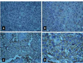
The expression of each marker was evalu-ated by a single pathologist (KRML). Semi-quan-titative analysis was performed for all antibodies according to the intensity of staining: negative, weak, moderate and strong (Figure-1).
Statistical Analysis
Statistical analyses were performed using SPSS 16.0 for Windows (release 16.02). For com-parison between categories, we used an ANOVA and Student’s t-tests and significance was determined at p ≤ 0.05. For adhesion molecule expression pro-files with significant associations with recurrence risks, Kaplan-Meier survival curves were used to illustrate disease-free survival. We performed a log-rank test to show differences between curves.
RESULTS
All cases were usual urothelial carci-nomas without any other specification. Fifteen (75%) cases were high grade urothelial carci-nomas. Only two (10%) cases were staged pTa,
A
C
B
D
IBJU |ADHESION MOLECULES AND UPPER URINARY TRACT CARCINOMA
469
five (25%) were pT1, three (15%) pT2 and the remaining 10 (50%) were staged pT3.
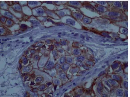
The pattern of staining was membrane for E-chaderin and cytoplasmatic for the other markers. Moderate and strong E-cadherin ex-pression was significantly related to tumor re-currence (p = 0.014) (Figure-2), while moderate
γ-catenin expression was correlated with a lower disease-free survival rate with marginal statisti-cal significance (p = 0.071)(Table-2). There was no relationship between β-catenin or α-Catenin expression with tumor recurrence, p = 0.999 and p = 0.465 respectively.
When CAM expression was analyzed in relation to the classical prognostic parameters of pathological stage, tumor grade, microvascu-lar invasion, multicentricity and tumor size, we found that α-catenin expression was negative or weak in 92.9% of tumors that were larger than 3 cm (p = 0.003) (Table-2).
DISCUSSION
When studying immune expression of E-cadherin and α-, β-, γ-catenin in UUC cells, we were able to show that moderate or strong
Kaplan-Meier curves showed that pa-tients with strong E-cadherin expression had a mean disease-free survival rate of 49.1 months. Patients with moderate expression levels had a mean survival of 83.9 months (p = 0.011) (Fig-ure-3). γ-catenin, β-Catenin and α-Catenin ex-pression levels did not demonstrate differences in disease-free survival length, p = 0.053; p = 0.951; p = 0.913, respectively.
Figure 2 - Micrography of an IHC reaction showing strong expression of E-cadherin.
expression levels of E-cadherin were related with both tumor recurrence (p = 0.014) and a shorter time before tumor recurrence (p = 0.011). Additionally, negative or weak expression of
IBJU
|
ADHESION MOLECULES AND UPPER URINARY TRA
CT CARCINOMA
4
70
E–chaderin invasion
F M pTa-1 pT2 pT3 Low High Absent Present No Yes ≤ 3 cm > 3 cm No Yes
Negative 0 1 0 0 1 0 1 0 1 0 1 0 1 0 1
Weak 1 3 1 1 2 1 3 2 2 3 1 2 2 4 0
Moderate 1 7 3 0 5 1 7 5 3 6 2 1 7 4 4
Strong 2 5 1 2 4 3 4 3 4 6 1 3 4 1 6
P value 0.774 0.626 0.493 0.565 0.354 0.348 0.014
γ-catenin
Weak 1 4 1 1 3 1 4 2 3 3 2 0 5 1 4
Moderate 2 8 2 1 7 1 9 4 6 7 3 4 6 7 3
Strong 1 4 2 1 2 3 2 4 1 5 0 2 3 1 4
P value > 0.999 0.612 0.119 0.282 0.170 0.120 0.071
α-catenin
Negative 1 10 4 1 6 3 8 6 5 9 2 0 11 4 7
Weak 2 3 0 2 3 2 3 3 2 3 2 3 2 2 3
Moderate 0 3 1 0 2 0 3 1 2 3 0 2 1 2 1
Strong 1 0 0 0 1 0 1 0 1 0 1 1 0 1 0
IBJU |ADHESION MOLECULES AND UPPER URINARY TRACT CARCINOMA
471
E-cadherin and γ-catenin were moder-ately or strongly expressed in most patients, but
α-catenin was absent or lost in the majority of cases.
Cadherins represent a family of trans-membrane glycoproteins that mediate homo-phile, calcium-dependent, intercellular adhe-sion. E-cadherin is the major cadherin molecule expressed by epithelial cells. Linkage between E-cadherin and intracellular catenins is neces-sary for the formation of strong intercellular adhesion. β-catenin and γ-catenin bind directly to the cytoplasmic domain of E-cadherin, and
α-catenin links the bound β-catenin to actin in the cytoskeleton (13,14).
Several studies have explored the ex-pression of cadherins and catenins and their role as prognostic factors in urothelial bladder car-cinomas. These studies suggested that aberrant expression of α-catenin, β-catenin, E-cadherin and γ-catenin correlates with higher tumor stag-es and worse patient prognosis (15-17). Kashibu-chi et al. showed a correlation between aberrant
β-catenin and γ-catenin expression with tumor
stage and a correlation between γ-catenin with tumor grade in urothelial bladder carcinomas. Furthermore, they found that abnormal expres-sion of E-cadherin was an independent predic-tor of disease-specific survival in this neoplasia (11). Different from them, we tried to add details to the pattern of immune expression, stratifying the intensity of staining in weak, moderate and strong patterns. The subjective quantification should be better explored in future studies by evaluating adhesion molecules gene expression profiles using quantitative real time PCR that could be proposed as markers to diagnose or fol-low patients that harbor upper urinary tract uro-thelial carcinomas.
with Balkan endemic nephropathy, lower E-cad-herin expression was correlated with higher tu-mor stages in both groups (18). However, some authors postulate that E-cadherin expression can be reversibly controlled by methylation (19). This methylation would be responsible for E-cadherin down-regulation during the epithelial-mesenchymal transition, which is an important stage in tumor invasion and dissemination (20). But for tumor establishment in the metastatic site there is a requirement for E-cadherin pression (21). In our study, we observed re-ex-pression of E-cadherin that was more expressed in pT3 local tumors (69.3%) and that were able to disseminate or recur locally. Interestingly, in agreement with our data, a recent study pub-lished by Lim et al. (22) found normal expression of E-cadherin in both usual and micropapillary urothelial bladder carcinomas, and that was in-dependent of tumor stage, tumor grade or pres-ence of microvascular invasion. In their study, they proposed that loss of E-cadherin expression may be a characteristic of a histologically spe-cial plasmocytoid or signed ring cell urothelial carcinoma.
Our study also showed that lower expres-sion of α-catenin was associated with tumors larger than 3 cm (p = 0.003). Tumor size is a very important prognostic parameter in UC. It is related to tumor recurrence, with 7-fold increase risk in finding tumors after transurethral resec-tion and BCG treatment. Tumor size has also been used as a criteria to proceed with more ag-gressive treatment in pT1-stage bladder cancer (23,24). Negative expression of this catenin has been related to worse prognosis, tumor stage, tu-mor grade and UC disease recurrence in other studies (11,25). However, there were no studies of α-catenin expression in UUCs.
Moderate γ-catenin expression was mar-ginally significantly related to tumor recurrence (p = 0.071) in our study. The same result was described by Clairotte et al. in cases of bladder cancer. They classified CAM expression as either normal, heterogeneous or absent. The percent-age of cases with absence of CAM expression was very low, as was in our series. They showed that heterogeneous expression of γ-catenin was
related to worse prognosis and shorter disease-free survival rate in patients with bladder can-cer. If our moderate expression category is simi-lar to their heterogeneous category, we observed similar findings in patients with UUCs (25).
Some authors have discussed the accu-racy of TMA immunohistochemistry in measur-ing protein expression (26), but the advantages of the method outperform the disadvantages in terms of standardization of reaction and analysis and have been applied in the evaluation of many prognostic markers in different tumors including urothelial carcinoma for more than 20 years (27).
We are aware that we have studied a small number of cases; however, UUCs are rare and it took a period of 10 years to collect the cases that were included in this study. A larger number of cases would allow analysis of differ-ent aspects, such as tumor location. Tumor loca-tion is important because it is well known that UCs of the renal pelvis and ureter have different behaviors. Additionally, it is difficult to compare different studies because each study establishes diverse criteria to evaluate immune expression of CAMs. A more standardized results evaluation would make it easier to discover the real role of CAMs in UCs.
However, this is the first study to explore immune expression of components of the cad-herin-catenin complex and their role in UUCs.
CONFLICT OF INTEREST
None declared.
REFERENCES
1. Munoz JJ, Ellison LM: Upper tract urothelial neoplasms: in-cidence and survival during the last 2 decades. J Urol. 2000; 164: 1523-5.
2. Park S, Hong B, Kim CS, Ahn H: The impact of tumor loca-tion on prognosis of transiloca-tional cell carcinoma of the upper urinary tract. J Urol. 2004; 171(2 Pt 1):621-5.
3. Catto JW, Yates DR, Rehman I, Azzouzi AR, Patterson J, Si-bony M, et al.: Behavior of urothelial carcinoma with respect to anatomical location. J Urol. 2007; 177: 1715-20.
IBJU |ADHESION MOLECULES AND UPPER URINARY TRACT CARCINOMA
473
5. Hashimoto H, Sue Y, Saga Y, Tokumitsu M, Yachiku S: Roles of p53 and MDM2 in tumor proliferation and determination of the prognosis of transitional cell carcinoma of the renal pelvis and ureter. Int J Urol. 2000; 7: 457-63.
6. Inoue K, Kamada M, Slaton JW, Fukata S, Yoshikawa C, Tamboli P, et al.: The prognostic value of angiogenesis and metastasis-related genes for progression of transitional cell carcinoma of the renal pelvis and ureter. Clin Cancer Res. 2002; 8: 1863-70. 7. Takeichi M: Cadherin cell adhesion receptors as a
morphoge-netic regulator. Science. 1991; 251: 1451-5.
8. Näthke IS, Hinck LE, Nelson WJ: Epithelial cell adhesion and development of cell surface polarity: possible mechanisms for modulation of cadherin function, organization and distribution. J Cell Sci Suppl. 1993; 17: 139-45.
9. Ozawa M, Nuruki K, Toyoyama H, Ohi Y: Cloning of an alterna-tive form of plakoglobin (gamma-catenin) lacking the fourth armadillo repeat. J Biochem. 1995; 118: 836-40.
10. Wakatsuki S, Watanabe R, Saito K, Saito T, Katagiri A, Sato S, Tomita Y: Loss of human E-cadherin (ECD) correlated with in-vasiveness of transitional cell cancer in the renal pelvis, ureter and urinary bladder. Cancer Lett. 1996; 103: 11-7.
11. Kashibuchi K, Tomita K, Schalken JA, Kume H, Takeuchi T, Kitamura T: The prognostic value of E-cadherin, alpha-, beta- and gamma-catenin in bladder cancer patients who underwent radical cystectomy. Int J Urol. 2007; 14: 789-94.
12. Kononen J, Bubendorf L, Kallioniemi A, Bärlund M, Schraml P, Leighton S, et al.: Tissue microarrays for high-throughput mo-lecular profiling of tumor specimens. Nat Med. 1998; 4: 844-7. 13. Charalabopoulos K, Tsambals S, Syrigos K, Giannakopoulos
C, Kalfakakou V, Kiortsis D, et al.: Correlation of E-cadherin expression with clinicopathological data in patients suffering from transitional cell carcinoma of the bladder. Exp. Oncol. 2003; 25: 180-5.
14. Koksal IT, Ates M, Danisman A, Sezer C, Ciftcioglu A, Karpu-zoglu G, et al.: Reduced E-cadherin and alpha-catenin expres-sions have no prognostic role in bladder carcinoma. Pathol Oncol Res. 2006; 12: 13-9.
15. Shimazui T, Schalken JA, Giroldi LA, Jansen CF, Akaza H, Koiso K, et al.: Prognostic value of cadherin-associated molecules (alpha-, beta-, and gamma-catenins and p120cas) in bladder tumors. Cancer Res. 1996; 56: 4154-8.
16. Garcia del Muro X, Torregrosa A, Muñoz J, Castellsagué X, Condom E, Vigués F, et al.: Prognostic value of the expression of E-cadherin and beta-catenin in bladder cancer. Eur J Cancer. 2000; 36: 357-62.
17. Syrigos KN, Harrington K, Waxman J, Krausz T, Pignatelli M: Al-tered gamma-catenin expression correlates with poor survival in patients with bladder cancer. J Urol. 1998; 160: 1889-93. 18. Velickovic LJ, Hattori T, Visnjic M, Dimov I, Stojanovic M,
Ste-fanovic V: E-cadherin expression in upper urothelial carcinoma in Balkan Endemic Nephropathy and non-endemic regions. Pathol Res Pract. 2009; 205: 682-9.
19. Ribeiro-Filho LA, Franks J, Sasaki M, Shiina H, Li LC, Nojima D, et al.: CpG hypermethylation of promoter region and inacti-vation of E-cadherin gene in human bladder cancer. Mol Car-cinog. 2002; 34: 187-98.
20. Ke XS, Qu Y, Goldfinger N, Rostad K, Hovland R, Akslen LA, et al.: Epithelial to mesenchymal transition of a primary prostate cell line with switches of cell adhesion modules but without malignant transformation. PLoS One. 2008; 3: e3368. 21. De Marzo AM, Knudsen B, Chan-Tack K, Epstein JI: E-cadherin
expression as a marker of tumor aggressiveness in routinely processed radical prostatectomy specimens. Urology. 1999; 53: 707-13.
22. Lim MG, Adsay NV, Grignon DJ, Osunkoya AO: E-cadherin ex-pression in plasmacytoid, signet ring cell and micropapillary variants of urothelial carcinoma: comparison with usual-type high-grade urothelial carcinoma. Mod Pathol. 2011; 24: 241-7. 23. Pieras E, Frontera G, Ruiz X, Vicens A, Ozonas M, Pizá P: Con-comitant carcinoma in situ and tumour size are prognostic fac-tors for bladder recurrence after nephroureterectomy for upper tract transitional cell carcinoma. BJU Int. 2010; 106: 1319-23. 24. Orsola A, Cecchini L, Raventós CX, Trilla E, Planas J, Landolfi
S, et al.: Risk factors for positive findings in patients with high-grade T1 bladder cancer treated with transurethral resection of bladder tumour (TUR) and bacille Calmette-Guérin therapy and the decision for a repeat TUR. BJU Int. 2010; 105: 202-7. 25. Clairotte A, Lascombe I, Fauconnet S, Mauny F, Félix S, Algros
MP, et al.: Expression of E-cadherin and alpha-, beta-, gam-ma-catenins in patients with bladder cancer: identification of gamma-catenin as a new prognostic marker of neoplastic pro-gression in T1 superficial urothelial tumors. Am J Clin Pathol. 2006; 125: 119-26.
26. Gudjónsson S, Bendahl PO, Chebil G, Höglund M, Lindgren D, Lundberg LM, et al.: Can tissue microarray-based analysis of protein expression predict recurrence of stage Ta bladder cancer? Scand J Urol Nephrol. 2011; 45: 270-7.
27. Fromont G, Rouprêt M, Amira N, Sibony M, Vallancien G, Vali-dire P, et al.: Tissue microarray analysis of the prognostic value of E-cadherin, Ki67, p53, p27, survivin and MSH2 expression in upper urinary tract transitional cell carcinoma. Eur Urol. 2005; 48: 764-70.