INTRODUCTION
Patients with benign prostatic hyperpla-sia (BPH) constitute a large number of urological patients. Management of these patients relies on history taking, clinical examination and inves-tigational modalities which are additional tools for accurate diagnosis and later follow-up. Ultra-sound examination opened the door for further modalities to follow (1). Since the introduction
of transrectal ultrasonography (TRUS) by Wanta-nabe et al. in 1968, it became a valuable tool for diagnosis of most prostatic diseases (2). Over the last two decades this technology has been used successfully in evaluating prostatic disease (1). The color Doppler Ultrasound (CDUS) was later followed by power Doppler Ultrasound (PDUS) that allowed us to assess the vascular architecture of the prostate through a non-invasive modal-ity (3). PDUS has high sensitivmodal-ity for displaying Objective: The aim of this work is to study the resistive index (RI) of prostatic
blood fl ow by transrectal power Doppler sonography in benign prostatic hyperpla-sia (BPH) to determine its correlation with other parameters of BPH.
Materials and Methods: Eighty-two male patients aged 52-86 years with lower urinary tract symptoms (LUTS) due to BPH were included in the study. Patients with prostate cancer, neurogenic bladder, or with other pathology (e.g. prostatitis, bladder stone) were excluded from the study. All patients were evaluated by full history including Internatinoal Prostate Symptoms Score (IPSS), general and local examination (DRE), neurologic examination, urofl owmetry, laboratory investiga-tions including urine analysis, routine laboratory tests and serum prostate specifi c antigen (PSA). Transrectal ultrasonography was used to calculate the total prostatic volume. Transrectal Power Doppler Ultrasound (PUD) was used to identify the cap-sular and urethral arteries of the prostate and to measures the RI value.
Results: The mean prostate volume was 75.1 ± 44.7 g. The mean RI of the right and left capsular arteries were 0.76 ± 0.06 and 0.76 ± 0.07, respectively. The mean RI of the urethral arteries was 0.76 ± 0.08. There was a high signifi cative correlation between the increase of the RI of the right and left capsular and urethral arteries and the degree of obstruction (P value < 0.001), severity of symptoms (P value < 0.001) and also the prostatic volume (P value < 0.001).
Conclusion: Resistive index of the prostatic blood fl ow can be applied as an easy and non-invasive tool to evaluate the lower urinary tract obstruction due to BPH.
Evaluation of the resistive index of prostatic blood fl ow
in benign prostatic hyperplasia
_______________________________________________
Osama Abdelwahab, Ehab El-Barky, Mostafa Mahmoud Khalil, Ahmad Kamar
Urology Departments, Benha Faculty of Medicine (OA, EEB, MMK), Egypt Mubarak Al-kabir Hospital (AK), Kuwait
ABSTRACT
ARTICLE
INFO
_______________________________________________________________ _____________________
Key words:
prostatic hyperplasia; prostate; regional blood fl ow; ultrasonography
Int Braz J Urol. 2012; 38: 250-7
________________
Submitted for publication: January 01, 2011
________________
slow blood flow, besides the capability to depict the number, course and continuity of prostatic vessels (4). The resistance index (RI) of the pros-tate vasculature was able to discriminate patients with normal prostate and those with benign pros-tatic hyperplasia (BPH) and be useful as a new hemodynamic parameter (5). This study aimed to evaluate the relation between RI of prostatic blood flow by PDUS in BPH and its correlation with other parameters of BPH.
MATERIALS AND METHODS
Eighty-two male patients aged 52-86 years with lower urinary tract symptoms (LUTS) due to BPH were included in this study. Patients were randomly selected on the basis of their pre-sentation to the outpatient clinic of Benha Uni-versity Hospital, Egypt. Patients with prostati-tis, prostatic abscess, histologically confirmed prostate cancer, neurogenic bladder and bladder stones were excluded from the study. All patients were assessed by full history taking with spe-cial emphasis on International Prostate Symp-tom Score (IPSS). Physical examination included general examination, digital rectal examination (DRE), neurological examination especially bul-bocavernosus reflex (BCR), cremasteric reflex and anal reflex to screen for possible neurological in-sult. Uroflowmetry was done for all the patients at least twice. Urine analysis, routine laboratory tests and serum prostate specific antigen (PSA) were done for all patients. Imaging studies in-cluded plain X-ray KUB, abdomen and pelvic ultrasound to assess post-voided volume and to exclude other associated pathologies. Transrec-tal ultrasound (TRUS) of the prostate was done to estimate prostatic volume followed by power Doppler ultrasonography (PDUS) to identify the capsular arteries (Figure-1) and urethral arteries (Figure-2) of the prostate and to measure the RI in both right and left capsular arteries and ure-thral arteries. All sonographic examinations were done by the same radiologist.
Informed consent was given by all par-ticipants and the study protocol was approved by the research ethical committee of Benha Faculty of Medicine. Doppler indices were used to obtain
information involving blood flow and vascular impedance in prostatic vessels. The indices de-pend on ratios involving the peak systolic veloc-ity (PSV), the end diastolic velocveloc-ity (EDV) and mean velocity (MV) through one cycle. RI is one of the primary indices used clinically and is cal-culated through the following equation (6).
All data were collected, tabulated and statistically analyzed by SPSS software version 0.16. P-value less than 0.05 was considered sta-tistically significant.
RI = (PSV) - (EDV) PSV
Figure 1 - PDUS examination of the prostatic vasculature showing capsular arteries.
TRUS examination: The procedure was performed after a preliminary cleaning enema in left lateral decubitus position with the knees bent and the bladder partially filled. TRUS examina-tion was performed using a Toshiba machine with end-fire probe with the sonoline Elegra system. The power Doppler imaging was done with a cen-tre probe frequency of 6.5 MHz and the wall filter set to low. The end of the transrectal probe was sheathed in a thin latex balloon (usually a con-dom), containing scanning gel.
RESULTS
Total number of patients enrolled in this study was 82. Age range was 52-86 years with mean age 66.8 ± 10.2. Seven parameters were stud-ied and the values of these parameters are
sum-marized in (Table-1). According to uroflowmetry results and pelvic and abdominal U/S, patients were classified into two groups: obstructed (Qmax < 15 mL/s and PVR > 100 mL) and non-obstructed (Qmax > 15 mL/s). Sixty two patients (76%) were ob-structed and 20 (24%) patients were non-obstruct-ed. There was a highly significant increase in the RI of right and left capsular arteries and urethral arteries correlated to increased obstructive pattern of the patients (Table-2).
According to IPSS, our patients were classi-fied into 2 groups. First group included 21 patients that had mild and moderate obstructive symptoms (mild 0-7 and moderate 8-19 on IPSS) and the second group included 61 patients with severe ob-structive symptoms (20-35 IPSS score). Mean RI of right & left capsular arteries and urethral arteries were measured in both groups and summarized in
Table 1 - Summary of results.
Parameters Mean ± SD Range
AGE (years) 66.8 ± 10.2 52-86
IPSS 26.5 ± 10.7 5-35
Prostate volume (gm) 75.1 ± 44.7 14-253
Qmax(mL/second) 13.3 ± 4.6 4.3-21.4
RI of right capsular artery 0.76 ± 0.06 0.62-0.90
RI of left capsular artery 0.76 ±0.07 0.60-0.90
RI of urethral arteries 0.76 ±0.08 0.57-0.94
Table 2 - Comparison of RI of Rt. & Lt. capsular and urethral arteries between obstructed and non-obstructed groups of patients (according to uroflowmetry).
Non-obstructed N = 20
Obstructed N = 62
t (P-value)
Mean ± SD (range)
Mean ± SD (range)
RI of right capsular artery 0.68 ± 0.03 (0.62-0.73)
0.79 ± 0.05 (0.63 - 0.90)
-10.3 < 0.001
RI of left capsular artery 0.67 ± 0.04 (0.60 - 0.74)
0.79 ± 0.05 (0.64 - 0.90)
-10.5 < 0.001
RI of urethral arteries 0.67 ±0.04
(0.57 - 0.79)
0.79 ±0.06 (0.67-0.94)
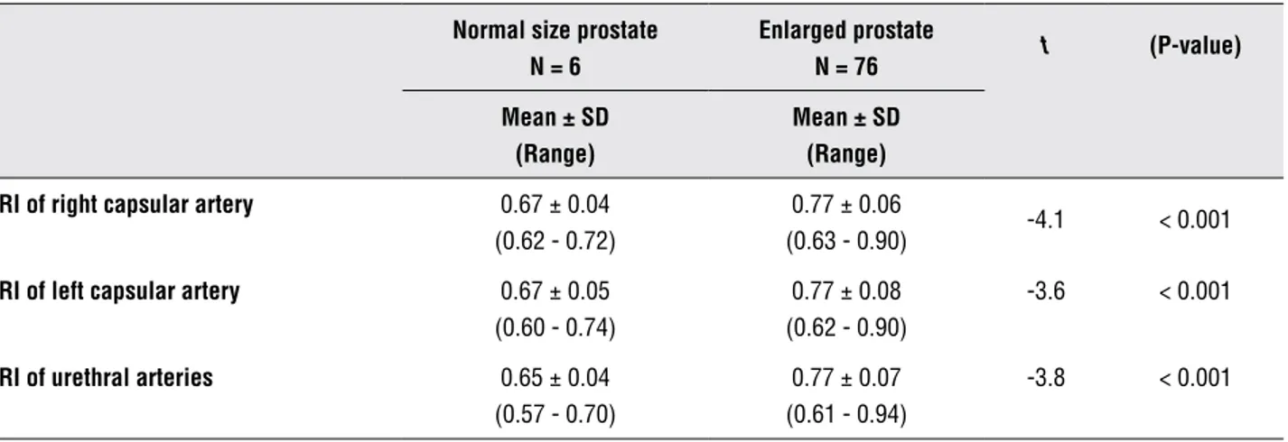
(Table-3). There was a highly significant increase in the RI of right and left capsular and urethral arter-ies correlated to the increasing severity of symp-toms of the patients according to IPSS. The size of the prostate was measured by TRUS and was con-sidered normal when < 25g and enlarged if > 25g. Six patients were considered to have normal pros-tate and 76 patients had enlarged prospros-tate. There was a significant increase in the RI of right and left capsular and urethral arteries correlated to the increase of the prostatic volume (Table-4).
DISCUSSION
With the advent of power Doppler imaging, RI measurement in patients with LUTS has become
a promising parameter for the diagnosis of BPH (1). The definite mechanism through which the RI is elevated in BPH is still unclear (1). It was as-sumed that a hyperplastic prostate tissue pushed the capsule out as it growed thus increasing the intraprostatic pressure as well as RI. The increase of the intraprostatic pressure is equally distribut-ed throughout the whole prostate, so the increase of RI was found in both peripheral and transition zones (7).
There is a relative difference in the RI of prostatic vasculature in different prostatic pa-thologies. The mean RI value of 0.579 (range, 0.45 – 0.80) for cancer cases was slightly lower than the mean values for cases with atypia, inflamma-tion, and benign disease, which were 0.601 (range,
Table 3 - Comparison of the RI of Rt & Lt capsular arteries and urethral arteries between patients with mild to moderate symptoms, and with severe symptoms according to IPSS.
Mild & moderate N= 21
Severe symptoms N = 61
t (P-value)
Mean ± SD (range)
Mean ± SD (range)
RI of Rt. capsular artery 0.68 ± 0.03 (0.62 - 0.73)
0.79 ± 0.04 (0.70 - 0.90)
10.8 < 0.001
RI of Lt. capsular artery 0.66 ± 0.03 (0.60 - 0.74)
0.79 ± 0.03 (0.68 - 0.90)
11.2 < 0.001
RI of Urethral arteries 0.67 ± 0.04 (0.57 - 0.79)
0.79 ± 0.06 (0.69 - 0.94)
8.6 < 0.001
Table 4 - Comparison of parameters between patients with a normal prostate size and those with enlarged prostate.
Normal size prostate N = 6
Enlarged prostate
N = 76 t (P-value) Mean ± SD
(Range)
Mean ± SD (Range) RI of right capsular artery 0.67 ± 0.04
(0.62 - 0.72)
0.77 ± 0.06
(0.63 - 0.90) -4.1 < 0.001
RI of left capsular artery 0.67 ± 0.05 (0.60 - 0.74)
0.77 ± 0.08 (0.62 - 0.90)
-3.6 < 0.001
RI of urethral arteries 0.65 ± 0.04 (0.57 - 0.70)
0.77 ± 0.07 (0.61 - 0.94)
0.49 – 0.86), 0.621 (range, 0.54 – 0.77) and 0.616 (range, 0.40 – 1.00), respectively. It is noteworthy that the subgroup of patients with the highest RI value measured at the relevant site was the one with inflammation compared with those with can-cer, atypia, and benign disease (8).
The parameters used by different research groups to correlate RI with BPH were different. Hayami et al. assessed the value of power Doppler imaging in predicting the histological components of BPH and demonstrated that RI was the most reliable factor for predicting the ratio of glandu-lar lumen to stromal elements. There values were considered useful for predicting the therapeutic response to different treatment options (9). Others pushed their research one step further. Turgut et al. used PDUS and RI in a trial to differentiate pros-tate cancer from BPH. The authors reported that the mean RI for cancer cases was slightly lower than the mean RI value for benign conditions de-spite the fact that the difference was statistically insignificant (P > 0.05) (8).
In the present study, out of 82 patients, 62 (76%) were diagnosed to be obstructed and the re-maining 20 (24%) unobstructed based on uroflow-metric studies. There was a significant difference in RI of right capsular arteries between the obstructed and non obstructed groups (P < 0.001). There was a significant difference in RI of left capsular arteries between the obstructed and non obstructed groups (P < 0.001). Similarly, there was a significant dif-ference in RI of urethral arteries between the ob-structed and non obob-structed groups (P < 0.001). There was a highly significant increase in the RI correlated to the increase of obstructive patterns of flow rates of the patients. Although there was a significant statistical value between RI of both right and left capsular arteries and urethral arteries when correlated to IPSS, uroflowmetry results and prostate volume, there was not a significant dif-ference in mean RI among the individual arteries.
Kojima et al. showed a significant correla-tion between RI and urodynamic parameters ob-tained in peak flow rate (Q-max) of uroflowmetry and IPSS in their study. Out of 33 patients with ob-struction, 28 (85%) had a RI of 0.7 or more, while 11 out of 24 patients (46%) without obstruction had a RI less than 0.7 (7).
In the study of Tsuru et al. (10), the RI of cap-sular arteries was (0.73 + 0.08) and that of urethral arteries was (0.69 + 0.08), and there was a significant correlation between IPSS and RI of capsular arteries. These results are similar to our results in capsular ar-teries but there was a noted difference from that ob-tained in urethral arteries and this may be explained by different methods of assessing intra-prostatic ar-teries and the great improvement of machine tech-nology that we used. Shinbo et al. studied the RI as a risk for acute urinary retention in patients with BPH; they stated that RI is increased in patients with BPH and is related to the severity of bladder outlet obstructive symptoms (11). Other researchers didn’t observe this strong correlation. Hayami and associ-ates showed that there was a weak difference in RI between the obstructed and non-obstructed groups (0.73 ± 0.1 vs. 0.71 ± 0.03) (9).
Frauscher et al. showed that the RI was sig-nificantly elevated in BPH patients in comparison to the normal group (12). These results are similar to that obtained by the study done by Kojima et al. who found that there was a significant increase of the RI in 40 cases of BPH (0.72 ± 0.05) compared to 37 cases with a healthy prostate (0.64 ± 0.04), (P < 0.0001) (5). Kojima et al. demonstrated that the RI value of patients with normal prostatic volume was significantly lower than that of patients with an enlarged prostate (0.64 ± 0.04 vs. 0.72 ± 0.06; P < 0.0001) (7).
Jamal and Khadr showed that the RI in-creases significantly correlated to the increase in prostatic volume, and that there was a significant difference in RI between patients with normal pros-tate and those with BPH (0.64 ± 0.04 vs. 0.72 ± 0.06; P < 0.0001) (13). This result is in agreement with previous studies and similar to our results.
It is noteworthy to mention that although most of the researchers - whether in total agree-ment, partial agreement or who even didn’t agree with our results - did not identify or correlate the individual capsular and urethral arteries, yet we analyzed signals from these vessels separately. This may be related to the advances in the machine technology.
correlate the RI of prostate vasculature with BPH symptoms, prostate size and degree of obstruction.
CONCLUSIONS
The RI measurement using PDUS can be added to the modalities available for investigat-ing BPH. Furthermore, its value can be correlated to the prostate size and degree of obstruction. Further research in this field will even allow the use of this modality to investigate other patholo-gies affecting the prostate and can be used also to evaluate the outcome of management.
CONFLICT OF INTEREST
None declared.
REFERENCES
1. Ozdemir H, Onur R, Bozgeyik Z, Orhan I, Ogras MS, Ogur E: Measuring resistance index in patients with BPH and lower urinary tract symptoms. J Clin Ultrasound. 2005; 33: 176-80.
2. Watanabe H, Kato H, Kato T, Morita M, Tanaka M: Diag-nostic application of ultrasonotomography to the prostate. Nihon Hinyokika Gakkai Zasshi. 1968; 59: 273-9.
3. Sedelaar JP, de la Rosette JJ, Debruyne FM: Progress in the imaging of the prostate gland. Curr Urol Rep. 2003; 4: 1-2. 4. Berger AP, Deibl M, Leonhartsberger N, Bektic J, Horninger
W, Fritsche G et al.: Vascular damage as a risk factor for benign prostatic hyperplasia and erectile dysfunction. BJU Int. 2005; 96: 1073-8.
5. Kojima M, Watanabe H, Watanabe M, Okihara K, Naya Y, Ukimura O: Preliminary results of power Doppler imag-ing in benign prostatic hyperplasia. Ultrasound Med Biol. 1997; 23: 1305-9.
6. Nelson TR, Pretorius DH: The Doppler signal: where does it come from and what does it mean? AJR Am J Roentgenol. 1988; 151: 439-47.
7. Kojima M, Ochiai A, Naya Y, Okihara K, Ukimura O, Miki T: Doppler resistive index in benign prostatic hyperplasia: correlation with ultrasonic appearance of the prostate and infravesical obstruction. Eur Urol. 2000; 37: 436-42. 8. Turgut AT, Olçücüoglu E, Koşar P, Geyik PO, Koşar U, Dogra
V: Power Doppler ultrasonography of the feeding arteries of the prostate gland: a novel approach to the diagnosis of prostate cancer? J Ultrasound Med. 2007; 26: 875-83. 9. Hayami S, Ushiyama T, Kurita Y, Kageyama S, Suzuki K,
Fujita K: The value of power Doppler imaging to predict the histologic components of benign prostatic hyperplasia. Prostate. 2002; 53: 168-74.
10. Tsuru N, Kurita Y, Masuda H, Suzuki K, Fujita K: Role of Doppler ultrasound and resistive index in benign prostatic hypertrophy. Int J Urol. 2002; 9: 427-30.
11. Shinbo H, Kurita Y, Takada S, Imanishi T, Otsuka A, Furuse H, et al.: Resistive index as risk factor for acute urinary re-tention in patients with benign prostatic hyperplasia. Urol-ogy. 2010; 76: 1440-5.
12. Frauscher F, Kreiter M, Frede T, Segner P, Strasser H, Reis-sigl, A, et al.: Color Doppler ultrasound of the prostate: As-sessment of the resistive index (RI). ECR 97 Presentation. 2007; 1159.
13. Jamal A, Khadr EA: Correlation between resistive index and prostatic volume in benign prostatic hyperplasia (BPH). Mansoura Med. J 2001; 32: 281-9.
14. Leventis AK, Shariat SF, Utsunomiya T, Slawin KM: Charac-teristics of normal prostate vascular anatomy as displayed by power Doppler. Prostate. 2001; 46: 281-8.
______________________
Correspondence address: Dr. Mostafa Mahmoud Khalil Benha Faculty of Medicine, Benha, Egypt Fareed Nada Street, Benha, Qalubiya Governorate, 13511,Arab Republic of Egypt Tel: + 965 9 960-5244 E-Mail: [email protected]
EDITORIAL COMMENT
The authors analyzed the resistive index (RI) of prostatic blood flow by transrectal power Dop-pler sonography as a method to evaluate the lower
The relative invasiveness of urodynamics and its indeterminate results in a significant num-ber of cases have stimulated the development of non invasive methods for the diagnosis of bladder outlet obstruction (BOO). In this context, the use of ultrasound (US) as a surrogate method to establish this diagnosis has constituted an interesting field of research. Prostate volume, post void residual volume, intra-vesical prostatic protrusion, bladder weight, detrusor wall thickness, urethral prostatic
angle and resistive index of prostatic arteries are the main forms to predict the risk of BOO through US analysis. The association of some of these mea-sures has led to accuracy rates over 90%. The lack of standardization of these measures is probably the main obstacle that has hampered the reproduction of the results in other urologic centers. Urologists should be aware of these facts in order to explore the maximum potential of this cheap, available, non invasive and innocuous diagnostic tool.
Dr. Alberto Azoubel Antunes
Chief of Prostate Section Division of Urology – University of Sao Paulo
Medical School Rua Barata Ribeiro, 490 / 76 Sao Paulo,SP, 01308-000, Brazil E-mail: [email protected]
EDITORIAL COMMENT
The authors present their experience with the use of prostatic resistive index (RI) to distin-guish between men with obstructive versus non obstructive prostatic hyperplasia. Their findings suggest ultrasound RI to be a validated auxiliary tool to identify patients suffering from significant obstructive urinary flow.
Since the obstructed group presented a significant higher RI than non obstructed group, selection criteria before transrectal ultrasound as-sessment seem to have been adequate (Qmax < 15
mL/s and PVR > 100 mL). However, although the mean RI difference was statistically significant for all measured parameters (right artery, left artery and urethral artery, P < 0.001) numbers were fre-quently overlapped (see Table-2). As such, identi-fying obstructive cases on an individual basis may be rather difficult.
Also, it is hypothesized that an enlarge-ment of the median lobe may not impact RI value as it does not impose increased resistance to cap-sular arteries. The association of intravesical pros-tatic protrusion (IPP) could add for a more precise diagnosis in this scenario (1,2).
REFERENCES
1. Shinbo H, Kurita Y, Takada S, Imanishi T, Otsuka A, Furuse H, et al.: Resistive index as risk factor for acute urinary re-tention in patients with benign prostatic hyperplasia. Urol-ogy. 2010; 76: 1440-5.
2. Chia SJ, Heng CT, Chan SP, Foo KT: Correlation of intra-vesical prostatic protrusion with bladder outlet obstruction. BJU Int. 2003; 91: 371-4.
Dr. Ricardo Miyaoka
Division of Urology State University of Campinas – UNICAMP Hospital das Clinicas UNICAMP
20 andar, A2 – sala 108 R. Vital Brazil – 250 Cidade universitária Zeferino Vaz, Distrito de
EDITORIAL COMMENT
TThe authors should be congratulated for this study about the resistive index of blood flow in the prostate of patients with BPH. There are few studies (1) describing the effects of the prostatic resistive blood flow in the urodynamic parameters, and the potential applications of this information can change our practice. The knowl-edge about it is still incipient and the resistive
index of blood flow of the prostate and bladder outlet can be a simple and useful tool to determi-nate infravesical obstruction. (2)
However, this study opens an opportu-nity for the development of more trials includ-ing necessarily a comparison with pressure/flow parameters that could reveal more information about the sensitivity, specificity and positive predictive value of this method regarding infra-vesical obstruction.
REFERENCES
1. Schuster A, Frauscher F, Strasser H, Recheis W, Pallwein L, Herwig R, et al.: Power Doppler ultrasound imaging for quantification of urinary bladder neck blood flow changes. Ultrasound Med Biol. 2004; 30: 1379-84.
2. Pinggera GM, Mitterberger M, Steiner E, Pallwein L, Frauscher F, Aigner F, et al.: Association of lower urinary tract symptoms and chronic ischaemia of the lower urinary tract in elderly women and men: assessment using colour Doppler ultrasonography. BJU Int. 2008; 102: 470-4.
Dr. Alexandre Fornari