Authors
Bruno Bordin Pelazza1
Cesar Augusto Saldanha Rosa2
Sebastião Rodrigues Ferreira Filho3
1 MSc in Health Sciences/ UFU (Intensive Care Unit Nurse Coronary/General ICU).
2 Degree in Medicine (In-tensive Care Unit Physician and MSc in Health Sciences at the Federal University of Uberlândia).
3 PhD in Nephrology/ UNIFESP (Full Professor of the Federal University of Uberlândia).
Submitted on: 02/27/2012. Approved on: 07/16/2012.
Correspondence to: Bruno Bordin Pelazza. Universidade Federal de Uberlândia/Instituto do Coração do Triângulo. Programa de Pós-graduação em Ciências da Saúde.
Rua Manoel Serralha, nº 1075, Santa Mônica. Uberlândia, MG, Brazil. CEP: 38408-246. E-mail: [email protected] Tel/Fax: (34) 3216-4403. Cel: (34)8853-6322. CAPES.
Introduction: Systolic blood pressure (SP) and pulse pressure (PP) rise gradu-ally during the aging process as a conse-quence of a reduction in arterial elasticity. The measure of systemic arterial pressure (SAP) taken at the root of the aorta has been considered an independent determi-nant of cardiovascular mortality supe-rior to the values of brachial SAP. Aim:
To compare the values of SAP central to those of braquial SAP in patients of dif-ferent age brackets who have systemic hypertension. Method: We evaluated the central SAP at the root of the aorta and the brachial SAP in the left arm using the ocillometric method 244 hypertensive patients who had been submitted to cine-angiocoronarography. Five groups of pa-tients were constituted: Group I, 39-49 years-old (y.o.), n = 36; Group II, 50-59 y.o., n = 67; Group III, 60-69 y.o., n = 69; Group IV, 70-79 y.o., n = 46; Group V,
≥ 80 y.o., n = 26. Results: When central SP was compared to brachial SP, it was possible to find significance in patients who were 50 y.o and upwards. It was not possible to find a statistical difference be-tween central diastolic pressure and bra-chial except in patients between the ages of 60-69 y.o. When comparing central to brachial PP, we observed that central PP was significantly greater (between 11 and 15 mmHg) in all patient above the age of 50 y.o. Conclusion: In older people, the values of SP and PP, taken directly at the root of the aorta, are superior to those obtained by indirect means from the bra-chial artery. These differences are signifi-cant from the age of 50 y.o. onwards.
Comparison between the central and brachial blood
pres-sure values in patients with hypertension undergoing
cineangiocoronarography
A
BSTRACTKeywords: health, hypertension, survivor-ship (public health).
I
NTRODUCTIONSystemic blood pressure (SBP) has impor-tant predictive value for cardiovascular events when measured by indirect meth-ods. The measurement of the detected SBP levels, usually conducted using a sphyg-momanometer connected to an upper extremity, has been used in both clinical practice and in research studies with large population samples.1-4 The use of other SBP reading methods such as oscillomet-ric devices with digital recording are also widespread in outpatient practice and in several clinical studies.4,5
Recently, some authors have shown that central SBP (cSBP) obtained from the root of the aorta is more strongly con-nected to cardiovascular diseases than the values obtained for brachial SBP (bSBP).6 Among SBP components, both central and brachial, central pulse pressure (cPP) has been shown to be an independent predictor of cardiovascular events.7,8 Benetos et al. verified that the role of pulse pressure (PP) is crucial in cardiovascular mortality and that values > 65 mmHg are accompanied by an increase in coronary risk, even with absolute values of systolic blood pressure (SBP) and diastolic blood pressure (DBP) within the normal limits.9-11
With aging, there is a progressive increase of SBP, and a higher increase of systolic pressure in relation to diastolic pressure. SBP continues to increase, even after the age of 60, while diastolic pressure tends to remain constant or decrease after the fifth or sixth decade of life.13,14
Therefore, PP increases with age due to the struc-tural changes in different components of the arterial wall, by decreasing the complacency of large arte-rial vessels due to a decrease in elastic fibers and an increase in the content of calcium ions and collagen fibers.15 Data suggest that cPP is closely correlated with left ventricular hypertrophy,16 to increase artery intima and media layer thickness,17 and constitutes an independent predictor of cardiovascular risk, surpass-ing brachial pulse pressure (bPP).18
O
BJECTIVEIn this context, the objective of this work was to com-pare the values of cSBP and bSBP across different age groups of systemic hypertension (SH) patients.
M
ETHODSThis cross-sectional study assessed 260 patients aged between 39 and 88 years, selected in the period from November 2009 to June 2011, who were electively admitted to the Heart Institute of the Triângulo Mineiro (ICT) in Uberlândia, Minas Gerais, to un-dergo coronary angiograms due to clinical signs and symptoms compatible with coronary insufficiency. Anthropometric and demographic data were collect-ed using a questionnaire that was completcollect-ed prior to the coronary angiogram.
Five patient groups were created: Group I, patients aged between 39 and 49 years, n = 34; Group II, patients aged between 50 and 59 years, n = 67; Group III, patients aged between 60 and 69 years, n = 69; Group IV, patients aged between 70 and 79 years, n = 46; and Group V, patients aged ≥ 80 years, n = 26. The variables studied in each group included the following: central systolic pressure (cSP) and brachial systolic pressure (bSP), central diastolic pressure (cDP) and brachial diastolic pressure (bDP), and cPP and bPP. All values have been expressed in mmHg.
Patients aged ≥ 18 years with SH who signed the free and informed consent form were included in this study. The patients showing the following
were excluded from the study: iodine allergy (7), hypertensive crisis (2), infected varicose ulcers (5), and mechanical failure of the hemodynamic system (4). The final sample included 242 patients, with 130 men and 112 women. All patients were hypertensive. Individuals who showed, at admission prior to the exam, SBP of ≥ 140 × 90 mmHg, with or without the use of antihypertensives, or SBP of < 140 × 90 mmHg with the use of antihypertensives, were considered to be hypertensive patients.
HEMODYNAMICSTUDY
In order to perform the cine coronary angiography, the patient was placed in the supine position (Siemens Coroskop T.O.P.) and the reading for the cSBP was conducted by positioning the catheter (Pig Tail 110 cm, caliber 5F) at the root of the aorta showing the values on a heart monitor (Siemens). The catheter was always introduced through the femoral or radial artery–the physician chose the best route. Confirmation of the exact catheter location was obtained by injection of contrast media (Pielograf [ionic] and Visipaque [non-ionic]). At a maximum of 60 s after reading the cSBP, the bSBP values were obtained via the oscillometric method using an Omron-HEM-431 digital device connected to the left upper extremity.
STATISTICAL ANALYSIS
The results of the variables have been expressed as mean ± standard deviation, and as mean ± standard error in the Figures, while categorical variables have been expressed as a ratio or percentage. Initially, each of the variables was assessed using analysis of vari-ance (ANOVA) to verify whether there was a signifi-cant difference between the groups. The differences between the groups were considered significant if the
p-value was < 0.05.
TABLE 1 CLINICALCHARACTERISTICSOFTHEPOPULATIONSANDNUMBEROFANTIHYPERTENSIVESBYAGEGROUP
Parameters Group I Group II Group III Group IV Group V p-value
(n = 34) (n = 67) (n = 69) (n = 46) (n = 26)
Age 44 ± 4.6* 54 ± 2.8* 64 ± 2.7* 74 ± 3.0* 84 ± 2.6* < 0.0001
Sex Male/Female 20/14 34/33 35/34 24/22 15/11 0.8639
DM (%) 26 27 23 41* 46* 0.0105
Smoking 12 40* 25 13 10 0.0009
Quantity of
antihypertensives (n) 2.09 ± 1.09 2.20 ± 0.97 2.25 ± 0.92 2.39 ± 0.91* 2.94 ± 1.16* 0.0284
Beta-adrenergic blockers
(%) 56 60 43 43 58 0.2492
ACEI (%) 33 28 22 28 31 0.7464
CCB (%) 22 27 29 30 27 0.9389
Diuretic (%) 22 27 23 28 23 0.9
* comparison between groups (p < 0.05). ACEI: angiotensin-converting enzyme inhibitor, CCB: calcium channel blocker, DM: diabetes mellitus. Group I, patients between 39 and 49 years of age; Group II, those aged between 50 and 59 years; Group III, those aged between 60 and 69 years; Group IV, those aged between 70 and 79 years; and Group V, those aged ≥ 80 years. The analysis of variance (ANOVA) and Tukey’s test were used for statistical analysis.
R
ESULTSSTUDYPOPULATION
The clinical characteristics of the patients and the number of antihypertensives used (n = 242) are shown in Table 1. The male/female sex ratio was similarly maintained, emphasizing the homogeneity of the sam-ple. The number of antihypertensives used in groups IV and V was greater than in those of groups I, II, and III, and the mean proportion of patients with diabetes mellitus in Groups IV and V was higher than those in Groups I, II, and III. In this study, we did not cor-relate the values of SBP with any other factors, such as comorbidities and the number of antihypertensives used; however, we performed a comparison between cSP and bSP.
When comparing cSP and bSP, we observed that cSP was significantly higher than bSP starting at age 50 (p < 0.05; Table 2 and Figure 1). We observed significant differences in these values between Group I and Group III; Group I and Group IV, and Group I and Group V. For bSP, we observed significant dif-ferences between Group I and Group IV and Group I
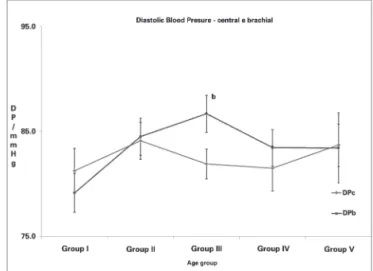
and Group V, with p < 0.05 for all comparisons. No significant variations in cDP were observed between any of the age groups. In the comparison of cDP versus bDP, significant differences were noted only in Group III (Table 3 and Figure 2).
In the comparison between cPP and bPP, we ob-served that cPP was higher than bPP starting at age 50 (p < 0.05; Table 4 and Figure 3). We observed
significant differences in cPP between Group I and
Group III, Group I and Group IV, and Group I and
Group V. Regarding bPP, we observed significant dif-ferences between Group I and Group IV and Group I
and Group V, with p < 0.05 for all comparisons.
D
ISCUSSIONThe diagnosis of SH is usually obtained through in-direct methods using oscillometric devices and/or auscultation placed in the upper extremities of pa-tients.19-22 Epidemiological studies assume that bra-chial SBP is proportional to cardiovascular risk.23 Recently, clinical trials have shown that cSBP is a better predictor of cardiovascular risk than bSBP24. In reality, cSBP and bSBP values may be significantly different.25,26 Our data showed that an increase in bSP was statistically significant starting at age 70, in con-trast to studies reported in the literature in which an increase of bSP is observed starting at age 5027,28.
Figure 2. Central and brachial diastolic blood pressure variance according to age group. - c = p < 0.05 (between the groups). Group I, patients between 39 and 49 years of age; Group II, patients aged between 50 and 59 years; Group III, those aged between 60 and 69 years; Group IV, those aged between 70 and 79 years; and Group V, those aged ≥ 80 years. Student's t test and Mann-Whitney tests were used for statistical analysis.
TABLE 2 SYSTOLICBLOODPRESSUREACCORDINGTOAGEGROUP
Parameters mmHg Group I Group II Group III Group IV Group V p-value
(n = 34) (n = 67) (n = 69) (n = 46) (n = 26)
Central systolic pressure (cSP) 136 ± 20 147 ± 27b 152 ± 28ab 160 ± 22ab 163 ± 22ab < 0.0001
Brachial systolic pressure (bSP) 132 ± 19 138 ± 23 142 ± 26 147 ± 24a 150 ± 21a 0.0061
a cSP: Group V = Group IV > Group III > Group I; Group I = Group II and Group II = Group III; b cSP > bSP in Groups II, III, IV, and V. Group I, patients between 39 and 49 years of age; Group II, those aged between 50 and 59 years; Group III, those aged between 60 and 69 years; Group IV, those aged between 70 and 79 years; and Group V, those aged ≥ 80 years.
Figure 1. Central and brachial systolic blood pressure variance according to age group. -a vs. a = p > 0.05; b vs. b = p > 0.05; a vs. b = p < 0.05; c = p < 0.05 (between the groups). Group I, patients aged between 39 and 49 years of age; Group II, those aged between 50 and 59 years; Group III, those aged between 60 and 69 years; Group IV, those between 70 and 79 years; and Group V, those aged ≥ 80 years. Student’s t test and Mann-Whitney tests were used for statistical analysis.
When the cSP was compared to the bSP, we ob-served higher levels of cSP starting at age 50 (147 ± 27 vs. 138 ± 23 mmHg), and this difference was maintained in the other groups assessed. This finding demonstrates that the drugs used to treat SH are often ineffective in reducing cSP levels.
The Conduit Artery Functional Endpoint (CAFE) study, which compared atenolol to amlodipine, con-cluded that the beta-adrenergic blocker was less effec-tive in reducing central pressure. In this study, 50% of the patients made use of beta blockers,29,30 a percent-age similar to that of the population assessed in our sample. As shown in Table 1, in all age groups, the use of beta-adrenergic blockers was > 40%. This find-ing may justify the differences observed between cSP
and bSP. Other studies have previously shown that beta-adrenergic blockers are less effective in decreas-ing cSP than other hypotensives.30-32
bDP is elevated in adults until the fifth or sixth de-cade of life, and these values decrease in older individ-uals.33 However, in our study, bDP remained similar across the groups, and the same result occurred with cDP levels. The use of antihypertensive drugs and the consequent maintenance of SBP within normal limits, appear to have restricted the increase of DP for the different age groups.
If we consider cPP < 50 mmHg as a normal level, the means of all the age groups in our study were above this value. When compared to the younger age groups, we observed that cPP was higher starting
TABLE 3 DIASTOLICBLOODPRESSUREACCORDINGTOAGEGROUP
Parameters mmHg Group I Group II Group III Group IV Group V p-value
(n = 34) (n = 67) (n = 69) (n = 46) (n = 26)
Central diastolic pressure (CDP) 81 ± 13 84 ± 14 82 ± 12b 81 ± 15 84 ± 10 0.6288
TABLE 4 PULSEPRESSUREACCORDINGTOAGEGROUP
Parameters mmHg Group I Group II Group III Group IV Group V p-value
(n = 34) (n = 67) (n = 69) (n = 46) (n = 26)
Central pulse pressure (cPP) 53 ± 19 64 ± 24b 70 ± 23ab 78 ± 23ab 79 ± 23ab < 0.0001
Brachial pulse pressure (bPP) 52 ± 17 53 ± 18 55 ± 17 64 ± 20a 66 ± 21a 0.0003
a cPP: Group V = Group IV > Group III > Group I; Group I = Group II and Group II = Group III; b cPP > bPP in Groups II, III, IV, and V. Group I, patients between 39 and 49 years of age; Group II, those aged between 50 and 59 years; Group III, those aged between 60 and 69 years; Group IV, those aged between 70 and 79 years; and Group V, those aged ≥ 80 years.
Figure 3. Central and brachial pulse pressure variance according to age group. -a vs. a = p > 0.05; b vs. b = p > 0.05; a vs. b =
p < 0.05; c = p < 0.05 (between the groups). Group I, patients between 39 and 49 years of age; Group II, those aged between 50 and 59 years; Group III, those aged between 60 and 69 years; Group IV, those aged between 70 and 79 years; and Group V, those aged ≥ 80 years. Student's t test and Mann-Whitney tests were used for statistical analysis.
at age 60 (70 ± 23 mmHg, p < 0.0001), while bPP increased starting at age 70 (64 ± 20 mmHg, p < 0.0003). If we analyze cPP versus bPP, we observe higher levels for cPP starting with Group II (64 ± 24
vs. 53 ± 18 mmHg), and this difference remains for the other groups assessed. This finding shows that an-tihypertensive drugs have not been able to maintain the same values for cPP and bPP; once again, the use of beta-adrenergic blockers may have interfered with a more effective reduction of cPP.
STUDYLIMITATIONS
This cross-sectional study had limitations inherent to this type of design. Thus, it is possible that the cen-tral and brachial pressure differences observed may have been specific to the population analyzed in our study, which showed undertreated coronary lesions and high pressure levels. On the other hand, the study did not analyze hypertension time, and some of the patients may have shown sharp increases of blood pressure due to examination-related stress.
C
ONCLUSIONWith aging, the pulse and systolic pressure values, measured directly at the root of the aorta, are higher than those obtained using an indirect method at the brachial artery. These differences are significant start-ing at age 50.
R
EFERENCES1. Pickering TG, Hall JG, Apple LJ, Falkner B, Graves J, Hill M, et al.; Subcommittee of Professional and Public Education of the American Heart Association Council on High Blood Pressure Research. Recommendations for blood pressure measurement in humans and experimental animals: Part 1: blood pressure measurement in humans: a statement for professionals from the Subcommittee of Professional and Public Education of the American Heart Association Council on High Blood Pressure Research. Hypertension 2005;45:142-61.
2. European Society of Hypertension-European Society of Cardio-logy Guidelines Committee. 2003 European Society of Hyper-tension-European Society of Cardiology guidelines for the mana-gement of arterial hypertension. J Hypertens 2003;21:1011-53. 3. Palota L, Cordella MP, Oliveira SM, Cesarino CB. A verifica-ção da calibraverifica-ção dos manômetros e condições dos esfigmo-manômetros aneróides: um programa de educação continuada para enfermeiros supervisores do Hospital de Base. Arq Cienc Saúde 2004;11:2-6.
4. Keavney B, Bird R, Caiazza A, Casadei B, Conway J. Measure-ment of blood pressure using the auscultatory and oscillometric methods in the same cuff deflation: validation and field trial of the A&D TM2421 monitor. J Hum Hypertens 2000;14:573-9. 5. Ni H, Wu C, Prineas R, Shea S, Liu K, Kronmal R, et al. Com-parison of Dinamap PRO-100 and mercury sphygmomanome-ter blood pressure measurements in a population-based study. Am J Hypertens 2006;19:353-60.
6. Roman MJ, Devereux RB, Kizer JR, Lee ET, Galloway JM, Ali T, et al. Central pressure more strongly relates to vascular disease and outcome than does brachial pressure: the Strong Heart Study. Hypertension 2007;50:197-203.
7. Safar ME, Blacher J, Pannier B, Guerin AP, Marchais SJ, Guyonvarc'h PM, et al. Central pulse pressure and mortality in end-stage renal disease. Hypertension 2002;39:735-8.
8. Safar ME. Pulse pressure, arterial stiffness, and cardiovascular risk. Curr Opin Cardiol 2000;15:258-63.
9. Benetos A, Safar M, Rudnichi, A, Smulyan H, Richard JL, Ducimetieère P, et al. Pulse pressure: a predictor of long-term cardiovascular mortality in a French male population. Hyper-tension 1997;30:1410-5.
10. Franklin S, Khan S, Wong N, Larson MG, Levy D. Is pulse pressure useful in predicting risk for coronary heart disease? The Framingham heart study. Circulation 1999;100:354-60. 11. Millar J, Lever A, Burke V. Pulse pressure as a risk factor for
12. Chae CU, Pfeffer MA, Glynn RJ, Mitchell GF, Taylor JO, Hen-nekens CH. Increased pulse pressure and risk of heart failure in the elderly JAMA 1999;281:634-9.
13. Franklin SS, Gustin W 4th, Wong ND, Larson MG, Weber MA, Kannel WB, et al. Hemodynamic patterns of age-related changes in blood pressure. The Framingham Heart Study. Cir-culation 1997;96:308-15.
14. Ogihara T, Hiwada K, Morimoto S, Matsuoka H, Matsumoto M, Takishita S, et al. Guidelines for treatment of hypertension in the elderly 2002 revised version. Hypertens Res 2003;26:1-36. 15. Nichols WW, O’Rourke MF, eds. McDonald’s Blood Flow in
Arteries: Theoretical, Experimental and Clinical Principles. Fif-th Edition. Oxford: Hodder Arnold; 2005. p.193-213. 16. Covic A, Goldsmith DJ, Panaghiu L, Covic M, Sedor J.
Analy-sis of the effect of haemodialyAnaly-sis on peripheral and central arte-rial pressure waveforms. Kidney Int 2000;57:2634-43. 17. Laurent S, Boutouyrie P, Asmar R, Gautier I, Laloux B, Guize
L, et al. Aortic stiffness is an independent predictor of all-cause and cardiovascular mortality in hypertensive patients. Hyper-tension 2001;37:1236-41.
18. McEniery CM, Yasmin, McDonnell B, Munnery M, Wallace SM, Rowe CV, et al; Anglo-Cardiff Collaborative Trial Inves-tigators. Central pressure: variability and impact of cardio-vascular risk factors: the Anglo-Cardiff Collaborative Trial II. Hypertension 2008;51;1476-82.
19. Yarows SA, Qian K. Accuracy of aneroid sphygmomanome-ters in clinical usage: University of Michigan experience. Blood Press Monit 2001;6:101-6.
20. Canzanello VJ, Jensen PL, Schwartz GL. Are aneroid sphyg-momanometers accurate in hospital and clinic settings? Arch Intern Med 2001;161:729-31.
21. van Montfrans GA. Oscillometric blood pressure measurement: progress and problems. Blood Press Monit 2001;6:287-90. 22. Amoore JN, Scott DH. Can simulators evaluate systematic
differences between oscillometric non-invasive blood-pressure monitors? Blood Press Monit 2000;5:81-9.
23. Turnbull F; Blood Pressure Lowering Treatment Trialists’ Colla-boration. Effects of different blood-pressure-lowering regimens on major cardiovascular events: results of prospectively-desig-ned overviews of randomised trials. Lancet 2003;362:1527-35. 24. Roman MJ, Okin PM, Kizer JR, Lee ET, Howard BV, Deve-reux RB. Relations of central and brachial blood pressure to left ventricular hypertrophy and geometry: the Strong Heart Study. Hypertension 2010:28:384-8.
25. Boutouyrie P, Bussy C, Lacolley P, Girerd X, Laloux B, Laurent S. Association between local pulse pressure, mean blood pressu-re, and large-artery remodeling. Circulation 1999;100:1387-93. 26. Blacher J, Guerin AP, Pannier B, Marchais SJ, Safar ME, Lon-don GM. Impact of aortic stiffness on survival in end-stage re-nal failure. Circulation 1999;99:2434-9.
27. Franklin SS, Larson MG, Khan SA, Wong ND, Leip EP, Kannel WB, et al. Does the relation of blood pressure to coronary heart disease risk change with aging? The Framingham Heart Study. Circulation 2001;103:1245-9.
28. Franklin SS, Jacobs MJ, Wong ND, L'Italien GJ, Lapuerta P. Predominance of isolated systolic hypertension among middle--aged and elderly US hypertensives: analysis based on Natio-nal Health and Nutrition Examination Survey (NHANES) III. Hypertension 2001;37:869-74.
29. Williams B, Lacy PS, Thom SM, Cruickshank K, Stanton A, Collier D, et al.; CAFE Investigators; Anglo-Scandinavian Car-diac Outcomes Trial Investigators; CAFE Steering Committee and Writing Committee. Differential impact of blood pressure--lowering drugs on central aortic pressure and clinical outco-mes: principal results of the Conduit Artery Function Evalua-tion (CAFE) study. CirculaEvalua-tion 2006;113:1213-25.
30. Williams B, Lacy PS; CAFE and the ASCOT (Anglo-Scandi-navian Cardiac Outcomes Trial) Investigators. Impact of heart rate on central aortic pressures and hemodynamics: analysis from the CAFE (Conduit Artery Function Evaluation) study: CAFE-Heart Rate. J Am Coll Cardiol 2009;54:705-13. 31. Morgan T, Lauri J, Bertram D, Anderson A. Effect of different
antihypertensive drug classes on central aortic pressure. Am J Hypertens 2004;17:118-23.
32. Mackenzie IS, McEniery CM, Dhakam Z, Brown MJ, Cock-croft JR, Wilkinson IB. Comparison of the effects of antihyper-tensive agents on central blood pressure and arterial stiffness in isolated systolic hypertension. Hypertension 2009;54:409-13. 33. Sesso HD, Stampfer MJ, Rosner B, Hennekens CH, Gaziano