323
Ulisses Alexandre CROTI, Domingo Marcolino BRAILE, Moacir Fernandes de GODOY, Airton Camacho MOSCARDINI
Correspondence: Ulisses Alexandre Croti
Hospital de Base – FAMERP – Av. Brigadeiro Faria Lima, 5416 CEP 15090-000 – São José do Rio Preto – São Paulo
E-mail: uacroti@uol.br
Clinical-Surgical Correlation
Braz J Cardiovasc Surg 2004; 19(3): 323-324
CLINICAL DATA
A four and a half-year-old white male patient weighing 14 kg was admitted for a physical examination. The patient complained of tiredness after moderate effort over the previous seven months and had been hospitalized with pneumonia on two occasions. He was in a good general state with a rosy complexion and acyanotic. The thorax presented with left anterior bulging, palpable ictus cordis with fremitis at the fifth intercostal space, fixed split S2, systolic murmur of +++/6, ejective at the left sternal border. The lungs had a symmetrical vesicular murmur without adventitious sounds. No alterations were evidenced in the abdomen and the peripheral pulses were palpable and symmetrical.
ELECTROCARDIOGRAM
The electrocardiogram evidenced sinusal rhythm with a heart rate of 145 beats/minute and electrical axis of the QRS complex
RBCCV 44205-702
Article received on June, 2004 Article accepted on July, 2004
Case 5/2004 – Pediatric Heart Surgery Service – Hospital de Base, Medical
School, São José do Rio Preto
324
+ 120º. An ample R-wave at V1 and a great P-wave at D2 were observed suggesting right atrial and ventricular overload.
RADIOGRAM
The cardiothoracic index was 0.47 without evidence of increased heart chambers. A slight pulmonary vascular prominence was seen.
ECHOCARDIOGRAM
Situs solitus was seen at levocardia. The veno-atrial, atrioventricular and ventriculoarterial connections were concordant. There were two interatrial shunts, one, an ostium secundum type was 6 mm in diameter and the other was in the coronary sinus region with 4 mm. There was also a slight reduction in the caliber of the pulmonary branch immediately above the valve. A Doppler echocardiogram demonstrated a turbulent, accelerated blood flow with a maximum gradient of 33 mmHg.
DIFFERENTIAL DIAGNOSIS
The electrocardiogram was compatible with pulmonary stenosis. The radiogram suggested normality or some congenital heart disease with hyper flow however this did not cause any great hemodynamic effect, similar to an interatrial shunt, partial anomalous drainage of the pulmonary veins or partial defect of the atrioventricular septum.
DIAGNOSIS
After identifying the interatrial shunt in the region of the coronary sinus and pulmonary stenosis, catheterization was
CROTI, UA ET AL - Clinical-Surgical Correlation - Case 5/2004 Braz J Cardiovasc Surg 2004; 19(3): 323-324
used to further study the case. Although catheterization did not determine the exact location of the interatrial shunts, it demonstrated a significant shunt between the coronary sinus and the left atrium. This confirmed the suspected diagnosis of unroofed coronary sinus syndrome with an ostium secundum-type interatrial shunt and the presence of supravalvar pulmonary stenosis with a gradient of 30 mmHg.
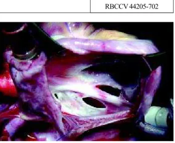
OPERATION