ABSTRACT: The purpose of this research was to study the growth of the nasopharynx and adenoid development. Lateral cephalometric radiographs obtained from 320 white Brazilian subjects between 4 and 16 years of age were used. All the participants were nose breathers and none of them had previously undergone adenoidectomy. Tracings were made from the radiographs and cephalometric measurements were performed. The results showed that adenoid sagital thickness is larger in the age group 4 – 5 years and decreases progressively. There is a slight increase in the age group 10 – 11 years, but afterwards the decrease continues. However, the nasopharyngeal free airway space does not decrease in the age group 10 – 11 years, despite the increasing thickness of the adenoid. This is attributable to the downward displacement of the hard palate, resulting in an increase of the free airway space due to growth. Although the nasopharynx follows a growth pattern similar to that of the rest of the body, adenoid tissue does not. Adenoidal development seems to differ from that of other lymphatic tissues, showing a peculiar pattern that can be revealed when hypertrophy due to infections and allergies is eliminated.
DESCRIPTORS: Nasopharynx; Adenoids; Radiography; Mouth breathing.
RESUMO: O objetivo da pesquisa foi estudar o crescimento da nasofaringe e o desenvolvimento da adenóide. Foram utilizadas as radiograias cefalométricas de peril obtidas de 320 indivíduos brasileiros brancos, cujas idades varia -vam entre 4 e 16 anos. Todos os participantes apresenta-vam respiração predominantemente nasal e não haviam sido submetidos previamente à adenoidectomia. A partir das radiograias foram feitos traçados cefalométricos, sobre os quais foram realizadas medições. Os resultados revelaram que a espessura sagital da adenóide é maior na faixa etária de 4 a 5 anos, regredindo, então, progressivamente, até a faixa etária de 10 a 11 anos, quando ocorre um leve aumento, voltando a diminuir em seguida. O espaço aéreo livre nasofaríngeo, entretanto, não diminui na faixa etária de 10 a 11 anos, mesmo diante do aumento da espessura da adenóide. Esse fato se deve ao desloca-mento do palato duro para baixo, o que determina a ampliação do espaço aéreo livre em razão do crescidesloca-mento. Apesar de a nasofaringe seguir um padrão de crescimento similar ao do resto do corpo, a adenóide não o segue. O desenvolvimento da adenóide parece ser diferente do dos demais tecidos de origem linfóide, apresentando um padrão peculiar que pode ser percebido quando se elimina a hipertroia causada por infecções e alergias.
DESCRITORES: Nasofaringe; Adenóides; Radiograia; Respiração bucal.
INTRODUCTION
The posterior nasopharyngeal wall is covered by lymphoid tissue that often undergoes hypertro-phy (adenoid) during the period prior to puberty. This adenoidal enlargement increases the chance of nasopharyngeal airway obstruction, which is particularly important, considering that mouth breathing resulting from nasal obstruction does not offer normal environmental conditions for the growth and development of the nasomaxil-lary complex1-3,11,13,14,17-19,21, pointing to an
asso-ciation between mouth breathing and dentofacial deformities. Therefore, the relationship between the nasopharynx size and the adenoid size is cru-cial7,9,20.
For many years, it has been believed that the adenoid would present a growth pattern similar to that of other lymphoid tissues such as the appen-dix, the spleen and the thymus, reaching its maxi-mum size just before puberty, showing subsequent atrophy16. Meanwhile, some authors have assured
* PhD, Private Practice.
** PhD, Department of Orthodontics, School of Dentistry, Fluminense Federal University. *** PhD, Department of Radiology, School of Medicine, Federal University of Rio de Janeiro.
Growth of the nasopharynx and adenoidal development in
Brazilian subjects
Crescimento da nasofaringe e desenvolvimento da adenóide
em brasileiros
Beatriz de Souza Vilella*
that adenoid tissue seems to have a specific growth potential9,15, and it is on this potential that hyper-trophic reactions of nasorespiratory infections and allergies may be superimposed15.
Subtelny, Koepp-Baker15 (1956) concluded that its growth peak may be reached as early as 9 to 10 years of age and is sometimes evident as late as 14 to 15 years of age, showing atrophy after this time. Linder-Aronson, Leighton9 (1983) verified that adenoid tissue is larger at the age of 5 and, therefore, it does not follow the classic lymphoid growth curve.
In relation to sexual dimorphism, some au-thors5,9 stated that boys and girls show different patterns of nasopharyngeal growth and adenoid development.
Considering that a systematic abnormal way of breathing can lead to a succession of events that are detrimental to the developing child and that there are contradictory conclusions based on the results of previous studies, it would be important to do a research in order to study the nasopharynx growth and the adenoid development.
MATERIAL AND METHODS
The present retrospective study was conducted in accordance with the October 10th, 1996, National Health Council’s resolution 196, and monitored by the Ethic Research Committee, Fluminense Fed-eral University.
The sample consisted of 320 white Brazilian children (160 boys and 160 girls) from the Depart-ment of Orthodontics, School of Dentistry, Flu-minense Federal University (UFF), Niterói City, RJ, Brazil. These patients were divided into six groups: 4 – 5 years (30), 6 – 7 years (50), 8 – 9 years (60), 10 – 11 years (60), 12 – 13 years (60), and 14 – 15 years (60). Subjects that had systematically used either topical or systemic medication for the nose were excluded from the sample, as well as those who had undergone adenoidectomy. As a selection criterion, subjects should breathe predominantly through the nose.
Clinical evaluation
Children were submitted to history taking and clinical examination. The clinical examination consisted of three tests to identify mouth breath-ers. The first test was performed in the following way: the patient was asked to close the lips and breathe deeply through the nose. Nose breathers
normally demonstrate a good reflex control of the alar muscles, which regulate the size and con-tour of the external part of the nostrils that dilate while inspiring. Nose breathers, even with tem-porary nasal congestion, will demonstrate reflex alar contraction and dilation of the nostrils during voluntary inspiration. Mouth breathers, even when able to breathe through the nose, do not alter the size and contour of the nostrils, and occasionally, contract nose openings when they inspire10.
Mouth or nose breathing was recorded by the second test by holding a cold dental mirror alter-nately in their front. The patient was seated in a relaxed position with the head upright. Ability to breathe through the nose was tested for each nasal passage separately, by observing whether the subject could breathe calmly for 30 seconds when one of the nostrils was closed by pressure, with the mouth closed8.
The third test was a functional test. Patients were asked to bend their knees 10 times in rapid succession. If, directly afterwards, they were able to continue breathing without difficulty, calmly, through the nose for 30 seconds, they were clas-sified as pure nose breathers8.
Cephalometric evaluation
Subjects were evaluated with standard lat-eral cephalometric radiographs. These radiographs were taken with the child’s head immobilized in a wall-mounted cephalostat and oriented to the Frankfort horizontal plane.
The head was fixed so that the median plane was parallel to the film. All the measurements were performed by the first author.
Reference points
The studied cephalometric reference points were as follows:
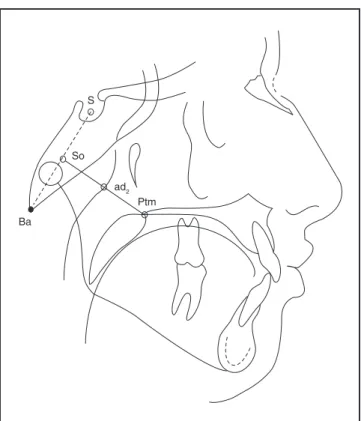
• Ba (Basion) – the most posteroinferior point on the clivus of os occipitale;
• Ptm (Pterygomaxillare) – intersection between the nasal floor and the posterior contour of the maxilla;
• S (Sella) – centre of the sella turcica; • So – midpoint on the line joining S and Ba; • ad2 – intersection of the posterior
nasopha-ryngeal wall and the Ptm-So line. Measurements
• Ptm-ad2 – linear distance from the Ptm point to the ad2 point, in mm, representing the na-sopharyngeal airway space;
• ad2-So – linear distance from the ad2 point to the So point, in mm, representing adenoid thickness;
• Ptm-So – linear distance from the Ptm point to the So point, in mm, representing the sagital depth of the bony nasopharynx in a higher level;
• ad2-So/Ptm-So – ratio between adenoid thick-ness and the bony nasopharynx depth, ob-tained by dividing the ad2-Someasurement by the Ptm-So value;
Linear distances (Figure 1) were measured with a digital caliper (Starret, São Paulo, Brazil), serial number 001296.
Statistical analysis
The method error (o’) for intra-individual measurements was calculated with the following formula:
o’ =
n
d
2
2
∑
where d is the difference between two meas-urements and n is the number of double determi-nations.
Calculations were also made for arithmetic means (c) and standard deviations (SD).
Student’s t test was used for testing the dif-ferences between means. A probability level of 1% was adopted (p < 0.01).
RESULTS
The intra-examiner error in determining the four measurements values used in this study was calculated using 30 randomly selected cases. It was found that the method error was 0.71 mm for the Ptm-ad2 measurement, 0.78 mm for the ad2-So measurement, 0.02 for the ad2-So/Ptm-So ratio and 0.98 mm for the Ptm-So measurement. Therefore, the intra-examiner method error was of little importance in this study.
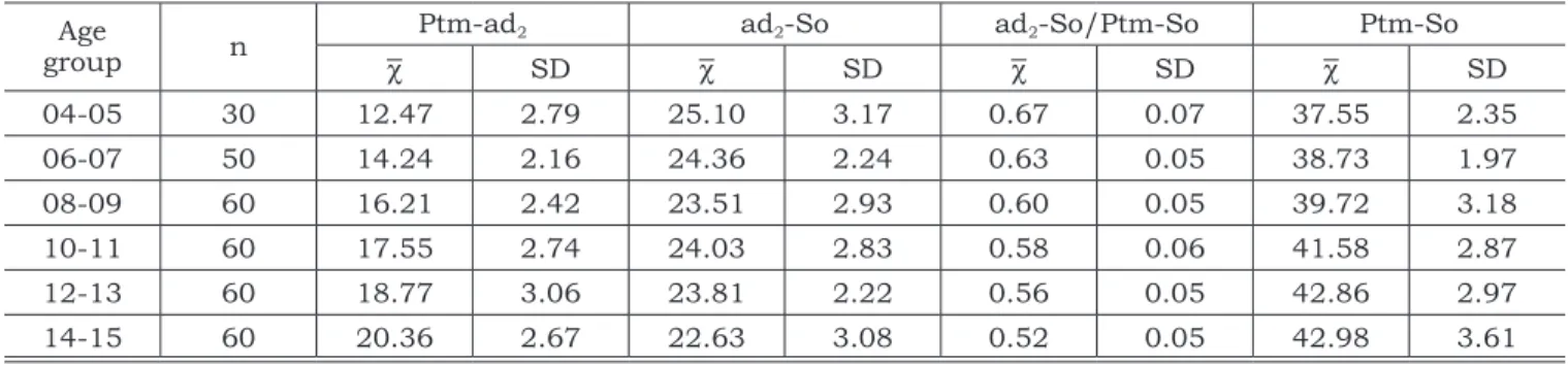
Means and standard deviations for Ptm-ad2, ad2-So, ad2-So/Ptm-So and Ptm-So variables for the six age groups are presented in Table 1. The nasopharyngeal airway space expressed by the Ptm-ad2 linear distance presented increasing val-ues between 4 and 16 years of age. Adenoid
thick-ness expressed by the ad2-So linear distance was larger in the age group 4 – 5 years and decreased subsequently until the age group of 8 – 9 years. There was a slight increase in the age group 10 – 11 years and the decrease continued from then on. The ratio between adenoid thickness and the bony nasopharynx depth decreased from 4 to 16 years of age. The bony nasopharynx depth ex-pressed by the Ptm-So linear distance presented increasing values between 4 and 16 years of age, which suggests that the nasopharynx becomes larger with growth.
In Table 2, it can be seen that the arithmetic means of the ad2-So and Ptm-So variables are sig-nificantly higher for boys in comparison to girls in the age group 14 – 15 years.
DISCUSSION
For orthodontists, it is of utmost importance to assess the patient’s ability to breathe through the nose, since the damaging consequences of mouth breathing have already been appropri-ately reported in literature1-3,11,13,14,17-19,21. Never-theless, there are few and disputed studies about
S
So
Ba
Ptm ad2
FIGURE 1 - Cephalometric tracing with the Ba, Ptm, S,
So and ad2 reference points and the Ptm-ad2, ad2-So
the growth pattern of the nasopharynx and about the adenoid growth cycle. In the present investi-gation, both were studied with the aid of lateral cephalometric radiographs. The sample selected consisted of nose-breathing white Brazilian, aged between 4 and 16 years, and was divided according to participants’ age. Two measurements proposed by Linder-Aronson, Leighton9 (1983) were used to-gether with the ad2-So/So ratio and the Ptm-So linear distance in order to allow the authors to achieve the research’s purposes.
The Ptm-ad2 measurement’s mean values show that the nasopharyngeal airway space is nar-rower in the age group 4 – 5 years, and that an increase occurs subsequently. A previous study20 has shown that this measurement presents a perfect agreement with the endoscopic diagnosis. Therefore, it can be routinely used to evaluate na-sopharyngeal airway obstruction. In other works, some authors9,12 verified that its smallest value was found at 5 years of age, which is in conso-nance with the current findings. However, they also found decreasing values between 8 and 10
TABLE 1 - Arithmetic means (c) and standard deviations (SD) for Ptm-ad2 (mm), ad2-So (mm), ad2-So/Ptm-So and
Ptm-So(mm) measurements according to age group.
Age
group n
Ptm-ad2 ad2-So ad2-So/Ptm-So Ptm-So
c SD c SD c SD c SD
04-05 30 12.47 2.79 25.10 3.17 0.67 0.07 37.55 2.35
06-07 50 14.24 2.16 24.36 2.24 0.63 0.05 38.73 1.97
08-09 60 16.21 2.42 23.51 2.93 0.60 0.05 39.72 3.18
10-11 60 17.55 2.74 24.03 2.83 0.58 0.06 41.58 2.87
12-13 60 18.77 3.06 23.81 2.22 0.56 0.05 42.86 2.97
14-15 60 20.36 2.67 22.63 3.08 0.52 0.05 42.98 3.61
TABLE 2 - Arithmetic means (c), standard deviations (SD) and Student’s t test between the means of Ptm-ad2 (mm),
ad2-So (mm), ad2-So/Ptm-So and Ptm-So (mm) measurements for male (M) and female (F) subjects, according to
age group.
Ptm-ad2 ad2-So
M F
p M F p
c SD c SD c SD c SD
04-05 12.29 3.09 12.64 2.65 0.742 25.65 3.73 24.56 2.52 0.356
06-07 14.62 1.98 14.21 2.37 0.509 24.33 2.29 24.39 2.24 0.926 08-09 16.29 2.29 16.13 2.57 0.799 24.30 2.53 22.72 3.13 0.015
10-11 17.66 2.52 17.44 2.98 0.759 24.35 2.47 23.71 3.16 0.386
12-13 19.13 3.48 18.42 2.57 0.372 23.70 2.42 23.91 2.04 0.718 14-15 20.56 2.56 20.16 2.80 0.566 23.68 3.06 21.57 2.76 0.007*
ad2-So/Ptm-So Ptm-So
M F
p M F p
c SD c SD c SD c SD
04-05 0.67 0.08 0.66 0.06 0.701 37.94 2.25 37.16 2.46 0.373
06-07 0.63 0.06 0.63 0.05 1.000 38.66 1.92 38.80 2.06 0.402
08-09 0.60 0.05 0.59 0.05 0.442 40.59 2.95 38.84 3.21 0.994
10-11 0.58 0.05 0.58 0.06 1.000 42.01 2.15 41.15 3.42 0.248
12-13 0.55 0.05 0.57 0.05 0.127 43.38 3.25 42.34 2.62 0.178
14-15 0.53 0.05 0.51 0.06 0.166 44.21 4.12 41.75 2.54 0.007*
years of age, which could not be noticed in the present research.
Examining ad2-So measurement’s mean val-ues, it could be observed that thickness of the adenoid is larger in the age group 4 – 5 years, showing subsequent decrease. This is in accord-ance with several authors’ findings4-6,9,12.
The present findings, however, do not sup-port Subtelny, Koepp-Baker’s15 conclusions (1956) that the adenoidal growth peak may be reached between 9 and 15 years of age, atrophying sub-sequently.
Still in relation to the ad2-So measurement, it could be noticed that the adenoid showed a slight increase in the age group 10 – 11 years, and then the decrease continued. This phenomenon had already been described by other researchers9,12, who imputed it to the influence of sexual hormones at puberty.
The ad2-So/Ptm-So ratio was introduced be-cause the adenoidal size in relation to the bony nasopharynx can be considered more important than its absolute size for respiratory pattern evalu-ation. This ratio was larger in the age group 4 – 5 years, then it diminished progressively until the age group 14 – 15 years. Fujioka et al.4 (1979) found the largest adenoid/nasopharyngeal space ratio at 4 years and 6 months, and the smallest at 15 years and 6 months of age, which is quite in accordance with the present findings.
The bony nasopharynx depth expressed by the Ptm-So linear distance was considered relevant to this research as it allows mensuration at the same higher nasopharyngeal level in which the airway space and the adenoidal thickness are measured. This distance increased from the age group 4 – 5 years to the age group 14 – 15 years of age. The increasing depth of the bony nasopharynx can help to explain the apparent contradiction between the nasopharyngeal free airway space increase as well as that of the adenoid thickness in the same age group 10 – 11 years. From 8 – 9 to 10 – 11 years, the bony nasopharynx depth increased 1.86 mm,
while the adenoidal thickness increased only 0.52 mm. The difference between the two values, therefore, represents the airway space increase. Indeed, it has already been reported that, due to growth, the hard palate and, as a consequence, the Ptm point is displaced downward, resulting in an adequate airway space for nasorespiratory needs, even when the adenoidal thickness is in-creasing10,15.
When the sample was divided according to the subjects’ gender, it was noticed that for boys the bony nasopharynx was significantly higher in comparison to girls in the age group 14 – 15 years. It could be seen that male subjects demonstrated growth until the age group 14 – 15 years, while the maximum nasopharyngeal capacity in females was achieved in the age group 12 – 13 years. These findings seem to be in accordance with what was stated by some authors5,9,12 that the nasopharynx growth follows a sexually determined pattern, simi-lar to that of the rest of the skeleton.
Results also show that, for boys, the adenoidal thickness was significantly higher in comparison to that of girls in the age group 14 – 15 years. It is very interesting to observe that the adenoidal tis-sue continues decreasing, even after the maximum nasopharyngeal capacity is achieved by females. Since male subjects demonstrate growth until the age group 14 – 15 years, the last period analysed, it is not known whether the same phenomenon occurs in boys.
CONCLUSION
The present findings support the statement that although the nasopharynx follows a growth pattern similar to that of the rest of the body, ad-enoidal tissue does not. Adad-enoidal development seems to differ from that of other lymphatic tis-sues, showing a peculiar pattern that can be re-vealed when hypertrophy due to infections and allergies is eliminated.
REFERENCES
1. Alcazar NMPV, Freitas MR, Janson G, Henriques JFC, Frei-tas KMS. Estudo cefalométrico comparativo dos espaços naso e bucofaríngeo nas más oclusões Classe I e Classe II, divisão 1, sem tratamento ortodôntico, com diferentes padrões de crescimento. Rev Dent Press Ortodon Ortop Facial 2004;9:68-76.
2. Bresolin D, Shapiro PA, Shapiro GG, Chapko MK, Dassel S. Mouth breathing in allergic children: its relationship to dentofacial development. Am J Orthod 1983;83:334-9. 3. Faria PTM, Ruellas ACO, Matsumoto MAN, Anselmo-Lima
4. Fujioka M, Young LW, Girdany BR. Radiographic evaluation of adenoidal size in children: adenoidal-nasopharyngeal ratio. AJR Am J Roentgenol 1979;133:401-4.
5. Handelman CS, Osborne G. Growth of the nasopharynx and adenoid development from one to eighteen years. Angle Orthod 1976;46:243-59.
6. Handelman CS, Pruzansky S. The size of the adenoids in normal and C. P. I. children. Presented at the IADR, Washington, DC; 1967.
7. Linder-Aronson S. Adenoids – Their effect on mode of breathing and nasal airflow and their relationship to char-acteristics of the facial skeleton and the dentition. A bio-metric, rhino-manometric and cephalometro-radiographic study on children with and without adenoids. Acta Otolar-yngol Suppl 1970;265:1-132.
8. Linder-Aronson S, Bäckström A. A comparison between mouth and nose breathers with respect to occlusion and facial dimensions. Odontol Revy 1960;11:343-75. 9. Linder-Aronson S, Leighton BC. A longitudinal study of the
development of the posterior nasopharyngeal wall between 3 and 16 years of age. Eur J Orthod 1983;5:47-58. 10. Moyers RE. Ortodontia. 4a ed. Rio de Janeiro:
Gua-nabara Koogan; 1991.
11. Netta MLSS, Maruo H, Vieira SR, Saga AY. Estudo cefalométrico comparativo das dimensões craniofaciais entre crianças respiradoras nasais e bucais, com malo-clusão Classe II, divisão 1. J Bras Ortodon Ortop Facial 2004;9:41-7.
12. Preston CB, Tobias PV, Salem OH. Skeletal age and growth of the nasopharynx in the sagittal plane: a cepha-lometric study. Semin Orthod 2004;10:16-38.
13. Solow B, Kreiborg S. Soft-tissue stretching: A possible control factor in craniofacial morphogenesis. Scand J Dent Res 1977;85:505-7.
14. Solow B, Siersbaek-Nielsen S, Greve E. Airway ad-equacy, head postures and craniofacial morphology. Am J Orthod 1984;86:214-23.
15. Subtelny JD, Koepp-Baker H. The significance of ad-enoid tissue in velopharyngeal function. Plast Reconstr Surg 1956;17:235-50.
16. Tanner JM. The human growth curve. In: Harrison GA, Weiner JS, Tanner JM, Barnicot NA, editors. Human biology. Oxford: Oxford University Press; 1964. 310 p. 17. Tarvonen PL, Koski K. Craniofacial skeleton of
seven-year-old children with enlarged adenoids. Am J Orthod Dentofacial Orthop 1987;91:300-4.
18. Tourne LPM, Schweiger J. Immediate postural re-sponses to total nasal obstruction. Am J Orthod Dentofa-cial Orthop 1996;110:606-11.
19. Vargervik K, Miller AJ, Chierici G, Harvold EP, Tower B. Morphologic response to changes in neuromuscular patterns experimentally induced by altered mode of respi-ration. Am J Orthod 1984;85:115-24.
20. Vilella OV, Vilella BS, Ianni Filho D, Karsten A, Mon-teiro AA, Koch HA et al. Evaluation of the nasopharyngeal free airway space based on lateral cephalometric radio-graphs and endoscopy. Orthodontics 2004;1(3):215-23. 21. Yamada T, Tane K, Miyamoto K, Yamauchi K.
In-fluences of nasal respiratory obstruction on craniofacial growth in young Macaca fuscata monkeys. Am J Orthod Dentofacial Orthop 1997;111:38-43.