Bruno Vieira Caputo(a) Gabriela Cineze Traversa- Caputo(b)
Claudio Costa(c)
Elcio Magdalena Giovani(a)
(a) Center for Study and Care of Special Patients - CEAPE, Dental School, Univ Paulista - Unip, São Paulo, SP, Brazil. (b) Private practice, São Paulo, SP, Brazil. (c) Stomatology Department, Dental School,
Univ de São Paulo, São Paulo, SP, Brazil.
Corresponding Author: Bruno Vieira Caputo E-mail: [email protected]
jaws of HIV-infected menopausal
women
Abstract: The advent of highly active antiretroviral therapy (HAART) has caused a reduction in mortality, thus contributing to an increase in the number of women with HIV/AIDS who reach the climacteric period, experience decline in ovarian function, and develop complications of vi-ral infection and HAART, which can accelerate bone loss. The aim of this study was to detect possible alterations in the jaws of HIV-infected women by panoramic radiography. The study comprised a total of 120 women above 40 years of age who were divided into the following two groups: women who are HIV positive (Group I) and women with no known HIV infection (Group II). Measurement of the following three radiomorphometric indexes was performed by panoramic radiography: Mental Index (MI), Panoramic Mandibular Index (PMI) and Antegonial Depth (AD). A total of 70% of women in the control group and 50% of women in the HIV group were in the postmenopausal period, and the av-erage values of both MI (p = 0.0054) and AD (p < 0.0001) for this period were lower in the HIV group than in the control group. For patients who were in the premenopausal period, the average AD was lower in the HIV group than in the control group (p = 0.0003). Despite the difference in the average age between groups, greater bone resorption in the mandible was found in the group of HIV-positive women.
Descriptors: HIV; Menopause; Radiography, Panoramic; Bone Resorption.
Introduction
The incidence of human immunodeiciency virus (HIV) infection is increasing among women. Over the years, the gender ratio for HIV in-fection has decreased gradually, and in recent years, there has also been an increase in the percentage of cases in the population over 40 years. A trend toward increased numbers of women with HIV has been observed in Brazil, and there has been an increased incidence of HIV infection among socioeconomically vulnerable populations.1
The advent of highly active antiretroviral therapy (HAART) has con-tributed to a signiicant increase in the life expectancy of these patients, thus providing these women with an opportunity to reach the climacteric stage. At this stage, these women experience a decline in ovarian func-tion and may develop complicafunc-tions related to both HIV and HAART.2
The menopausal period is divided into the following three phases:
Declaration of Interests: The authors certify that they have no commercial or associative interest that represents a conflict of interest in connection with the manuscript.
Submitted: Sep 27, 2012
Premenopause begins with the decline of ovarian function, and then the phase transition (i.e., peri-menopause) occurs, which is followed by meno-pause. Postmenopause is the period after the cessa-tion of ovarian funccessa-tion, which causes a reduccessa-tion in estrogen and progestin levels.2,3
Women tend to experience a loss of bone mass at an accelerated rate after menopause; however, this demineralization in HIV-infected patients can be at-tributed to an important group of drugs known as antiretroviral protease inhibitors, which are known osteopenic agents. These drugs may increase the risk of bone fractures in people with HIV/AIDS.4-6
The factors that may contribute to the reduction in bone density in HIV-infected women include men-strual dysfunction (hormonal imbalance), weight loss (alterations in nutritional status), decreased body mass, increased bone resorption, and Cauca-sian ethnicity. These risk factors are associated with an increase in the incidence of osteopenia and osteo-porosis.7
Bone densitometry is considered the gold stan-dard among imaging methods used for disease diag-nosis.8,9 However, the cost and lack of access to this
exam both hinder its use as a method for diagnosing the pathology. Considering this dificulty, research-ers have been seeking new ways to assist in the early diagnosis of osteoporosis. Studies have shown a cor-relation between radiomorphometric indexes in den-tal radiographs and the bone mineral density of the lumbar spine, femoral neck and jaw.10-13
Panoramic radiography is a low-cost exam that is routinely performed in dental clinics, and a large number of tests are performed annually to aid in the diagnosis of oral pathologies, such as dental caries. Hence, it would be beneicial if radiographs could be used to identify menopausal women with un-detected osteoporosis. Dentists could evaluate the mandibular alveolar ridge and make necessary re-ferrals for patients who require a more speciic diag-nosis and treatment.14,15
The erosion in the lower jaw cortex, which is detected on panoramic radiographs, may be an im-portant indicator for identifying older individuals or postmenopausal women with low bone mineral den-sity.15-17 Because of the possible alterations in bone
structure of the jaws in general and especially in HIV patients who are being treated with HAART, our aim was to identify these bone alterations in women with HIV who are 40 years of age or older and are experiencing pre-, peri- or postmenopause.
Methodology
A randomized cross-sectional study of HIV-positive menopausal women who were undergoing dental care was conducted by surveying data on the signs, symptoms, and characteristics of menopause and performing an analysis of panoramic radiogra-phy. The study was approved by the Ethics Commit-tee of Paulista University (220/10).
A total of 120 women were included in the study; they were divided into the following two groups:
• Group I: Sixty women were selected from among those referred to the Center for Study and Care of Special Patients (CEAPE) at Paulista University in Brazil from June 2010 to February 2011. The inclusion criteria for this group were as follows:
-
40 years of age or older,-
experiencing at least three symptoms of meno-pause,-
HIV-positive status (ELISA/Western blot), and-
using HAART.• Group II (control group): This group included 60 women who attended the Integrated Clinic at Paulista University in Brazil from June 2010 to February 2011 and were not HIV-positive. Inclu-sion criteria, which were similar to those in the other group, were as follows:
-
40 years of age or older and-
experiencing at least three symptoms of meno-pause.The patients were classiied based on the period of menopause according to the presence or absence of bleeding, as in the study by Ferreira et al. 2
base of the mandible, the height of the center of the mental foramen, the distance between the bottom of the mental foramen and the base of the mandible (the normal value is greater than or equal to 0.3 mm) (Figure 2).
Measurements were obtained in millimeters bi-laterally. Panoramic radiographs were randomly se-lected from a sample to collect measurements, and a blind observer made these measurements for each group. The data were statistically analyzed such that the quantitative variables were compared be-tween groups using Student’s t-test. For the qualita-tive variables, we used the chi-square test of tradi-tional homogeneity. In addition, group comparisons were performed in terms of the radiomorphometric degree indexes using the Mann-Whitney test to con-irm the results. For variables with only two levels, we used Fisher’s exact test, and for the other vari-ables, we used the chi-square test. For all of the tests, the signiicance level was 5%.
There was intraexaminer agreement for the mea-surements that were repeated 14 days after the ini-tial measurements, and the results were compared with the irst measurements.
Results
There was a signiicant difference in the mean of 70 kVp.
Subsequently, the subjects were scanned using a Scanner HP Scanjet G4050 (Hewlett-Packard De-velopment Company, L.P., Palo Alto, USA) in posi-tive mode using the transparent materials adapter (TMA) at a resolution of 300 dpi and 100% gray-scale. The scanned image was downloaded and opened in ImageJ software, version 1.44 for Mac OS X operating system (Public domain image pro-cessing program, National Institutes of Health, Bethesda, USA), in which readings of the entire bone structure were taken, particularly of the trabecular bone, to observe bone integrity (or the loss thereof) and possible resorption at the points of interest. The following linear measurements were also performed:
• The Mental Index (MI)17 is the marking of the
route that parallels the lower border of the man-dible and is perpendicular from the center of the mental foramen (MF) to the cortical lower jaw. This index measures the height of the mandibu-lar inferior cortex (the normal value is greater than or equal to 3.1 mm) (Figure 1).
• Antegonial Depth (AD)18 measures the distance
along the perpendicular line from the deepest point of the concave antegonial notch depth to the lower cortical line that is parallel to the edge of the mandible (Figure 1).
• The Panoramic Mandibular Index (PMI)19 is
the ratio of the mandibular cortical thickness as measured on a line that is perpendicular to the
Figure 1 - A cropped panoramic radiograph showing the
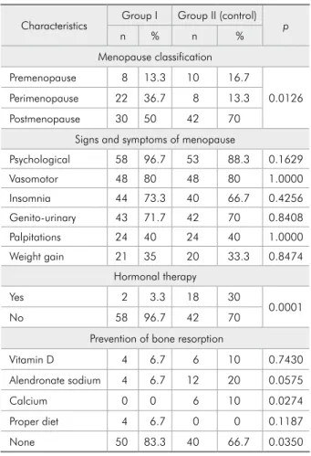
age of the patients between groups; the mean age of the patients in the control group was 56.1 years, and that of the patients in the HIV group was 46.9 years. Both groups were predominantly Caucasian (66.7% of the control group and 60.0% of the HIV group). There was a predominance of postmeno-pausal women in both groups (Table 1). There was no signiicant difference in any of the signs and symptoms between groups (p > 0.16).
The proportion of patients who were taking hor-mone replacement therapy was higher in the control group than in the HIV group (p = 0.0001). In both groups, most of the women did not engage in any type of preventive practice for osteoporosis (66.7% of women in the control group and 83.3% of women in the HIV group). In the control group, 20.0% of women were taking alendronate sodium, 10.0% of women were taking vitamin D, and 10.0% of wom-en were taking calcium.
Regarding the HIV patients, Table 2 shows the frequency distributions of the CD4 count, viral load, and HAART variables.
Table 3 shows the average results for the ra-diomorphometric index measures. Comparing the groups according to the stage of menopause, the AD was lower in the HIV group than in the control group (p = 0.0003) in the premenopausal period. In postmenopausal women, the values of both MI (p = 0.0054) and AD (p < 0.0001) were lower in the HIV group (Figure 3) than in the control group (Fig-ure 4).
Discussion
The results for the mean age and ethnicity were in accordance with those of another study conduct-ed by Ferreira et al.2 in which the authors studied
women with HIV and controls. According to Kojic
et al.,20 women with HIV present with early
meno-pause (between 47 and 48 years of age), which explains the age discrepancies between the study group and the control group.
The decisions regarding the sample size ac-counted for the dificulty in identifying HIV-posi-tive women who are at a more advanced age, need medical care and are willing to proceed with dental
Table 1 - Prevalence (%) of menopausal symptoms accord-ing to the characteristics of the studied population.
Characteristics Group I Group II (control) p
n % n %
Menopause classification
Premenopause 8 13.3 10 16.7
0.0126
Perimenopause 22 36.7 8 13.3
Postmenopause 30 50 42 70
Signs and symptoms of menopause
Psychological 58 96.7 53 88.3 0.1629
Vasomotor 48 80 48 80 1.0000
Insomnia 44 73.3 40 66.7 0.4256
Genito-urinary 43 71.7 42 70 0.8408
Palpitations 24 40 24 40 1.0000
Weight gain 21 35 20 33.3 0.8474
Hormonal therapy
Yes 2 3.3 18 30
0.0001
No 58 96.7 42 70
Prevention of bone resorption
Vitamin D 4 6.7 6 10 0.7430
Alendronate sodium 4 6.7 12 20 0.0575
Calcium 0 0 6 10 0.0274
Proper diet 4 6.7 0 0 0.1187
None 50 83.3 40 66.7 0.0350
Table 2 - Characteristics of women according to HIV status.
Characteristics Prevalence
n %
CD4 count (cells/mm³)
0–199 12 20
200–499 22 36.7
≥ 500 26 43.3
HIV status (number of copies)
Undetectable 46 76.7
0–50000 8 13.3
> 50000 6 10
HAART
NRTI 56 93.3
PI 38 63.3
NNRTI 20 33.3
II 2 3.3
treatment. The division of the periods of menopause is in agreement with that used in previous studies.2
A considerable number of patients (43.3%) had CD4 T counts that were equal to or greater than 500 cells/mm³, while a slightly lesser number of pa-tients (36.7%) had CD4 T counts between 200 cells/ mm³ and 499 cells/mm³. Regarding the viral load, most of the patients had undetectable loads (76.7%), while 13.3% of the patients had a load of less than 50000 copies. The lower the cell counts of CD4 T lymphocytes circulating in the blood, the more im-munocompromised the patient and the more likely it is that the patient may be affected by opportunistic infections, complications, and alterations in bone structure. These complications are likely to occur if the patient has a CD4 count below 200 cells/mm³, and the CD4 count is considered an important indi-cator for monitoring HIV patients in general.1
In both groups, menopausal symptoms were
mostly psychological symptoms, followed by vaso-motor symptoms; this inding is in agreement with the existing literature.2,21 Studies have shown that
menopausal symptoms are common among HIV-in-fected women, even when they have not yet reached menopause. Several topics, such as the use of hor-mone replacement therapy by HIV-positive women, have not been discussed in the literature, but these topics have been studied in menopausal HIV-nega-tive women.2,22 Some authors have emphasized that
middle-aged women with HIV infection have re-duced bone mineral density independent of the use of HAART and that the presence of osteopenia and osteoporosis cannot be attributed solely to the use of antiretroviral therapy.23,24
Several studies have shown that HAART may be associated with an increased prevalence of osteope-nia and osteoporosis, which can increase the risk of bone fractures in people with HIV. Furthermore, protease inhibitors are known osteopenic agents; however, a more consistent link between this group of drugs and bone loss remains to be proven.4-6,25-28
Panoramic radiography can be considered an auxiliary tool for dentists in the detection of possible alterations and bone fractures in the jaw, especially in postmenopausal women.13,14,29 Measurements of
MI, PMI, and AD have already been used and have been validated in the scientiic literature.10,17-19,29 In
the postmenopausal subjects in this study, the values of both MI (p = 0.0054) and AD (p < 0.0001) were lower in the HIV group compared with those in the control group.
The mandibular cortical thickness below the mental foramen correlates with the mineral den-sity of the lumbar spine and the proximal femur. A mandibular cortical thickness below the men-tal foramen of less than or equal to 3 millimeters could be considered a parameter for the diagnosis of low bone mineral density, and patients meeting this criterion could be referred for a bone densi-tometry examination. The authors also found that the low value of MI in postmenopausal women can be used to identify women with osteoporosis because this index is quantitative and has a moder-ate correlation with the BMD of the hip and lum-bar regions, with high accuracy in intra and
inter-Table 3 - Mean, standard deviation and p value of indexes (in mm) in patients.
Indexes Group Mean SD p
Premenopause
MI Control 4.01 0.56 0.4349
HIV 3.86 0.12
PMI Control 0.36 0.06 0.8882
HIV 0.36 0.06
AD Control 1.24 0.29 0.0003
HIV 0.68 0.20
Perimenopause
MI Control 4.06 0.45 0.8957
HIV 4.10 0.89
PMI Control 0.33 0.05 0.9891
HIV 0.33 0.06
AD Control 1.04 0.32 0.3049
HIV 1.26 0.58
Postmenopause
MI Control 3.84 0.73 0.0054
HIV 3.36 0.63
PMI Control 0.33 0.07 0.1723
HIV 0.31 0.06
AD Control 1.34 0.37 < 0.0001
examination.10,12,17,29
Due to the lack of related studies involving HIV patients, we do not have suficient data with which to compare our results for the radiomorphometric indexes of the HIV group, but other studies with HIV-negative patients have found a decrease in the
AD.18,30 However, a recent study reported no
signif-icant differences in the mean AD or in the gonial angles compared to the bone mineral density values of the lumbar spine and hip.29
Conclusion
Panoramic radiography was demonstrated to be
an eficient method for performing jaw measure-ments correlated to bone density. Despite the dif-ference in the average age between groups, greater bone resorption in the mandible was found in the group of HIV-positive women. Further studies are required to identify and establish links between HIV-infected women, HAART, and menopause.
Acknowledgements
To Coordenação de Aperfeiçoamento de Pesso-al de Nível Superior - Programa de Suporte à Pós-Graduação de Instituições de Ensino Particulares (CAPES-PROSUP).
Figure 3 - A panoramic radiograph of a 47-year-old HIV-infected postmenopausal woman with
lower values of the indexes.
Figure 4 - A panoramic radiograph of a 57-year-old postmenopausal woman (control group) with
higher values of the indexes.
References
1. Joint United Nations Program on HIV/AIDS. Report on the global HIV/AIDS epidemic 2008: executive summary. Geneva: UNAIDS; 2008. 36 p.
2. Ferreira CE, Pinto-Neto AM, Conde DM, Costa-Paiva L, Morais SS, Magalhães J. Menopause symptoms in women
infected with HIV: prevalence and associated factors. Gynecol Endocrinol. 2007 Apr;23(4):198-205.
4. Walker Harris V, Brown TT. Bone loss in the HIV-infected patient: evidence, clinical implications, and treatment strate-gies. J Infect Dis. 2012 Jun;205 Suppl 3:S391-8.
5. Pinto Neto LF, Ragi-Eis S, Vieira NF, Soprani M, Neves MB, Ribeiro-Rodrigues R, et al. Low bone mass prevalence, thera-py type, and clinical risk factors in an HIV-infected Brazilian population. J Clin Densitom. 2011 Oct-Dec;14(4):434-9. 6. Wang MWH, Wei S, Faccio R, Takeshita S, Tebas P, Powderly
WG, et al. The HIV protease inhibitor ritonavir blocks os-teoclastogenesis and function by impairing RANKL-induced signaling. J Clin Invest. 2004 Jul;114(2):206-13.
7. Dolan SE, Carpenter S, Grinspoon S. Effects of weight, body composition, and testosterone on bone mineral density in HIV-Infected women. J Acquir Immune Defic Syndr. 2007 Jun 1;45(2):161-7.
8. Thomas J, Doherty SM. HIV infection – a risk factor for osteo-porosis. J Acquir Immune Defic Syndr. 2003 Jul 1;33(3):281-91.
9. Montessori V, Press N, Harris M, Akagi L, Montaner JS. Adverse effects of antirretroviral therapy for HIV infection. CMAJ. 2004 Jan;170(2):229-38.
10. Dagistan S, Bilge OM. Comparison of antegonial index, mental index, panoramic mandibular index and mandibular cortical index values in the panoramic radiographs of normal males and male patients with osteoporosis. Dentomaxillofac Radiol. 2010 Jul;39(5):290-4.
11. Passos JS, Gomes Filho IS, Sarmento VA, Sampaio DS, Gon-çalves FP, Coelho JM, et al. Women with low bone mineral density and dental panoramic radiography. Menopause. 2012 Jun;19(6):704-9.
12. Taguchi A, Tsuda M, Ohtsuka M, Kodama I, Sanada M, Nakamoto T, et al. Use of dental panoramic radiographs in identifying younger postmenopausal women with osteoporo-sis. Osteoporos Int. 2006 Dec;17(3):387-94.
13. Geraets WG, Verheij JG, van der Stelt PF, Horner K; Lindh C; Nicopoulou-Karayianni K, et al. Prediction of bone mineral density with dental radiographs. Bone. 2007 May;40(5):1217-21.
14. Chalazonitis AN, Koumarianos D, Tzovara J, Chronopoulos P. How to optimize radiological images captured from digital cameras, using the Adobe Photoshop 6.0 program. J Digit Imaging 2003 Jun;16(2):216-29.
15. Taguchi A, Ohtsuka M, Nakamoto T, Naito K, Tsuda M, Kudo Y, et al. Identiication of postmenopausal women at risk of osteoporosis by trained general dental practitioners using panoramic radiographs. Dentomaxillofac Radiol. 2007 Mar;36(3):149-54.
16. Klemetti E, Kolmakov S, Kröger H. Pantomography in as-sessment of the osteoporosis risk group. Scand J Dent Res. 1994 Feb;102(1):68-72.
17. Taguchi A, Suei Y, Ohtsuka M, Otani K, Tanimoto K, Ohtaki M. Usefulness of panoramic radiography in the diagnosis
of postmenopausal osteoporosis in women. Width and mor-phology of inferior cortex of the mandible. Dentomaxillofac Radiol. 1996 Nov;25(5):263-7.
18. Dutra V, Yang J, Devlin H, Susin C. Mandibular bone re-modeling in adults: evaluating on panoramic radiographs. Dentomaxillofac Radiol. 2004 Sep;33(5):323-8.
19. Benson BW, Prihoda TJ, Glass BJ. Variations in adult cortical bone mass as measured by a panoramic mandibular index. Oral Surg Oral Med Oral Pathol. 1991 Mar;71(3):349-56. 20. Kojic EM, Wang CC, Cu-Uvin S. HIV and menopause: a
re-view. J Womens Health (Larchmt). 2007 Dec;16(10):1402-11. 21. Miller SA, Santoro N, Lo Y, Howard AA, Arnsten JH, Floris-Moore M, et al. Menopause symptoms in HIV-infected and drug-using women. Menopause. 2005 May-Jun;12(3):348-56. 22. Conde DM, Silva ET, Amaral WN, Finotti MF, Ferreira RG,
Costa-Paiva L, et al. HIV, reproductive aging, and health implications in women: a literature review. Menopause. 2009 Jan-Feb;16(1):199-213.
23. Arnsten JH, Freeman R, Howard AA, Floris-Moore M, San-toro N, Schoenbaum EE. HIV infection and bone mineral density in middle-aged women. Clin Infect Dis. 2006 Apr 1;42(7):1014-20.
24. Fausto A, Bongiovanni M, Cicconi P, Menicagli L, Ligabò EV, Melzi S, et al. Potential predictive factors of osteoporosis in HIV-positive subjects. Bone. 2006 Jun;38(6):893-7.
25. Schambelan M, Benson CA, Carr A, Currier JS, Dubé MP, Gerber JG, et al. Management of metabolic complications associated with antirretroviral therapy for HIV-1 infection: recommendations of an international Aids society– USA panel. J Acquir Immune Defic Syndr. 2002 Nov 1;31(3):257-75. 26. Grund B, Peng G, Gibert CL, Hoy JF, Isaksson RL, Shlay
JC, et al. Continuous antiretroviral therapy decreases bone mineral density. AIDS. 2009 Jul 31;23(12):1519-29. 27. Carr A, Hoy J. Low bone mineral density with tenofovir: does
statistically significant mean clinically significant? Clin Infect Dis. 2010 Oct;51(8):973-5.
28. Rivas P, Górgolas M, García-Delgado R, Díaz-Curiel M, Go-venechea A, Fernández-Guerrero ML. Evolution of bone min-eral density in AIDS patients on treatment with zidovudine/ lamivudine plus abacavir or lopinavir/ritonavir. HIV Med. 2008 Feb;9(2):89-95.
29. Leite AF, Figueiredo PT, Guia CM, Melo NS, de Paula AP. Correlations between seven panoramic radiomorphometric índices and bone mineral density in postmenopausal women. Oral Surg Oral Med Oral Pathol Oral Radiol Endod. 2010 Mar;109(3):449-56.