Arq Neuropsiquiatr 2004;62(3-A):571-574
1MD, Master in Neurology, Department of Neurology, School of Medicine of Ribeirão Preto, University of São Paulo, Ribeirão Preto SP, Brazil (SMRP, USP) Supported by CAPES; 2MD, PhD, Associate Professor of Neurology, Department of Neurology, SMPR/USP; 3M.D, PhD, Associate Professor of Dermatology, Department of Internal Medicine, SMRP/USP; 4MD, Department of Rehabilitation Medicine and Laboratory of Clinical Neurophysiology, Instituto Lauro de Souza Lima, Bauru SP, Brazil; 5MD, PhD, Plastic Surgeon, PqC-6, Instituto Lauro de Souza Lima, Bauru SP, Brazil; 6MD, PhD, Professor of Neurology, Department of Neurology, SMRP/USP.
Received 19 January 2004. Accepted 12 April 2004.
Dr. Wilson Marques Jr. - Departamento de Neurologia / Faculdade de Medicina de Ribeirão Preto - Av. Bandeirantes 3900 - 14049-900 Ribeirão Preto SP - Brasil. E-mail: [email protected]
NEAR NERVE POTENTIAL OF
SURAL NERVE IN LEPROSY
Ana Paula M. Arruda
1, Wilson Marques Jr
2, Norma T. Foss
3,
José A. Garbino
4, Marcos Virmond
5, Amilton A. Barreira
6ABSTRACT - Leprosy neuropathy is characterized by initial involvement of the small nerve fibers, later follo-wed by involvement of the large fibers, when routine nerve conduction studies become abnormal. To increase the diagnostic yield and precocity of these studies, we applied the near nerve technique to the sural nerve of 8 leprosy patients. Contrary to our expectations, the main component of the sural nerve sensory action potential was abnormal in all patients, but the minimum conduction velocity originating from small 3-6 µm
fibers was normal or only mildly involved in three patients. Also, although Schwann cells are the first to be involved in leprosy, the results are suggestive of axonal degeneration instead of demyelination. To bet-ter understand the neurophysiology and physiology of leprosy and to increase the accuracy and precocity of the diagnosis, it will be necessary to investigate patients in the very early stages of the disease and to cor-relate these findings with the corresponding nerve pathology.
KEY WORDS: leprosy neuropathy, minimum conduction velocity, near nerve technique, sensory nerve ac-tion potential.
Estudo da condução do nervo sural com a técnica de eletrodos justa-nervo em pacientes com moléstia de Hansen
RESUMO - Na hanseníase, as fibras de pequeno calibre são acometidas antes que as fibras mais grossas, ocasião em que o estudo da condução torna-se anormal. Neste estudo, utilizou-se a técnica de registro com eletrodos justa-nervo com a finalidade de aumentar a precocidade e a acurácia diagnóstica, devido à sua capacidade de detectar potenciais oriundos de fibras com cerca de 3-6 µm. Contrário às nossas
expec-tativas, o componente principal do potencial sensitivo do nervo sural foi anormal em todos os pacientes, enquanto a velocidade de condução mínima foi normal ou discretamente alterada em 3 pacientes. Além disso, os resultados são sugestivos de degeneração axonal e não mielinopatia, como seria esperado em uma doença que compromete inicialmente a bainha de mielina. Para um melhor entendimento da fisiolo-gia e fisiopatolofisiolo-gia, e para aumentar a precocidade diagnóstica, é fundamental estudar casos bem preco-ces e correlacionar os dados neurofisiológicos com a respectiva anatomia patológica.
PALAVRAS-CHAVE: estudo da condução sensitiva, moléstia de Hansen, potencial de ação sensitivo, técni-ca de registro com eletrodos justa-nervo.
The main target of Mycobacterium leprae is the peripheral nerve1. In this neuropathy, the small nerve
fibers conducting pain and temperature sensations are affected significantly before the large myelina-ted fibers that conduct vibration sense, position sen-se, and motor impulses2,3. This selective sequential
involvement of the nerve fibers impairs the detec-tion of leprosy neuropathy at the initial stages of the disease by neurophysiological evaluation since
routine nerve conduction studies only record poten-tials originating from fibers wider than 7 µm in dia-meter4.
We have recently applied the near nerve tech-nique to lepromatous patients to study the senso-ry nerve action potential (SNAP) of the median ner-ve at the wrist5in order to assess the contribution
improve the earlier detection of nerve injury in lep-rosy. As the median nerve is not suitable for biop-sy, after the previous study we decided to apply the same technique to investigate the late componen-ts of the SNAP of the sural nerve, where nerve biop-sies are usually carried out, allowing correlative stu-dies between morphology and physiology. We be-lieve these studies will not only improve our knowl-edge about neurophysiological findings in leprosy and its clinical applications, but will also improve our understanding of the physiology of this neuro-pathy.
METHOD
We studied the sural SNAP from 8 patients with lep-rosy and 10 normal controls. The disease was classified as multibacillary in 5 patients and paucibacillary in the remaining 3. Both patients and controls denied alcohol or drug abuse, diabetes mellitus and hereditary condi-tions. A metabolic screening for the most common causes of neuropathies was also peformed.
The near nerve technique was performed as described by Shefner et al.6, with the modifications introduced by
Marques and Barreira7. Briefly, the patient was
comforta-bly positioned in lateral or ventral decubitus, with the leg in complete relaxation. The area to be examined was carefully cleaned with water, soap and ether. The stimu-lating electrodes were positioned at the ankle, with the cathode being proximal to the anode. The needle recor-ding electrodes were positioned 14 cm proximally in a unipolar recording arrangement. The position of the ac-tive recording electrode was carefully adjusted near the nerve, so that when the electrode was used to stimula-te, an intensity as low as 0.5 to 1 mA was able to evoke a response at the ankle, implying that the distance bet-ween the needle and the nerve is 0.5 to 1 mm, conside-red to be the ideal position for the method. Filter set-tings were 200 and 3000 Hz, analysis time was 40 ms, the number of averaging ranged from 500-2000 sweeps per trial, and at least 2 trials were executed. A sensitivi-ty of 5-20 µV was used to measure the amplitude and
latency of the main component, while the late compo-nent was recorded with a sensitivity of 5 µV and a gain
of 0.1 to 1 µV/division was used to measure its latency
and amplitude. Temperature was kept above 33οC. Thie study was approved by the Ethics Committee of our University Hospital and of the Institute Lauro de Sou-za Lima. All patients and controls gave informed consent.
RESULTS
Mean age was 35.8 years (range: 19-58 years) for the controls and 48.5 (range: 28-72 years} for the patients. SNAP data for the sural nerves of controls and patients are presented in Tables 1 and 2, res-pectively. The main component and the late
compo-572 Arq Neuropsiquiatr 2004;62(3-A)
nents were recorded in only 3 and 5 patients, res-pectively. The mean amplitude of the main compo-nent was 21.5 µV (SD 8.71) for the controls and 2.8 µV (SD 2.07) for the patients, while the values for conduction velocity were 55.0 m/s (SD 6.67) and 46.13 m/s (SD 8.43). The mean conduction veloci-ty of the late component, minimum conduction ve-locity (MCV), was 16.16 m/s (SD 2.30) for the con-trols and 12.25 m/s (SD 8.55) for the patients; the limits, however, ranged from 11.1 to 18.4 in the first group and from 3.1 to 23.6 in the second group, consi-dering only the recorded potentials. Amplitudes we-re similar.
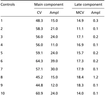
Table 1. Electyrophysiological evaluation of the sural nerve using the near nerve technique in normal controls.
Controls Main component Late component
CV Ampl MCV Ampl
1 48.3 15.0 14.9 0.3
2 58.3 21.0 11.1 0.1
3 56.0 24.0 17.1 0.2
4 56.0 11.0 16.9 0.1
5 59.1 24.0 15.7 0.2
6 64.3 39.0 17.3 0.2
7 57.1 30.0 17.9 0.1
8 45.2 15.0 18.4 1.2
9 44.8 12.0 18.3 0.1
10 60.9 24.0 14.0 0.1
Ampl, amplitude (µV); CV, conduction velocity (m/s); MCV, minimum con-duction velocity.
Table 2. Electyrophysiological evaluation of the sural nerve using the near nerve technique in patients with leprosy.
Patients Main component Late component
CV Ampl MCV Ampl
1 NR NR 8.6 0.1
2 NR NR NR NR
3 38.2 1.6 12.7 0.1
4 45.2 1.6 3.1 0.2
5 55.0 5.2 23.6 0.1
6 NR NR NR NR
7 NR NR 15.1 0.1
8 NR NR NR NR
No response was recorded in the sural nerves of patients 2, 6 and 8; in patients 1 and 7 the main component was not recorded, but the late compo-nent was normal; the main compocompo-nent of patients 3, 4 and 5 had normal or mildly decreased conduc-tion velocities and severely decreased amplitudes, while the behavior of the MCV was extremely vari-able, being normal in patient 3, extremely reduced in patient 4 and too high in patient 5.
DISCUSSION
Shefner et al.6applied the near nerve technique
to patients with peripheral neuropathy and found that in 31% of them the only electrophysiological abnormality present was an abnormal late compo-nent of the SNAP, demonstrating the efficacy of this technique in the study of potentials arising from nerve fibers ranging from 3 to 6 µm in diameter, that are not recorded in routine nerve conduc-tion studies4. Considering that in the initial
phas-es of leprosy there is a predominant involvement of unmyelinated fibers, following impairment of small myelinated nerve fibers2,8, we decided to
investigate the contribution of the near nerve technique to increase the diagnostic yield of nerve conduction studies in leprosy, since an early diagno-sis results in early treatment, the best measure to avoid the feared consequences of leprosy neuropa-thy, including painless ulcerations, and muscle atro-phy and weakness3.
Recently, Marques et al.5applied the near nerve
technique to study the sensory fibers of the medi-an nerve is newly diagnosed leprosy patients. Unfor-tunately, the median nerve is not accessible to bio-psy, preventing morphological analysis in those ca-ses with detected neurophysiological abnormali-ties. This is not the case for the sural nerve, an exclu-sively sensory nerve whose morphology has been extensively studied in peripheral neuropathies, in-cluding leprosy.
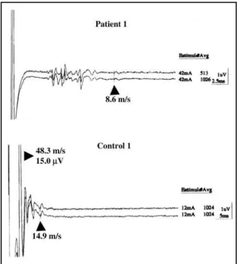
All nerves analyzed in this series were abnormal, but the findings were not homogeneous. In patients 2, 6 and 8 no SNAP was recorded, suggesting com-plete axonal degeneration of all nerve fibers. Pa-tients 1 (Fig 1) and 7 did not have the main compo-nent, but the late component was present, with mildly decreased and normal conduction velocities, respectively, suggesting degeneration of the large fibers but preservation of those fibers that give ori-gin to the observed late components. Patients 3, 4 and 5 had a low amplitude main component with preserved or only mildly decreased conduction
ve-Arq Neuropsiquiatr 2004;62(3-A) 573
locities, suggesting loss of the large fibers that gi-ve origin to these components. The MCV of these nerves were highly different, being normal, extre-mely reduced or moderately increased, respective-ly suggesting preservation of the fibers that give origin to the late component, regeneration or de-myelination, and loss of the small fibers.
The abnormalities found in the main component were relatively homogeneous, always suggesting axonal loss, a finding already reported by many au-thors5,8-13, but is not in perfect agreement with the
proposed physiology of this neuropathy since Schwann cells are the first to be involved by the ba-cilli14,15. This theoretically implyies that
demyelina-tion should be the first abnormality detected, as was reported by Tzourio et al.16for the superficial
radi-al nerve from recently diagnosed patients.
Confirming our previous study5, the analysis of
the late component, paradoxically, did not add too much to the neurophysiological evaluation of these patients since the main component was always ab-normal, even in the two patients (3 and 6) with pre-served MCV. As discussed in that paper, the origin of these normal late components must be investiga-ted. We suppose that they may be produced by
574 Arq Neuropsiquiatr 2004;62(3-A)
normal fibers whose electrophysiological behavior is similar to that of normal 4-6 µm fibers. We also hy-pothesize that the findings observed in patients 3, 4 and 5 may represent the sequence of events invol-ving the small fibers in this neuropathy. Initially, the-re is loss of the these fibers, and the last the-recorded com-ponents have an MCV proportional to the diame-ter of the remaining fibers, higher than the normal values for a late component, as observed in patient 5. Regeneration may then occur, giving origin to late components with extremely reduced conduc-tion velocities (patient 4). Eventually, these regen-erating components may show a behavior electro-physiologically similar to that of normal 3-6 µm fi-bers, with the occurrence of an electrophysiolog-ical “normal” late component originating from ab-normal nerve fibers.
As an affected nerve may function normally in leprosy17, it is possible that all patients studied in
this series had advanced disease, even those with mild manifestations. If this assumption is true, we may be studying nerves that show a significant de-gree of axonal degeneration, regeneration and even remyelination. The definitive understanding of all these findings and assumptions will probably be reached with the evaluation of patients in the very early stages of the disease, studied not only dis-tally but also at the topography were the initial at-tack occurs, and with studies analyzing both mor-phology and electrophysiology.
REFERENCES
1. Minauchi Y, Igata A. Leprous neuritis. In Matheus WB. Handbook of clinical neurology: neuropathies. Amsterdam Elsevier 1987;215-238. 2. Gibbels E, Henke-Lübke U, Klingmüller G. Unmyelinated nerve fibers
in leprosy: a quantitative and qualitative study of sural nerve biopsies in two cases of lepromatous leprosy. Lepr Rev 1988;59:153-162. 3. Sabin TD, Swift TR, Jacobson RR. Leprosy. In Dyck PJ, Thomas PK, Griffin
JW, Low PA, Poduslo JF, (EDS). Peripheral neuropathy, 3rd ed. Philadelphia: Saunders, 1993;1354-1379.
4. Buchthal R, Rosenfalck A, Behse F. Sensory potentials of normal and diseased nerves. In Dyck PJ, Thomas PK, Lambert EH, Bunge R, (EDS). Peripheral neuropathy, 2nd ed. Philadelphia: Saunders, 1984;981-1015. 5. Marques W Jr, Foss NT, Arruda AP, Barreira AA. Near-nerve in
lepro-matous leprosy. Muscle Nerve 2003;28:460-463.
6. Shefner JM, Buchthal F, Krarup C. Slowly conducting myelinated fibers in peripheral neuropathy. Muscle Nerve 1991;14:534-542.
7. Marques W Jr, Barreira AA. Normal median near nerve potential. Braz J Med Biol Res 1997;30:1431-1435.
8. Anthia NH, Mehta LN, Shetty VP, Irai PF. Clinical, electrophysiologi-cal, quantitative, histological and ultrastructural studies of the index branch of the radial cutaneous nerve in leprosy: 1. Preliminary report. Int J Lepr 1975;43:106-113.
9. Dastur DK, Ramamohan Y, Shah JS. Ultrastructure of lepromatous nerves: neural pathogenesis in leprosy. Int J Lepro 1973;41:77-80. 10. Ramakrishnan AG, Srinivasan TM. Electrophysiological correlates of
hanseniasis. Int J Leprosy 1995;63:395-408.
12. Freitas MR, Nascimento OJ, Quaglino EA, Hanh MD. Small-fiber polyneuropathy in leprosy without skin changes: study of 17 cases. Arq Neuropsiquiatr 2003;61:542-546.
13. Jardim MR, Antunes SL, Santos AR, et al. Criteria for diagnosis of pure neural leprosy. J Neurol 2003;250:806-809.
14. Rambukana A. How does Mycobacterium leprae target the peripher-al nervous system? Trends Mycrobiol 2000;8:156-157.
15. Rambukana A, Zanazzi G, Tapinos M, Salzer JL. Contact-dependent demyelination by Mycobacterium leprae in the absence of immune cells. Science 2002;296:862-863.
16. Tzourio C, Said G, Millan J. Asymptomatic nerve hypertrophy in lep-romatous leprosy: a clinical, electrophysiological and morphological study. J Neurol 1992;239:367-342.