Dysregulation of Autonomic Nervous System
in Chagas
’
Heart Disease Is Associated with
Altered Adipocytokines Levels
João Marcos Barbosa-Ferreira1*, Charles Mady1, Barbara Maria Ianni1, Heno Ferreira Lopes2,3, Felix José Alvarez Ramires1, Vera Maria Cury Salemi1, Cesar José Grupi4, Denise Tessariol Hachul5, Fábio Fernandes1
1Cardiomyopathy Unit of the Heart Institute (InCor), do Hospital das Clínicas da Faculdade de Medicina da Universidade de São Paulo, São Paulo, Brazil,2Hypertension Unit of the Heart Institute (InCor), do Hospital das Clínicas da Faculdade de Medicina da Universidade de São Paulo, São Paulo, Brazil,3Universidade Nove de Julho—UNINOVE, São Paulo, Brazil,4Electrocardiology Unit of the Heart Institute (InCor), do Hospital das Clínicas da Faculdade de Medicina da Universidade de São Paulo, São Paulo, Brazil,5Clinical Arrhythmia Unit of the Heart Institute (InCor), do Hospital das Clínicas da Faculdade de Medicina da Universidade de São Paulo, São Paulo, Brazil
*jmbemfica@hotmail.com
Abstract
Background
Chagas disease (CD) induces autonomic dysfunction and inflammatory activity, which may promote metabolic abnormalities. We studied metabolism and his correlation with Auto-nomic Nervous System (ANS) and inflammation in CD.
Methods and Results
Sixty subjects were divided into 4 groups: control group (CG), IF (indeterminate form) group; ECG group (ECG abnormalities and normal left ventricular systolic function), and LVD group (left ventricular sistolic dysfunction). Levels of adiponectin, leptin, insulin, inter-leukin-6 (IL-6), and tumor necrosis factor-alpha (TNF-alpha) were assayed in serum sam-ples by ELISA. ANS was assessed by heart rate variability in frequency domain in 24-hour Holter and postural tilt test (rest and orthostatic position). High frequency (HFr) component values were used to estimate parasympathetic activity and low frequency (LFr) component, sympathetic activity. Analyzes were made of the correlations of each of the metabolic parameters (leptin and adiponectin) with the inflammatory cytokines (interleukin-6 and TNF- alpha) and with the ANS assessment measurements. No significant differences were observed in leptin and insulin levels. Adiponectin was higher in ECG and LVD groups: [CG = 4766.5 (5529.5), IF = 4003.5 (2482.5), ECG = 8376.5 (8388.5), LVD = 8798 (4188.0) ng/ mL, p<0.001)]. IL-6 and TNF-alpha were higher in LVD group: [IL-6: CG = 1.85 (6.41); IF = 1.58 (1.91); ECG = 1.0 (1.57); LVD= 31.44 (72.19) pg/ml; p = 0.001. TNF-alpha: CG = 22.57 (88.2); IF = 19.31 (33.16); ECG = 12.45 (3.07); LVD = 75.15 (278.57) pg/ml; p = 0.04]. Adi-ponectin levels had a positive association with the HFr component (r = 0.539; p = 0.038)
OPEN ACCESS
Citation:Barbosa-Ferreira JM, Mady C, Ianni BM, Lopes HF, Ramires FJA, Salemi VMC, et al. (2015) Dysregulation of Autonomic Nervous System in Chagas’Heart Disease Is Associated with Altered Adipocytokines Levels. PLoS ONE 10(7): e0131447. doi:10.1371/journal.pone.0131447
Editor:Herbert B. Tanowitz, Albert Einstein College of Medicine, UNITED STATES
Received:October 25, 2014
Accepted:June 2, 2015
Published:July 6, 2015
Copyright:© 2015 Barbosa-Ferreira et al. This is an open access article distributed under the terms of the
Creative Commons Attribution License, which permits unrestricted use, distribution, and reproduction in any medium, provided the original author and source are credited.
Data Availability Statement:Data have been deposited to Figshare:http://dx.doi.org/10.6084/m9. figshare.1384885.
and an inverse association with the LFr component (r = - 0.539; p = 0.038) in ECG group. Leptin levels had a negative association with the HFr component (r= - 0.632; p = 0.011) and a positive association with the LFr component (r = 0.632; p = 0.011) in LVD group.
Conclusions
We found increased adiponectin levels in Chagas’heart disease with systolic dysfunction and in patients with ECG abnormalities and normal systolic function at rest. Adipocytokines levels (adiponectin and leptin) were associated with ANS parameters in Chagas’heart disease.
Introduction
Chagas’disease (CD) occurs from the southern United States to Patagonia and affects around
8 million people in Latin America [1]. Moreover, due to the intensification of the migratory
flow, CD is becoming more relevant in nonendemic countries, such as the United States, Can-ada, some European countries, Japan, and Australia. In the United States, it is estimated that 300 thousand legal immigrants may be infected with the disease. Spain has the second largest
prevalence with around 40 to 60 thousand infected immigrants [2]. The natural history of
Cha-gas’disease is summarized in the acute and chronic phases[3]. In the chronic phase, about 70%
of the patients have no symptoms and routine examinations do not show any abnormalities.
This stage is called indeterminate form (IF). The patients of IF form of Chagas’disease, in
eral, have a very good prognosis. Survival in this group appears comparable to that of the
gen-eral population [4]. The remaining 30% have the chronic digestive and/or cardiac form, and
10% of these patients may progress to severe forms of heart disease. The progression to
myo-cardial dysfunction represents the leading cause of morbidity and mortality in Chagas’disease
[3]. Therefore, investigations regarding the pathophysiology of the development and
progres-sion of cardiomyopathy are of fundamental importance in the proposed new therapies in an attempt to minimize morbidity and mortality.
Myocardial damage directly related to parasite persistence, immunologic mechanisms, microvascular disturbances, and autonomic dysfunction are involved in the pathophysiological
mechanism of chagasic cardiomyopathy [5]. Cardiac dysautonomia is a well-established
fea-ture of Chagas disease, in which anatomic denervation and functional abnormalities have been extensively described. Neuronal depopulation occurs in cardiac parasympathetic ganglia in
Chagas’heart disease associated with scattered sympathetic denervation [6]. Several methods
are currently available to evaluate autonomic function such as Valsalva maneuver, deep breath-ing, orthostatic test and heart rate variability in the time domain or in the frequency domain
[5]. Previous studies suggest that autonomic dysfunction may precede left ventricular systolic
dysfunction [7,8]. The mechanisms of Chagas’cardiomyopathy can influence other
pathophys-iological pathways, such as metabolic impairment.
Recent research has led to a growing appreciation of the complexity of metabolic aspects of heart failure (HF) pathophysiology. Not only the myocardium, but also peripheral tissues and organs are affected by metabolic failure, resulting in a global imbalance between catabolic and anabolic signals. Metabolic feedback signals from muscle and fat actively contribute to disease
progression [9]. The adipocytokines are bioactive mediators produced by adipose tissue. The
main adipocytokines are adiponectin and leptin. Adiponectin has beneficial anti-inflammatory
and anti-atherogenic effects as well as insulin-sensitizing action [10–14]. However, the role of
adiponectin in cardiovascular disease is still a controversy. Previous studies have demonstrated that adiponectin is increased in systolic HF patients, even predicting morbidity and mortality
[15,16]. Leptin promotes pro-inflammatory and pro-thrombotic activities, neointimal
prolifer-ation, endothelial dysfunction and induction of insulin resistance [17,18]. In CD patients, we
have previously found reduced leptin levels in Chagas’cardiomyopathy when compared with a
control group and other forms of CD [19]. Our speculation is that chagasic patients have
altered sympathetic activity resulting in decreased leptin synthesis.
So, the basic hypothesis is that chronic Chagas’cardiomyopathy may serve as a model in
which severe inflammatory activity and early involvement of the autonomic nervous system (ANS) influence metabolism. Alterations in metabolism such as insulin resistance and the role of adipose tissue can lead to a systemic inflammatory state that contributes to vasculopathy
and cardiovascular risk [14]. This two-way mechanism can bring chronic metabolic
complica-tions of HF to the fore and gradually shift its clinical presentation. The study of this association can promote emerging therapeutic concepts with specific metabolic targets. The aim of this
study was to evaluate the metabolic parameters in the different forms of Chagas’disease and its
association with inflammatory activity and with measures of autonomic nervous system function.
Materials and Methods
Patient selection
Sixty subjects were divided into 4 groups: control group (CG), IF (indeterminate form) group of Chagas disease; ECG group (Chagas heart disease with ECG abnormalities and normal left ventricular systolic function), and LVD group (Chagas heart disease with left ventricular sys-tolic dysfunction). The control group consisted of 15 healthy individuals. IF group comprised subjects with 2 positive serologic reactions for Chagas' disease and no cardiac involvement as defined by chest X-rays, 12-lead ECG, and 2-dimensional echocardiography. All patients had normal barium studies of the esophagus and the colon. ECG group consisted of patients with normal left ventricular (LV) systolic function showing right or left bundle-branch block, left anterior fascicular block, diffuse ST changes, ventricular premature beats that may be multi-form or runs of non-sustained ventricular tachycardia registered on the ECG. LVD group, comprised patients with LV dysfunction demonstrated by a left ventricular ejection fraction less than 40% in echocardiography. All of these groups were matched according to sex, age (± 2
years intervals), and body mass index (± 1 kg/m2intervals). The inclusion criteria were patients
with 2 positive serologic reactions for Chagas' disease and age over 18 years. The exclusion cri-teria were myocardial infarction (evaluated by Q waves on an electrocardiogram (ECG) or seg-mental left ventricular (LV) dysfunction by 2-dimensional echocardiography), moderate or severe valvar heart disease (evaluated by clinical examination and by Doppler echocardiogra-phy), arterial hypertension, smoking, diabetes mellitus, current use of statins, atrial fibrillation, advanced atrioventricular block, pacemaker, thyroid diseases, chronic obstructive pulmonary disease, and heart failure functional classes III and IV by the New York Heart Association (NYHA) classification. All subjects signed a written consent form, and the Ethics Committee of the Heart Institute (InCor), University of São Paulo Medical School approved the study.
Antropometric measurements
Echocardiographic study
Comprehensive transthoracic echocardiographic studies were performed with a Sequoia 512 ultrasound machine (Acuson, Montain View, CA USA). Two-dimensional guided M-mode measurements of the left ventricle (LV) in short axis view and LV, left atrium and right atrium end diastolic and end systolic volumes were taken from apical 4-chamber view. LV ejection fraction (LVEF) was calculated using a modified Simpson biplane method. LV regional wall motion was evaluated based on 17-segment model segmentation, and each segment was con-firmed in multiple views as follows: normal or hyperkinesis, hypokinesis, akinesis, dyskinesis, and aneurysmal. Diastolic function was assessed from pulsed-wave Doppler of the transmitral inflow velocities, with the sample volume at the mitral valve leaflet tips, and from tissue Dopp-ler imaging of the septal and lateral mitral annulus, both at apical 4-chamber view. Mitral, tri-cuspid, aortic, and pulmonary regurgitations were qualitatively evaluated.
Laboratory measurements
Fasting glucose, total cholesterol, HDL-cholesterol, LDL-cholesterol and tryglicerides were measured using standard assays.
The subjects underwent serum adiponectin, insulin, leptin, interleukin-6 (IL-6), and tumor necrosis factor-alpha (TNF-alpha) measurements by ELISA. Serum insulin was measured with a commercially available kit (Millipore, St. Charles, Missouri, USA). The sensitivity of the kit
was 1μU/mL, and the reference interval was from 31 to 65μU/ml. The measurements were
made in duplicate with a coefficient of variation of 4.3%. Serum leptin was measured with a commercially available kit (Millipore, St. Charles, Missouri, USA). The sensitivity of the kit was 0.78 ng/mL, and the reference interval was from 12.9 to 26.8 ng/mL. The measurements were made in duplicate with a coefficient of variation of 7.3%. Serum adiponectin levels were measured with a commercially available kit (Millipore, St. Charles, Missouri, USA). The sensi-tivity of the kit was 100 ng/mL, and the reference interval was from 7000 to 14,500 ng/mL. The measurements were made in duplicate with a coefficient of variation of 3.1%. Interleukin-6 was measured with a commercially available kit (USCNK, Life Science Inc., Wuhan, China). The sensitivity of the kit was 5.6 pg/mL, and the reference interval was from 75 to 175 pg/mL. The measurements were made in duplicate with a coefficient of variation of 10.3%. Tumor necrosis factor-alpha was measured with a commercially available kit (USCNK, Life Science Inc., Wuhan, China). The sensitivity of the kit was 5.9 ng/mL, and the reference interval was from 150 to 175 pg/mL. The measurements were made in duplicate with a coefficient of varia-tion of 6.4%.
Evaluation of autonomic nervous system function
The autonomic nervous system (ANS) function was evaluated by heart rate variability (HRV) in the frequency domain using Fast Fourier Transform model in 24-hour Holter and postural tilt test (rest and orthostatic position). The following indices were calculated: Total power, Low
frequency (LFr) component in absolute values of power (ms2) and in normalized units (n.u.),
High frequency (HFr) component in absolute values of power (ms2) and in normalized units
(n.u.), and the LFr/HFr ratio. We also calculated the changes in LFr and HFr components from rest to orthostatic position. The increase of the LF component and the LFr/HFr ratio were interpreted as a predominance of sympathetic activity. The increase in the HFr component and the reduction in the LFr/HFr ratio were interpreted as a predominance of parasympathetic activity[20].
- Component LFr or HFr in absolute values / (total power–very low frequency component) x 100
The patients were instructed not to take any stimulants, such as coffee, tea, soft drinks and alcoholic beverages, on the day prior to and on the day of the exams.
The“Task Force Monitor”(CNSystems Medizintechnik GmbH, Graz, Austria) was used for
analysis during postural tilt testing. The five minutes immediately prior (rest in supine posi-tion) and the five minutes immediately after inclination (orthostatic posiposi-tion) were assessed. The mean of the spectral components obtained in the established times was calculated. Cardiac cycles with a variation greater than 25% to a previous one were excluded as a way of abolishing the consequent alterations to the ventricular and supraventricular extrasystoles. Only the read-ings with at least 85% sinus beats were assessed.
Statistical analyses
Statistical analyses were performed using ANOVA test to evaluate differences in means among groups. If the homogeneity of the variances was not observed, the non-parametric Kruskal-Wallis test was used. When there is evidence of difference in at least one group, it was used Tukey tests for comparisons. Spearman coefficient was used for correlation analysis. We evalu-ated the correlations in each of the three groups of patients with Chagas disease separately (IF group, ECG group and LVD group). The correlations were made between each of the
adipocy-tokines (adiponectin and leptin) with the inflammatory cyadipocy-tokines (interleukin-6 and TNF-α)
and with the ANS assessment measurements (in normalized units). A P value<0.05 was
con-sidered statistically significant.
Results
Clinical characteristics
There were no significant differences in age, weight, and body mass index among groups. The ejection fraction was significantly lower in LVD group. Systolic and diastolic blood pressure was lower in the LVD group compared to other three groups. The fat mass was lower in the LVD group, with statistical significance for the absolute values. The drugs used by the individu-als of the LVD group were as follows: amiodarone in three patients (20%), beta-blockers in 15 patients (100%), angiotensin converting enzyme inhibitors in 13 patients (86.6%), angiotensin receptor blockers in two patients (13.3%), furosemide in nine patients (60%), spironolactone in 11 patients (73.3%) and digoxin in one patient (6.6%). Individuals from others groups did not use any drugs. Baseline physical and hemodynamic characteristics in Chagas' disease patients
and healthy controls are displayed inTable 1.
Laboratory measurements
There were no significant differences in fasting glucose, total cholesterol, HDL-cholesterol, LDL-cholesterol, tryglicerides, leptin, and insulin among groups. Adiponectin was significantly
increased in ECG and LVD groups compared to IF and control groups (p<0.001). The levels of
interleukin-6 (p = 0.001) and tumor necrosis factor-alpha (p = 0,04) were higher in the LVD group compared to the other three groups. The data from metabolic and inflammatory
param-eters are described inTable 2.
Evaluation of autonomic nervous system function
ECG group. The data related to the 24-hour Holter and postural tilt test in supine (rest) and in
orthostatic position are detailed in Tables3,4and5.
Correlations
There were significant correlations between adiponectin with some ANS assessment indexes in ECG group. Adiponectin level is associated negatively with the LFr component and positively with the HFr component in 24-hour Holter. No correlations between adiponectin and
inflam-matory cytokines were found. The values of correlations of adiponectin are shown inTable 6.
Table 1. Clinical and hemodynamic characteristics of the studied population.
Variable Control Group IF Group ECG Group LVD Group p value*
Age (years) 43.8 (±7.43) 42.33 (±7.30) 43.20 (±6.14) 42.67 (±6.72) 0.942
NYHA (I/II) - - - 4/11
-HR (bpm) 74.93 (±7.00) 73.47 (±9.20) 73.33 (±8.20) 69.47 (±10.91) 0.604
SBP (mmHg) 122.67 (±7.04) 120.67 (±9.61) 121.33 (±9.90) 101.33 (±9.90)** <0.001
DBP (mmHg) 70.00 (±7.56) 72.67 (±7.99) 74.00 (±7.37) 62.67 (±5.94)** 0.001
BMI (kg/m2) 23.9 (±1.20) 23.9 (±1.39) 23.7 (±1.20) 23.2 (±1.50) 0.602
Weight (kg) 70.4 (±8.50) 68.1 (±10.00) 67.8 (±8.50) 65.8 (±8.80) 0.595
Fat mass (kg) 15.7 (±4.40) 14.5 (±5.80) 14.3 (±6.20) 10.5 (±3.30)** 0.015
Fat mass (%) 22.9 (±5.50) 20.7 (±8.10) 21.4 (±9.20) 16.1 (±5.00) 0.071
LVEF (%) 74.67 (±4.61) 74.87 (±6.02) 69.40 (±8.97) 30.20 (±5.76)** 0.049
Values expressed as mean (±SD) *p values were calculated using ANOVA
**p<0.001 compared to control group using Tukey test
NYHA: New York Heart Association functional class; HR: heart rate; SBP: systolic blood pressure; DBP: diastolic blood pressure, BMI: body mass index; LVEF: left ventricular ejection fraction; kg: kilograms, %: percentage
doi:10.1371/journal.pone.0131447.t001
Table 2. Metabolic and inflammatory parameters.
Variable Control Group IF Group ECG Group LVD Group p value*
Insulin (μU/mL) 3.41 (1.98) 4.31 (2.85) 4.3 (3.06) 4.58 (2.88) 0.901
Leptin (ng/mL) 3.42 (7.43) 3.03 (6.53) 5.56 (6.20) 2.86 (2.67) 0.626
Adiponectin (ng/mL) 4766.5 (5529.50) 4003.5 (2482.50) 8376.5 (8388.50)** 8798.0 (4188.00)** <0.001
Interleukin-6 (pg/mL) 1.85 (6.41) 1.58 (1.91) 1.00 (1.57) 31.44 (72.19)** 0.001
TNF-alpha (pg/mL) 22.57 (88.20) 19.31 (33.16) 12.45 (3.07) 75.15 (278.57)** 0.040
Fasting glucose (mg/dL) 93.47 (±14.41) 91.07 (±9,04) 93.80 (±6.07) 93.80 (±8.18) 0.589 Total cholesterol (mg/dL) 190.00 (±28.03) 186.10 (±46.83) 190.60 (±31.09) 193.70 (±38.85) 0.956 LDL-cholesterol (mg/dL) 116.60 (±25.14) 114.20 (±42.00) 119.80 (±25.03) 123.10 (±34.38) 0.689 HDL-cholesterol (mg/dL) 53.07 (±17.05) 48.47 (±1.84) 45.67 (±8.76) 49.53 (±9.30) 0.571 Tryglicerides (mg/dL) 101.30 (±69.56) 112.4 (±63.61) 125.8 (±68.89) 105.3 (±81.80) 0.395
Values expressed as medians (interquartile range)) *p values were calculated using Kruskal-Wallis test **p<0.05 compared to the control group using Tukey test TNF-alpha: tumor necrosis factor alpha
Discussion
We found increased adiponectin levels in patients with Chagas’heart disease. Furthermore, we
observed a relationship in regard to adipocytokine levels and ANS function in Chagas’heart
disease. The levels of adiponectin are associated with reduced sympathetic activity and
increased parasympathetic activity and the levels of leptin are associated with increased
sympa-thetic activity and reduced parasympasympa-thetic activity in subjects with Chagas’disease and
car-diac involvement.
The levels of adiponectin have already been evaluated in animal models of acute infection withTrypanosoma cruzi. In 2005, Coombs et al conducted a study of infected rats during the
acute phase of Chagas’disease, demonstrating that the adipocytes infected withT.cruzi
dis-played changes in the secretion of adipocytokines. There were decreased levels of adiponectin
and leptin and increased levels of interleukin-6 and TNF-alpha [21]. Another study performed
by Nagajyothi et al. showed increased expression of pro-inflammatory cytokines, including
interleukin-6 and TNF-alpha in an adipocyte culture infected withT.cruzi.[22] Ferreira et al.
recently observed persistence ofT.cruziin human adipocytes with chronic Chagas’
cardiomy-opathy. However, the data are scarce in relation to adipocytokines levels in the chronic phase
of CD in humans.[23]
In heart failure, regardless of the cause, adiponectin has a biphasic behavior, with reduced
levels in diastolic HF and increased levels in systolic HF [24]. In our study, patients from IF
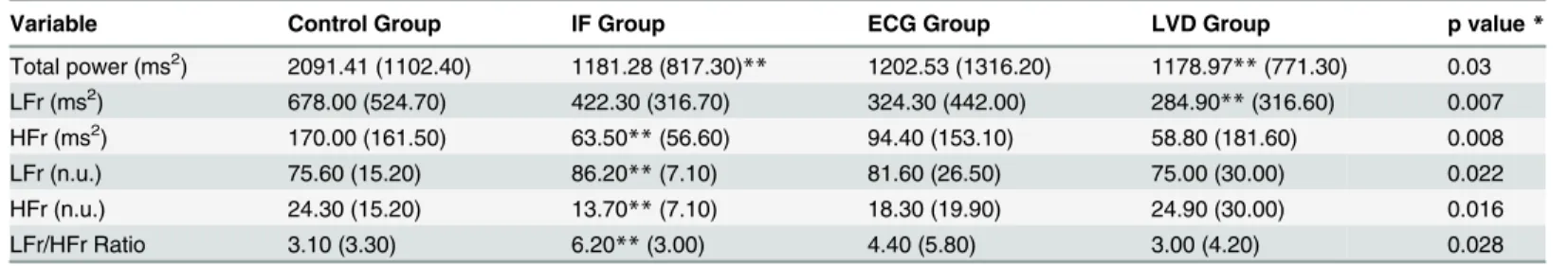
Table 3. Evaluation of autonomic nervous system indices using the 24-hour Holter.
Variable Control Group IF Group ECG Group LVD Group p value*
Total power (ms2) 2091.41 (1102.40) 1181.28 (817.30)** 1202.53 (1316.20) 1178.97**(771.30) 0.03
LFr (ms2) 678.00 (524.70) 422.30 (316.70) 324.30 (442.00) 284.90
**(316.60) 0.007
HFr (ms2) 170.00 (161.50) 63.50
**(56.60) 94.40 (153.10) 58.80 (181.60) 0.008
LFr (n.u.) 75.60 (15.20) 86.20**(7.10) 81.60 (26.50) 75.00 (30.00) 0.022
HFr (n.u.) 24.30 (15.20) 13.70**(7.10) 18.30 (19.90) 24.90 (30.00) 0.016
LFr/HFr Ratio 3.10 (3.30) 6.20**(3.00) 4.40 (5.80) 3.00 (4.20) 0.028
Values expressed as medians (interquartile range) *p values were calculated using Kruskal-Wallis test **p<0.05 compared to control group using Tukey test
LFr: low frequency; HFr: high frequency; ms2: milliseconds squared; n.u.: normalized units.
doi:10.1371/journal.pone.0131447.t003
Table 4. Evaluation of autonomic nervous system indices using the postural tilt test (supine position).
Variable Control Group IF Group ECG Group LVD Group p value*
Total power (ms2) 2182.99 (1308.00) 945.9 (1551.00) 646.37**(589.02) 1073.10 (2308.01) 0.002
LFr (ms2) 560.71 (364.42) 264.61
**(450.93) 104.75**(68.47) 178.46**(557.54) 0.001
HFr (ms2) 326.89 (178.71) 73.64
**(157.00) 72.60**(280.00) 362.60 (1005.11) <0.001
LFr (n.u.) 68.74 (12.35) 70.78 (21.18) 63.19 (18.26) 27.68**(37.57) 0.022
HFr (n.u.) 31.01 (11.61) 29.21 (21.18) 36.80 (18.26) 72.31**(39.36) 0.023
LFr/HFr Ratio 2.19 (1.36) 2.42 (2.42) 1.71 (1.10) 0.38**(1.10) <0.001
Values expressed as medians (interquartile range) *p values were calculated using Kruskal-Wallis test **p<0.05 compared to control group using Tukey test
LFr: low frequency; HFr: high frequency; ms2: milliseconds squared; n.u.: normalized units.
group had no significant difference in adiponectin levels when compared with control group.
This finding reinforces the good prognosis of IF form of Chagas’disease, similar to general
population [4]. We found higher levels of adiponectin in LVD and ECG groups, contrasting
with the reduced levels found in the studies using animal models of acute infection [21]. The
increase of adiponectin in the LVD group may be explained by the presence of systolic dysfunc-tion and emphasizing that this group had less fat mass, which can also increase the levels of
adi-ponectin.[25,26]
We should however draw attention to the increase of adiponectin in the ECG group. This group did not have a reduction in LVEF or clinical signs of HF and had similar weight, BMI, and fat mass than the IF and control groups. In the REDS-II study, a history of ECG abnormal-ities in asymptomatic chagasic patients was a prognostic factor for disease progression. The Table 5. Evaluation of autonomic nervous system indices using the postural tilt test (orthostatic position).
Variable Control Group IF Group ECG Group LVD Group p value*
Total power (ms2) 1891.20 (1325.30) 596.28**(558.82) 613.36**(896.44) 1001.77**(1122,88) 0.005
LFr (ms2) 666.73 (1018.90) 213.24
**(262.26) 260.53**(356.76) 84.65**(394,67) 0.009
HFr (ms2) 154.75 (157.22) 39.18
**(77.01) 98.02 (94.55) 329.95 (612,96) <0.001
LFr (n.u.) 83.21 (14.21) 82.37 (11.50) 77.80 (28.16) 35.97**(42.26) 0.007
HFr (n.u.) 16.78 (14.21) 17.62 (11.50) 22.19 (28.16) 64.02**(42.26) 0.006
LFr/HFr Ratio 4.95 (4.13) 4.67 (5.72) 3.50 (2.89) 0.56**(1.34) 0.019
Change LFr (n.u.) 21.58 (23.04) 14.14 (25.34) 8.26 (16.33) 4.93 (51.83)** 0,02
Change HFr (n.u.) -37.11 (33.97) -42.96 (46.60) -25.80 (25.63) -9,85 (18.18)** <0,001
Values expressed as medians (interquartile range) *p values were calculated using Kruskal-Wallis test **p<0.05 compared to control group using Tukey test
LFr: low frequency; HFr: high frequency; ms2: milliseconds squared; n.u.: normalized units.; Change LFr and HFr (n.u.): Change of each component from
rest to orthostatic position in normalized units
doi:10.1371/journal.pone.0131447.t005
Table 6. Spearman rank correlation coefficients of adiponectin with autonomic nervous system indices and inflammatory cytokines.
Variable IF group ECG group LVD group
LFr (n.u.) in 24-hour Holter 0.114 (p = 0.685) - 0,539 (p = 0.038)* - 0.075 (p = 0.791)
HFr (n.u.) in 24-hour Holter 0.079 (p = 0.781) 0.539 (p = 0.038)* 0.075 (p = 0.791)
LFr/HFr Ratio in 24-hour Holter - 0.114 (p = 0.685) - 0.518 (p = 0.048)* - 0.075 (p = 0.791) LFr (n.u.) in postural tilt test (supine position) 0.318 (p = 0.248) - 0.275 (p = 0.321) 0.339 (p = 0.216) HFr (n.u.) in postural tilt test (supine position) - 0.318 (p = 0.248) 0.275 (p = 0.321) - 0.364 (p = 0.182) LFr/HFr Ratio in postural tilt test (supine position) 0.318 (p = 0.248) - 0.293 (p = 0.289) 0.364 (p = 0.182) LFr (n.u.) in postural tilt test (orthostatic position) 0.100 (p = 0.723) 0.054 (p = 0.850) 0.300 (p = 0.277) HFr (n.u.) in postural tilt test (orthostatic position) - 0.100 (p = 0.723) - 0.054 (p = 0.850) - 0.300 (p = 0.277) LFr/HFr Ratio in postural tilt test (orthostatic position) 0.100 (p = 0.723) 0.054 (p = 0.850) 0.304 (p = 0.271)
Interleukin-6 - 0.061 (p = 0.830) - 0.496 (p = 0.329) - 0.003 (p = 0.934)
TNF-alpha 0.383 (p = 0.349) - 0.500 (p = 0.667) 0.429 (p = 397)
*P value<0,05
HFr: high frequency; LFr: low frequency; n.u.: normalized units; TNF-alpha: Tumor Necrosis Factor-alpha
There were significant correlations between leptin with some ANS assessment indexes in LVD group. Leptin level is associated positively with the LFr component and negatively with the HFr component in 24-hour Holter. No correlations between leptin and inflammatory cytokines were found. The values of correlations of leptin are shown inTable 7.
authors observed a moderate rate of progression to cardiomyopathy with systolic dysfunction
and HF in patients with ECG abnormalities (1.85% per year) [27].
Although the electrocardiogram is an established marker of severity of Chagas’disease, the
increased adiponectin levels found in patients with ECG abnormalities without systolic dys-function suggest that adiponectin may help to identify patients at higher risk of developing sys-tolic HF in this group.
It is not well established where adiponectin is predominantly secreted in HF patients and if its increased levels are only a compensation mechanism. Adiponectin is synthetized almost exclusively by adipocytes and its expression seems to be regulated by distinct signaling
path-ways [12]. Some studies suggest that adiponectin is over expressed in adipocytes from adipose
tissue of systolic HF patients [28,29]. Additionally, other studies indicate the presence of a
car-diac adiponectin system with independent regulation and with dysfunction in HF. Adiponectin is released from the cardiomyocytes into the peripheral circulation in proportion to extent of
systolic dysfunction irrespective of etiologies of HF [30,31]. Moreover, Nagajyothi et al. has
shown that adiponectin is considerably produced in the heart during Chagas’acute infection.
However, it is not well established the mechanism by which this occurs and the significance of
elevated adiponectin levels in the heart during acute and chronic Chagas’infection [32].
Many mechanisms have been proposed for the increase of adiponectin in systolic HF of any cause. The patients with cardiac cachexia showed higher adiponectin levels than the patients with systolic HF without cachexia. It is still not clear if the increased adiponectin levels in these
patients are a cause or effect of cachexia [25,26]. Bobbert et al. demonstrated that patients
with inflammatory cardiomyopathy have higher levels of adiponectin than patients with non-inflammatory cardiomyopathy and that, between the patients with non-inflammatory cardiomyop-athy, those that have higher levels of adiponectin had better outcomes. These data suggest that the increase of adiponectin could act as a compensation mechanism due its anti-inflammatory
action [33]. Other mechanisms proposed for the increase of adiponectin in systolic HF are a
compensatory effect for the resistance to insulin found in HF or a mechanism of resistance to
the action of adiponectin [34].
However, among the variables assessed in our study, the increased levels of adiponectin
was associated with ANS function in patients with early phase of Chagas’heart disease. There
was a negative association with the sympathetic branch and a positive association with the Table 7. Spearman rank correlation coefficients of leptin with autonomic nervous system indices and inflammatory cytokines.
Variable IF group ECG group LVD group
LFr (n.u.) in 24-hour Holter 0,396 (p = 0.143) - 0,182 (p = 0.869) 0.632 (p = 0.011)*
HFr (n.u.) in 24-hour Holter - 0.414 (p = 0.125) - 0.046 (p = 0.516) - 0.632 (p = 0.011)* LFr/HFr Ratio in 24-hour Holter 0.414 (p = 0.125) - 0.034 (p = 0.904) 0.632 (p = 0.011)* LFr (n.u.) in postural tilt test (supine position) - 0.236 (p = 0.398) 0.404 (p = 0.136) 0.329 (p = 0.232) HFr (n.u.) in postural tilt test (supine position) 0.236 (p = 0.398) - 0.404 (p = 0.136) - 0.361 (p = 0.187) LFr/HFr Ratio in postural tilt test (supine position) - 0.236 (p = 0.398) 0.400 (p = 0.140) 0.361 (p = 0.187) LFr (n.u.) in postural tilt test (orthostatic position) 0.068 (p = 0.810) 0.200 (p = 0.475) 0.168 (p = 0.550) HFr (n.u.) in postural tilt test (orthostatic position) - 0.068 (p = 0.810) - 0.200 (p = 0.475) - 0.168 (p = 0.550) LFr/HFr Ratio in postural tilt test (orthostatic position) 0.068 (p = 0.810) 0.200 (p = 0.475) 0.154 (p = 0.584)
Interleukin-6 0.118 (p = 0.676) - 0.371 (p = 0.468) - 0.214 (p = 0.467)
TNF-alpha 0.395 (p = 0.333) - 0.500 (p = 0.667) - 0.261 (p = 0.111)
*P value<0,05
HFr: high frequency; LFr: low frequency; n.u.: normalized units; TNF-alpha: Tumor Necrosis Factor-alpha
parasympathetic branch of ANS in ECG group. Our study does not allow conclusions on casual inference or the effect of one variable over the other. However, previous publications described possible pathways for this association.
Other studies, in different models, demonstrate the same inverse association between adipo-nectin and sympathetic activity found in our study. Studies in animals and humans, with cold exposure as a way to activate the sympathetic nervous system, showed reduced levels of
adipo-nectin [35,36]. An association has also been reported between hypoadiponectinemia and
sym-pathetic hyperactivity in patients with diabetes mellitus and obstructive sleep apnea [37–40].
This interaction between adiponectin and the sympathetic nervous system seems to be in a two-way. A study carried out by Tanida et al. showed that the intravenous infusion of
adipo-nectin reduced renal sympathetic nerve activity in animal models [41]. On the other hand,in
vitroandin vivostudies conducted using beta-adrenergic agonists have shown decreased expression and secretion of adiponectin through a direct inhibitory effect on the adipocytes [42,43].
Adipose tissue is predominantly innervated by the sympathetic nervous system [44,45]. In
our study, we can speculate that the peripheral impairment of the sympathetic nervous system in chagasic patients could lead to a reduction in sympathetic activity in the adipose tissue and increase the secretion of adiponectin. This mechanism can be explained in two ways. The first mechanism is that the impairment of the sympathetic system could lead to the direct loss of the inhibitory effect of the adrenergic tone on the expression and secretion of adiponectin by the
adipocytes [42,43]. On the other hand, the main action of the sympathetic nervous system on
the adipose tissue is to stimulate lipolysis with increased production of free fatty acids [44,45].
The accumulation of free fatty acids in the adipose tissue reduces the secretion of adiponectin
[46,47]. Therefore, the reduction of sympathetic activity in the adipose tissue could reduce
lipolysis and reduce the production of free fatty acids, leading to increased levels of adiponectin.
With regard to others metabolic parameters, insulin, in the majority of studies, demonstrate
a reduction of its levels or a compromise of its function in Chagas’disease patients [48–50].
The data on leptin levels from previous studies vary in heart failure patients. Some studies have
shown raised levels, especially in more advanced functional classes [17,51], and other studies
have demonstrated reduced levels, particularly in patients with cachexia [18,52]. In patients
with Chagas’disease, we have previously found reduced levels in patients with HF when
com-pared with a control group and other forms of CD [19]. In the present study, we did not detect
significant differences in the levels of insulin and leptin between the different groups.
However, we observed a positive association between leptin levels and sympathetic activity
in LVD group. Leptin’s relationship with the sympathetic component of the ANS is two-way,
with increased secretion of leptin leading to increased sympathetic activity, and sympathetic
activity inhibiting the synthesis of leptin [53,54]. Paolisso et al. described, in healthy subjects,
the same positive association between leptin levels and sympathetic activity found in our study
[55]. Other studies demonstrated that acute and chronic increase in plasma leptin levels
trig-gers a remarkable increase in sympathetic activity in various organs including the adipose tis-sue [56,57].
With regard to associations observed in our study, we have to address that the small sample size, although homogeneous, may have reduced the power to detect other associations.
Our findings in ANS study were also observed in others publications. Autonomic involve-ment is a well-establishhed feature of CD, in which anatomic and functional abnormalities have been extensively described. The majority of studies observed parasympathetic dysautono-mia preceding left ventricular systolic dysfunction and a progressive involvement of
In our study, we observed HFr indexes in LVD group equivalent or higher when compared with control group. We did not interpret these findings as a normal vagal function in patients with systolic dysfunction. The reciprocal relationship or push-pull organization of LFr and HFr components of HRV, which can be better appreciated by using a normalization procedure, shows a view of a predominance of an ANS branch over the other, named as sympatho-vagal
balance [20]. So, our study suggests that there was a greater impairment of sympathetic
system and a predominance of parasympathetic activity in LVD group, although there was involvement of both. Another reason for this finding is that, for ethical reasons, we chose not to suspend the use of prescribed medications. So all patients in the LVD group were using beta-blockers, which can increase cardiac vagal modulation and lower sympathetic activity,
increasing HFr indexes [59].
Another issue to be discussed is the controversy about the use of LFr component as a marker of sympathetic modulation. Some authors consider that this component is a parameter that
includes both sympathetic, vagal and baroreflex influences, especially in rest [60]. We used the
concept of the international Task Force document of Heart Rate Variability which considers LFr component (especially when expressing it in normalized units) as an index of sympathetic
modulation [20].
Conclusions
In conclusion, we found increased adiponectin levels in Chagas’heart disease with and without
significant systolic dysfunction. Furthermore, adipocytokines levels were associated with some ANS parameters. Adiponectin levels were positively associated with HFr component and inversely associated with LFr component in ECG group and leptin levels were inversely associ-ated with HFr component and positively associassoci-ated with LFr component of HRV in LVD group.
This is the first study to investigate the influence of cardiac autonomic function, as
esti-mated by HRV, on serum adipocytokines in chagasic patients. We used Chagas’disease as a
model in which autonomic dysfunction influences adipocytokine levels, which are important metabolic parameters in cardiovascular function. A better understanding of the relationship between autonomic and adipose tissue functions can lead to further evaluation for determining which individuals are at the highest risk for cardiovascular disease and mortality, and can also lead to the development of new therapies for reducing that risk.
Author Contributions
Conceived and designed the experiments: JMB FF HFL BMI FJAR CM. Performed the experi-ments: JMB FF VMCS CJG DTH. Analyzed the data: JMB FF HFL BMI FJAR. Contributed reagents/materials/analysis tools: JMB FF VMCS CJG DTH. Wrote the paper: JMB FF HFL FJAR CM.
References
1. World Health Organization (2012) Research priorities for Chagas Disease, Human African Trypanoso-miasis and Leishmaniasis: Technical Report of the TDR Disease Reference Group on Chagas Dis-ease, Human African Trypanosomiasis and Leishmaniasis. Geneva, Switzerland: WHO. 2. Gascon J, Bern C, Pinazo MJ (2010) Chagas disease in Spain, the United States and other
non-endemic countries. Acta Tropica 115:22–7. doi:10.1016/j.actatropica.2009.07.019PMID:19646412
4. Ianni BM, Arteaga E, Frimm CC, Barretto ACO, Mady C (2001) Chagas’heart disease: Evolutive evalu-ation of eletrocardiographic and echocardiographic parameters in patients with the indeterminate form. Arq Bras Cardiol 77:59–62. PMID:11500748
5. Marin-Neto JA, Cunha-Neto E, Maciel BC, Simões MV (2007) Pathogenesis of chronic Chagas Heart Disease. Circulation 115:1109–23. PMID:17339569
6. Dávila DF, Inglessis G, Dávila CAM (1998) Chagas’heart disease and the autonomic nervous system. Int J Cardiol 66:123–127. PMID:9829322
7. Ribeiro ALP, Moraes RS, Ribeiro JP, Ferlin EL, Torres RM, Oliveira EO et al. (2001) Parasympathetic dysautonomia precedes left ventricular systolic dysfunction in Chagas disease. Am Heart J 141:260–
5. PMID:11174350
8. Molina RBG, Matsubara BB, Hueb JC, Zanati SG, Meira DA, Cassolato JL et al. (2006) Dysautonomia and ventricular dysfunction in the indeterminate form of Chagas disease. Int J Cardiol 113:188–93. PMID:16376440
9. Doehner W, Frenneaux M, Anker SD (2014) Metabolic impairment in heart failure: the myocardial and systemic perspective. J Am Coll Cardiol 64:1388–1400. doi:10.1016/j.jacc.2014.04.083PMID:
25257642
10. Lopes HF, Egam BM (2006) Autonomic dysregulation and the metabolic syndrome: Pathologic partners in an emerging global pandemic 87:538–47. PMID:17128327
11. Wajchenberg BL, Nery M, Cunha MR, Silva MER (2009) Adipose tissue at the crossroads in the devel-opment of the metabolic syndrom, inflammation and atherosclerosis. Arq Bras Endocrinol Metab 53:145–50.
12. Antoniades C, Antonopoulos AS, Tousoulis D, Stefanadis C (2009) Adiponectin: from obesity to cardio-vascular disease. Obesity Reviews 10:269–79. doi:10.1111/j.1467-789X.2009.00571.xPMID:
19389061
13. Guzik TJ, Mangalat D, Korbut R (2006) Adipocytokines- Novel link between inflamation and vascular function? J Physiol and Pharmacol 57:505–28.
14. Berg AH, Scherer PE (2005) Adipose tissue, inflamation and cardiovascular disease. Circulation Res 96:939–49. PMID:15890981
15. George J, Patal S, Wexler D, Sharabi Y, Pelg E, Kamari Y et al. (2006) Circulating adiponectin concen-trations in patients with congestive heart failure. Heart 92:1420–4. PMID:16621874
16. Dieplinger B, Gegenhuber A, Poelz W, Haltmayer M, Mueller T (2009) Prognostic value of increased adiponectin plasma concentrations in patients with acute destabilized heart failure. Clin Biochem 42:1190–3. doi:10.1016/j.clinbiochem.2009.02.023PMID:19272369
17. Schulze PC, Kratzch J, Linke A, Schoene N, Adams V, Gielen S et al (2003) Elevated serum levels of leptin and soluble leptin receptor in patients with chronic heart failure. Eur J Heart Fail 5:33–40. PMID:
12559213
18. Filippatos GS, Tsilias K, Venetsanou K, Karambinos E, Manolatos D, Kranidis A et al. (2000) Leptin serum levels in cachetic heart failure patients. Relationship with tumor necrosis factor-alpha system. Int J Cardiol 76:117–22. PMID:11104865
19. Fernandes F, Dantas S, Ianni BM, Ramires FJA, Buck P, Salemi VMC et al. (2007) Leptin levels in dif-ferent forms of Chagas’disease. Braz J Med Biol Res 40:1631–6. PMID:17713658
20. European Society of Cardiology (1996) Task force of the European Society of Cardiology and the North American Society of Pacing and Electrophysiology. Heart rate variability: standards of measurement, physiological, interpretation and clinical use. Circulation 93:1043–65. PMID:8598068
21. Coombs TP, Mukherjee Salmeida CJG, Jelicks LA, Schubert W, Lin Y, Jayabalan DS et al (2005) The adipocyte as an important target cell for infection withTrypanosoma cruzi. J Biol Chem 280:24085–94. PMID:15843370
22. Nagajyothi F, Desruisseaux MS, Thiruvur N, Weiss LM, Braunstein VL, Albanese C et al. (2008) Trypa-nosoma cruziinfection of cultured adipocytes results in an inflamattory phenotype. Obesity 16: 1992–
1997. PMID:19186325
23. Ferreira AVM, Segatto M, Menezes Z, Macedo AM, Gelape C, Andrade LO et al. (2011) Evidence for Trypanosoma cruzi in adipose tissue in human chronic Chagas disease. Microbes Infect 13:1002–5. doi:10.1016/j.micinf.2011.06.002PMID:21726660
24. Fu M, Zhou J, Qian J, Jin X, Zhu H, Zhong C et al. (2012) Adiponectin through its biphasic serum is a useful biomarker during transition from diastolic dysfunction to systolic dysfunction- an experimental study. Lipids Health Dis 11:106–15. doi:10.1186/1476-511X-11-106PMID:22935137
26. Araújo JP, Lourenço P, Rocha-Gonçalves F, Ferreira A, Bettencourt P (2009) Adiponectin is increased in cardiac cachexia irrespective of body mass index. Eur J Heart Fail 11:567–72. doi:10.1093/eurjhf/ hfp046PMID:19359328
27. Sabino EC, Ribeiro AL, Salemi VM, Di Lorenzo Oliveira C, Antunes AP, Menezes MM et al. (2013) Ten years incidence of Chagas cardiomyopathy among asymptomatic Trypanosoma cruzi-seropositive for-mer blood donors. Circulation 127:1105–15. doi:10.1161/CIRCULATIONAHA.112.123612PMID:
23393012
28. Tsukamoto O, Fujita M, Kato M, Yamazaki S, Asano Y, Ogai A et al. (2009) Natriuretic peptides enhance the production of adiponectin in human adipocytes and in patients with chronic heart failure. J Am Coll Cardiol 53:2070–7. doi:10.1016/j.jacc.2009.02.038PMID:19477358
29. Khan RS, Kato TS, Chokshi A, Chew M, Yu S, Wu C et al. (2012) Adipose tissue inflammation and adi-ponectin resistance in patients with advanced heart failure: Correction after ventricular assist device implantation. Circ Heart Fail 5:340–8. doi:10.1161/CIRCHEARTFAILURE.111.964031PMID:
22379072
30. Skurk C, Wittchen F, Suckau L, Witt H, Noutsias M, Fechner H et al. (2008) Description of a local car-diac adiponectin system and its deregulation in dilated cardiomyopathy. Eur Heart J 29:1168–80. doi:
10.1093/eurheartj/ehn136PMID:18390538
31. Takano H, Obata J, Kodama Y, Kitta Y, Nakamura T, Mende A et al.. (2009) Adipnectin is released from the heart in patients with heart failure. Int J Cardiol 132:221–6. doi:10.1016/j.ijcard.2007.11.040
PMID:18192035
32. Nagajhioti F,Weiss LM, Zhao D, Koba W, Jelicks LA, Cui M et al. (2014) High fat diet modulates Trypa-nosoma cruziinfection associated myocarditis. PLoS Negl Trop Dis 8:e3118. doi:10.1371/journal. pntd.0003118PMID:25275627
33. Bobbert P, Scheibenbogen C, Jenke A, Kania G, Wilk S, Krohn S et al. (2011) Adiponectin expression in patients with inflammatory cardiomyopathy indicates favourable outcome and inflammation control. Eur Heart J 32:1134–47. doi:10.1093/eurheartj/ehq498PMID:21278397
34. Berendoncks AMV, Conraads VM (2011) Functional adiponectin resistance and exercise intolerance in heart failure. Curr Heart Fail Rep 8:113–22. doi:10.1007/s11897-011-0056-6PMID:21424675
35. Imai J, Katagiri H, Yamada T, Ishigaki Y, Ogihara T, Uno K et al. (2006) Cold exposure supresses serum adiponectin levels through sympathetic nerve activation in mice. Obesity 14:1132–41. PMID:
16899794
36. Iwen KA, Wenzel ET, Ott V, Perwitz N, Wellhöner P, Lehnert H et al. (2011) Cold-induced alteration of adipokine profile in humans. Metabolism 60:430–7. doi:10.1016/j.metabol.2010.03.011PMID:
20423746
37. Wakabayashi S, Aso Y (2004) Adiponectin concentrations in sera from patients with type 2 diabetes are negatively associated with sympathovagal balance as evaluated by power spectral analysis of heart rate variation. Diabetes Care 27:2392–7. PMID:15451906
38. Takahashi N, Anan F, Nakagawa M, Yufu K, Shinohara T, Tsubone T et al. (2007) Hypoadiponectine-mia in type 2 diabetes mellitus in men is associated with overactity as evaluated by cardiac 123I-metaiodobenzylguanidine scintigraphy. Metabolism 56:919–24. PMID:17570253
39. Lieb DC, Parson HK, Mamikunian G, Vinik AI (2012) Cardiac autonomic imbalance in newly diagonsed and established diabetes is associated with markers of adipose tissue inflammation. Exp Diabetes Res 2012:1–7.
40. Lam JC, Xu A, Tam S, Khong PI, Yao TJ, Lam DC et al. (2008) Hypoadiponectinemia is related to sym-pathetic activation and severity of obstructive sleep apnea. Sleep 31:1721–7. PMID:19090328
41. Tanida MT, Shen J, Horii Y, Matsuda M, Kihara S, Funahashi T et al. (2007) Effects of adiponectin on the renal sympathetic nerve acitivity and blood pressure in rats. Exp Biol Med 232:390–7.
42. Fasshauer M, Klein J, Neumann S, Eszlinger M, Paschchke R (2001) Adiponectin gene expression is inhibited by ß-adrenergic stimulation via protein kinase A in 3T3-L1 adipocytes. FEBS 507:142–6. 43. Delporte ML, Funahashi T, Takahashi M, Matsuzawa Y, Brichard SM (2002) Pre- and post-translational
negative effect of ß-adrenoreceptor agonists on adiponectin secretion: in vitro and in vivo studies. Bio-chem J 367:677–85. PMID:12139486
44. Bartness TJ, Song CK (2007) Sympathetic and sensory innervation of white adipose tissue. J Lipid Res 48:1655–72. PMID:17460327
45. Bartness TJ, Shrestha YB, Vaughan CH, Schwartz GJ, Song CK (2010) Sensory and sympathetic ner-vous system control of white adipose tissue. Mol Cell Endocrinol 318–343.
47. Moro C, Klimcakova E, Lolmede K, Berlan M, Lafontan M, Stich V et al. (2007) Atrial natriuretic peptides inhibits the production of adipokines and citokines linked to inflammation and insulin resistance in human subcutaneous adipose tissue. Diabetologia 50:1038–47. PMID:17318625
48. Oliveira LCM, Juliano Y, Novo NF, Neves MM (1993) Blood glucose and insulin response to intrave-nous glucose by patients with chronic Chagas’Disease and alcoholism. Braz J Med Biol Res 26:1187–
90. PMID:8136719
49. Guariento ME, Saad MJA, Muscelli EOA, Gontijo JAR (1993) Heterogeneous insulin response to an oral glucose load by patients with indeterminate clinical form of Chagas’disease. Braz J Med Biol Res 26:491–5. PMID:8257938
50. Nagajyothi F, Kuliawat R, Kusminski CM, Machado FS, Desruisseaux MS, Zhao D et al. (2013) Alter-ations in glucose homeostasis in a murine model of Chagas disease. Am J Pathol 182:886–94. doi:10. 1016/j.ajpath.2012.11.027PMID:23321322
51. Schulze PC, Biolo A, Gopal D, Shahzad K, Balog J, Fish M et al.(2011) Dynamics in insulin resistance and plasma levels of adipokines in patients with acute descompensated and chronic stable heart fail-ure. J Cardiac Fail 17:1004–11.
52. Murdoch DR, Rooney E, Dargie HJ, Shapiro D, Morton JJ, McMurray JJV (1999) Inappropriately low plasma leptin concentration in the cachexia associated with chronic heart failure. Heart 82:352–6. PMID:10455089
53. Barbosa-Ferreira JM, Fernandes F, Dabarian A, Mady C (2013) Leptin in heart failure. Expert Opin Med Diagn 7:113–7. doi:10.1517/17530059.2013.735229PMID:23530847
54. Trayburn P, Duncan JS, Hoggard N, Rayner DV (1998) Regulation of leptin production: a dominant role for the sympathetic nervous system?. Proc Nutr Soc 57:413–9 PMID:9793999
55. Paolisso G, Manzella D, Montano N, Gambardella A, Varricchio M (2000) Plasma Leptin concentrations and cardiac autonomic nervous system in healthy subjects with different body weights. J Clin Endocri-nol Metab 85:1810–4. PMID:10843157
56. Grassi G, Elam M (2002) Leptin, sympathetic and baroreflex function: another step on the road to sym-pathetic differention. J Hypertens 20:1487–9. PMID:12172307
57. Hausberg M, Morgan DA, Chapleau MA, Sivitz WI, Mark AL, Haynes WG (2002) Differential modulation of leptin-induced sympathoexcitation by baroreflex activation. J Hypertens 20:1633–41. PMID:
12172326
58. Simões MV, Pintya AO, Bromberg-Marin G, Sarabanda AV, Antloga CM, Pazin-Filho A et al. (2000) Relation of regional sympathetic denervation and myocardial perfusion disturbance to wall motion impairment in Chagas’cardiomyopathy. Am J Cardiol 86:975–981. PMID:11053710
59. Bloom HL, Vinik AI, Colombo J (2014) Differential effects of adrenergic antagonists (carvedilol vs meto-prolol) on parasympathetic and sympathetic activity: a comparison of clinical results. Heart Int 9:15–21. 60. Goldstein DS, Bentho O, Park M, Sharabi Y (2011) LF power of heart rate variability is not a measure of