Article
J. Braz. Chem. Soc., Vol. 24, No. 4, 601-608, 2013. Printed in Brazil - ©2013 Sociedade Brasileira de Química 0103 - 5053 $6.00+0.00
A
*e-mail: [email protected]
Energy Transfer Processes in Tb(III)-Dibenzoylmethanate Complexes with
Phosphine Oxide Ligands
Francisco A. Silva Jr.,a Helenise A. Nascimento,a Dariston K. S. Pereira,a
Ercules E. S. Teotonio,*,a Hermi F. Brito,b Maria Cláudia F. C. Felinto,c
José Geraldo P. Espínola,a Gilberto F. Sád and Wagner M. Faustinoa
aDepartamento de Química, Universidade Federal da Paraíba, 58051-970 João Pessoa-PB, Brazil
bDepartamento de Química Fundamental, Instituto de Química, Universidade de São Paulo,
05508-900 São Paulo-SP, Brazil
cInstituto de Pesquisas Energéticas e Nucleares, Av. Prof Lineu Prestes, 2242,
Cidade Universitária, 05508-000 São Paulo-SP, Brazil
dDepartamento de Química Fundamental, Centro de Ciências Exatas e da Natureza (CCEN),
Universidade Federal de Pernambuco, 50670-90 Recife-PE, Brazil
Este trabalho relata a síntese, a caracterização e as propriedades luminescentes dos complexos de fórmulas [Tb(DBM)3L], [Tb(DBM)2(NO3)L2)] e [Tb(DBM)(NO3)2(HMPA)2)] (DBM = dibenzoilmetanato; L: TPPO = óxido de trifenilfosfina ou HMPA = óxido de hexametilfosforamida). Os compostos foram caracterizados por análise elementar (CHN), titulação complexométrica com EDTA e espectroscopia no infravermelho com transformada de Fourier (FTIR), e as propriedades de fotoluminescência foram avaliadas. As energias dos estados tripletos do ligante DBM foram determinadas experimentalmente a partir dos espectros de fosforescência resolvidos no tempo dos compostos análogos do íon Gd3+. As energias aumentam em função do número de ânions nitratos que substituem o ligante DBM nos complexos. Ademais, os espectros de luminescência e os tempos de vida dos níveis emissores revelaram que a eficiência de transferência de energia ligante-metal segue a mesma tendência. Ao contrário dos complexos tris-DBM, o bis- e o mono-DBM apresentaram elevada intensidade de luminescência, sendo candidatos promissores para camadas emissoras de luz em dispositivos moleculares conversores de luz (LCMD).
T h e T b3 +-β- d i ke t o n a t e c o m p l exe s [ T b ( D B M )
3L ] , [ T b ( D B M )2( N O3) L2] a n d [Tb(DBM)(NO3)2(HMPA)2] (DBM = dibenzoylmethanate; L: TPPO = triphenylphosphine oxide or HMPA = hexamethylphosphine oxide) were prepared and characterized by elemental analysis (CHN), complexometric titration with EDTA and Fourier transform infrared (FTIR) spectroscopy, and the photoluminescence properties evaluated. The triplet state energies of the coordinated DBM ligands were determined using time-resolved phosphorescence spectra of analogous Gd3+ complexes. The results show that the energies increase along with the number of coordinated nitrate anions replacing the DBM ligand in the complexes. The luminescence spectra and emission lifetime measurements revealed that the ligand-to-metal energy transfer efficiency follows the same tendency. Unlike the tris-DBM complexes, bis- and mono-DBM presented high luminescence, and may act as promising candidates for preparation of the emitting layer of light converting molecular devices (LCMDs).
Keywords: terbium(III), β-diketonate, phosphine oxide, energy transfer
Introduction
Several transition metal and lanthanide β-diketonate complexes have been reported in the literature.1-4 In
lanthanide compounds, the coordinated ligands play various roles that permit obtaining complexes with high
luminescence quantum yields. They can (i) relax the
Laport’s parity selection rule;5-7 (ii) protect the emitting
intraconfigurational-4f electronic transitions (ε ca. 0.01 to 3 L mol−1 cm−1), since the excited organic ligands can
transfer energy to a suitable excited energy level of the lanthanide ion.7-9
It is easy to find Ln3+-β-diketonate complexes
exhibiting quantum yields above 70% in the literature,2
in which the efficiency of the β-diketonate-to-Ln3+
intra-molecular energy transfer process is largely dependent on the energy difference between the donor state of the ligand and the acceptor state of the Ln3+ ion (∆E).10,11 The
more operative intra-molecular ligand-to-metal energy transfer is observed in compounds presenting ligand donor states above the lanthanide acceptor energy levels,
minimizing the back energy-transfer processes.1,10-12
Furthermore, β-diketonate ligands generally have their donor states localized on the chelating ring, shortening the donor-acceptor distance, RL, and thus enhancing the
luminescence quantum yield.1,12
After the pioneering study by Weissman,13 a great
number of authors have been concerned with elucidating the details of intra-molecular energy-transfer for different lanthanide coordination compounds, particularly Eu3+ and
Tb3+ ones.14-20 The main emitting level of the Tb3+
ion, 5D
4, is approximately 3250 cm−
1 above the main
emitting level of the Eu3+ ion, 5D
0. Therefore, a single
β-diketonate ligand is often not optimum to sensitize both ions, since the triplet states are not resonant for both of them.17
Energy levels of β-diketonate ligands remain essentially the same in tris and tetrakis-complexes such that the ligand-to-metal intra-molecular energy transfers in these complexes are very similar. In the absence of additional luminescence quenching routes, the luminescence quantum yields of tris and tetrakis-complexes are similar suggesting that they are independent of the number of coordinated β-diketonate ligands.15-17,21 However, recently
our group reported that the luminescence of the complex [Tb(TTA)2NO3(TPPO)] is much higher than in its tris and
tetrakis analogues.22-25
In this work, our group extends the investigation to
themono-, bis- and tris-DBM complexes, [Ln(DBM)3L],
[Ln(DBM)2(NO3)L2)] and [Ln(DBM)(NO3)2L2)] (DBM =
dibenzoylmethanate; L: TPPO = triphenylphosphine oxide or HMPA = hexamethylphosphine oxide; Ln = Tb3+ and Gd3+), and the study of the dependence of Tb3+
luminescence properties as a function of the number of coordinated β-diketonate ligands. The corresponding Gd3+
complexes were used to mimic the Tb3+ complexes and
determine the excited state energies of the coordinated ligand.
Experimental
Reagents and syntheses
Terbium oxide (Tb4O7), dibenzoylmethane, phosphine
oxide ligands (TPPO and HMPA) as well as the solvents (ethanol and acetone) were purchased from Aldrich Co., and used without any previous treatment. Terbium chloride and nitrate were synthesized as described in the literature.26 The terbium and gadolinium complexes were
synthesized in the same way; and the preparation of terbium complexes are given as representative.
Syntheses of the tris-diketonate complexes
Tb(DBM)3L (L = TPPO or HMPA)
To an ethanol solution containing 1.00 g (4.46 mmol) of DBM, and 0.41g (1.49 mmol) of TPPO (or 0.29 g of HMPA), a solution of 0.56 g (1.49 mmol) of terbium chloride in 30 mL of ethanol was added dropwise, under stirring. The pH value of the resulting solution was then adjusted to approximately 6.0 using NaOH (0.01 mol L−1)
in a 50/50 water/ethanol mixture. The resulting yellow solid was filtered, washed with ethanol and dried under vacuum.
Tb(DBM)3(TPPO): yield 1.50 g (90.8%); C63H48TbO7P:
calc. C 68.36, H 4.37, Tb 14.36; found C 68.15, H 4.52, Tb 13.97; FTIR (KBr) ν/cm−1 3055 (w), 2923 (w), 1597 (s),
1550 (s), 1519 (s), 1477 (s), 1454 (m), 1404 (s), 1311 (m), 1219 (w), 1168 (s), 1118 (s), 1091 (m), 1068 (w), 1026 (m), 999 (w), 941 (w), 852 (w), 817 (w), 783 (w), 744 (m), 721 (s), 690 (s), 609 (m), 540 (s).
T b ( D B M )3( H M PA ) : y i e l d 1 . 3 0 g ( 8 7 . 5 % ) ; C51H51TbN3O7P: calc. C 60.78, H 5.10, N 4.17, Tb 15.77;
found C 60.63, H 5.05, N 4.22, Tb 16.09; FTIR (KBr) ν/cm−1 3059 (w), 2927 (w), 1600 (s), 1550 (s), 1520 (s),
1477 (s), 1458 (m), 1411 (s), 1303 (m), 1220 (w), 1157 (s), 1068 (m), 1022 (w), 987 (s), 941 (w), 783 (w), 748 (s), 717 (s), 686 (m), 609 (m), 509 (w).
Gd(DBM)3(TPPO): yield 1.42 g (85.9%); C63H48GdO7P:
calc. C 68.46, H 4.38, Gd 14.23; found C 68.46, H 4.42, Gd 14.14; FTIR (KBr) ν/cm−1 3059 (w), 3024 (w), 1597
G d ( D B M )3( H M PA ) : y i e l d 1 . 3 8 g ( 9 1 . 7 % ) ; C51H51GdN3O7P: calc. C 60.48, H 5.11, N 4.18, Gd 15.63;
found C 60.53, H 5.15, N 4.02, Gd 15.58; FTIR (KBr) ν/cm−1 3059 (w), 2922 (w), 1597 (s), 1551 (s), 1519 (s),
1477 (s), 1458 (m), 1412 (s), 1304 (m), 1219 (w), 1180 (s), 1064 (m), 1022 (w), 988 (s), 941 (w), 783 (w), 748 (s), 718 (s), 687 (m), 609 (m), 509 (w).
Syntheses of the bis-diketonate complexes
[Tb(DBM)2(L)2(NO3)]
1.00 g (4.46 mmol) of DBM and 1.24 g (4.46 mmol) of TPPO (or 0.87 g of HMPA) were dissolved in 30 mL of ethanol and added dropwise under stirring to 30 mL of an ethanol solution containing 0.56 g (1.49 mmol) of terbium chloride. The pH value of the resulting mixture was adjusted to 1.0 with concentrated HNO3 solution, and then
to 6.0 using NaOH (0.01 mol L−1) in a 50/50 water/ethanol
mixture. The yellow solid was filtered, washed with ethanol and dried under vacuum.
[Tb(DBM)2(TPPO)2(NO3)]: yield 2.27 g (83.4%); C66H52TbNO9P2: calc. C 64.76, H 4.28, N 1.14, Tb 12.98;
found C 64.16, H 4.30 , N 1.84, Tb 13.26; FTIR (KBr) ν/cm−1 3055 (m), 2920 (w), 1597 (s), 1550 (s), 1520 (s),
1477 (s), 1454 (s), 1404 (s), 1311 (m), 1219 (w), 1165 (s), 1118 (s), 1092 (w), 1068 (w), 1026 (m), 999 (w), 941 (w), 852 (w), 817 (w), 783 (w), 744 (m), 721 (s), 690 (s), 609 (m), 540 (s).
[Tb(DBM)2(HMPA)2(NO3)]: yield 1.96 g (86.0%); C42H58TbN7O9P2: calc. C 49.18, H 5.70, N 9.56, Tb 15.49;
found C 49.72, H 5.90, N 8.76, Tb 15.52; FTIR (KBr) ν/cm−1 3059 (w), 2924 (m), 1597 (s), 1550 (s), 1516 (s),
1477 (s), 1458 (m), 1408 (s), 1300 (m), 1219 (w), 1161 (s), 1068 (m), 1026 (w), 987 (s), 941 (w), 783 (w), 744 (s), 721 (s), 686 (m), 609 (m), 509 (w), 478 (w).
[Gd(DBM)2(TPPO)2(NO3)]: yield 2.34 g (85.4%); C66H52GdNO9P2: calc. C 64.85, H 4.29, N 1.15, Gd 12.86;
found C 64.23, H 4.32 , N 1.62, Gd 12.61; FTIR (KBr) ν/cm−1 3055 (m), 2920 (w), 1593 (s), 1551 (s), 1520 (s),
1477 (s), 1453 (s), 1404 (s), 1312 (m), 1219 (w), 1168 (s), 1119 (s), 1092 (w), 1072 (w), 1026 (m), 999 (w), 941 (w), 852 (w), 814 (w), 783 (w), 745 (m), 721 (s), 691 (s), 609 (m), 540 (s), 447 (w).
[Gd(DBM)2(HMPA)2(NO3)]: yield 2.08 g (90.5%); C42H58GdN7O9P2: calc. C 49.26, H 5.71, N 9.57, Gd 15.35;
found C 49.12, H 5.66, N 9.67, Gd 15.93; FTIR (KBr) ν/cm−1 3059 (w), 2924 (m), 2897 (m), 2808 (m), 1597 (s),
1554 (s), 1516 (s), 1477 (s), 1454 (m), 1408 (s), 1304 (m), 1219 (w), 1192 (s), 1161(s), 1068 (m), 1026 (w), 987 (s), 941 (w), 818 (w), 783 (w), 748 (s), 721 (s), 687 (m), 610 (m), 505 (w), 478 (w).
Syntheses of the mono-diketonate complexes
[Tb(DBM)(HMPA)2(NO3)2]
Mono-dibenzoylmethanate complex was obtained only with HMPA as neutral ligand. The synthetic route for this complex was similar to that used for [Tb(DBM)2(HMPA)(NO3)], but using the 1:2:1 molar ratio
of DBM:HMPA:Tb3+. An ethanol solution (ca. 20 mL) of
terbium nitrate (1.66 g, 4.44 mmol) was added to 20 mL of an ethanol solution containing a mixture of 1.00 g (4.46 mmol) of DBM and 1.61 g (8.92 mmol) of HMPA. After ca. 24 h, the yellow single crystals were filtered, washed with ethanol and dried in a desiccator under vacuum.
[Tb(DBM)(HMPA)2(NO3)2]: yield 2.90 g (75.0%); C27H47TbN8O10P2: calc. C 37.51, H 5.48, N 12.96, Tb 18.38;
found C 37.28, H 5.31, N 10.48, Tb 18.32; FTIR (KBr) ν/cm−1 3062 (w), 2935 (m), 1597 (s), 1554 (s), 1523 (s),
1481 (s), 1384 (s), 1300 (s), 1230 (w), 1188 (s), 1165 (s), 1138 (s), 1068 (m), 1029 (w), 991 (s), 817 (w), 756 (s), 721 (m), 690 (w), 609 (w).
[Gd(DBM)(HMPA)2(NO3)2]: yield 3.15 g (81.4%); C27H47GdN8O10P2: calc. C 37.58, H 5.49, N 12.99, Gd 18.22;
found C 37.40, H 5.28, N 12.88, Gd 17.80; FTIR (KBr) ν/cm−1 3059 (w), 2997 (m), 2891 (m), 2854 (m), 2816 (m),
1601 (s), 1558 (s), 1524 (s), 1481 (s), 1458 (s), 1385 (s), 1300 (s), 1238 (w), 1161 (s), 1138 (s), 1061 (m), 1030 (w), 984 (s), 941 (w), 818 (w), 756 (s), 721 (m), 687 (w), 605 (w), 521 (w), 482 (w).
Apparatus
The elemental analyses of carbon, hydrogen and nitrogen in the tris-, bis- and mono-diketonate complexes were performed using a Perkin-Elmer model 2400
microanalyzer, whereas the Tb3+ ion contents were
determined by complexometric titration with EDTA.27
Infrared absorption spectra were recorded in the range of 400 up to 4000 cm−1 in KBr pellets using a Shimadzu FTIR
spectrophotometer model IRPRESTIGE-21.
source, and an R928P PMT photomultiplier as detector. All spectra were recorded using detector mode correction. The second-order diffraction of the source radiation was eliminated by using a cut-off filter. Time-resolved
luminescence spectra of the Gd3+-complexes and the
luminescence decay curves of the Tb3+-complexes were
recorded at 77 K using the same equipment, but operating in phosphorescence mode with a pulsed Xenon lamp as the excitation source. A time delay of 0.100 ms was applied. The luminescence instruments were fully controlled by the FluorEssence program. All luminescence data were obtained from samples contained in a 2 mm diameter quartz tube.
Results and Discussion
Characterization of the complexes
The elemental analysis (C, H and N) and the complexometric titration data indicated that complexes presenting the formulas [Ln(DBM)3L], [Ln(DBM)2(NO3)L2]
and [Ln(DBM)(NO3)2(HMPA)2] (Ln = Gd3+ and Tb3+;
DBM = dibenzoylmethanate; L: TPPO = triphenylphosphine oxide or HMPA = hexamethylphosphine oxide) were obtained. The tris- and bis-diketonate complexes of either HMPA or TPPO were easily obtained. However, it was not possible to obtain the mono-diketonate with TPPO, even after many attempts. This is probably due to the lower donor capacity and steric hindrance of TPPO when compared to HMPA, which allows coordination of more than one DBM ligand to the lanthanide ion.
The coordination modes of the dibenzoylmethanate, phosphine oxide and nitrate ligands were investigated based on their characteristic FTIR absorption bands (Figure S1 in the Supplementary Information (SI) section). The FTIR spectra exhibited strong bands at around 1600 cm−1
that might be assigned to ν(C=O) coupled with ν(C=C) of the DBM ligand. These bands are shifted to a lower wavenumber in comparison with the free ligand, indicating that DBM is coordinated to the metal ion in chelating
mode.28 The spectra also show strong bands around
1160 cm−1, which might be assigned to ν(P=O) of the
phosphine oxide ligands (TPPO and HMPA). These bands are also shifted to lower wavenumbers in comparison with the respective free ligands. Two absorption bands were also observed around 1180 and 1036 cm−1 which may be
assigned to νa(NO2) and νs(NO2) modes, indicating that the
NO3− is chelated and has C2v symmetry.28,29 Furthermore, the
two characteristic bands assigned to ν1 + ν4 combination
modes (1820 and 1767 cm−1) are separated about 55 cm−1,
reinforcing the implication that nitrate is coordinated as a bi-dentate ligand.28
Luminescent properties of the Tb3+-DBM complexes
According to the intra-molecular energy transfer mechanism suggested by the experimental data and theoretical models,1,12,14 the excited T
1 states of the ligands
play a critical role in defining the Ln3+ β-diketonate
complexes. In order to estimate these energy levels in the Tb3+ complexes, phosphorescence spectra of equivalent
Gd3+ complexes that do not present intraconfigurational-4f
transitions in the visible region were recorded.1
Figures 1a and 1b show the steady state emission spectra of the [Gd(DBM)3L], [Gd(DBM)2(NO3)L2] and
[Gd(DBM)(NO3)2(HMPA)2] complexes with excitation
at 370 nm. These spectra are characterized by one very low intensity broad band in the spectral range of 420-455 nm and the strongest bands are in the spectral range of 460-700 nm, which are assigned to the S1 → S0 and to
the T1 → S0 transitions of the DBM ligand, respectively.
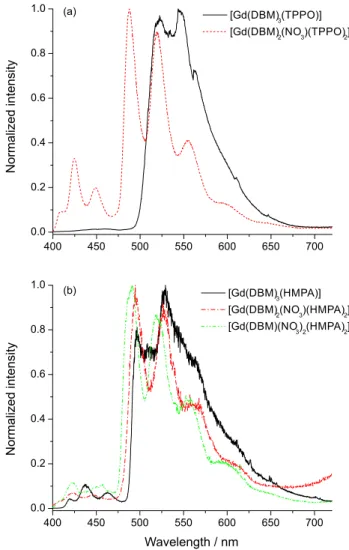
Figure 1. Steady state emission spectra of the tris-, bis- and mono-dibenzoylmethanate recorded at 77 K under excitation at 370 nm: (a) [Gd(DBM)3(TPPO)2] and [Gd(DBM)2(NO3)(TPPO)2], and (b) [Gd(DBM)3(HMPA)2], [Gd(DBM)2(NO3)(HMPA)2] and [Gd(DBM)
In order to determine the exact position of 0-0 phonon transitions, the time-resolved luminescence spectra (Figures 2a and 2b) were recorded using a time delay of 0.100 ms. As can be seen, these spectra present only those broad bands that may be attributed to the triplet to singlet transitions. The T1 state energies determined as the shortest
wavelength phosphorescence bands for the complexes are
[Gd(DBM)3(TPPO)] (20325 cm−
1), [Gd(DBM)
3(HMPA)]
(20660 cm−1), [Gd(DBM)
2(TPPO)2(NO3)] (21186 cm−1),
[ G d ( D B M )2( H M PA )2( N O3) ] ( 2 1 1 4 2 c m−
1) a n d
[Gd(DBM)(NO3)2(HMPA)2] (21231 cm−1).
A significant increase in the T1 state energies is
observed when changing the inner coordination sphere (around the lanthanide ion) from tris- to either bis- or mono-DBM complexes. This is in agreement with results previously observed for Gd(III)-TTA-phosphine oxide complexes22,26 and suggests that the inter-ligand interactions
play an important role in the energy level structures of Ln3+
diketonate complexes.
Figure 3 shows the excitation spectra of the
[Tb(DBM)2(NO3)L2] and [Tb(DBM)3L] complexes
recorded at 77 K in the 250-520 nm range, monitoring the emission from the 5D
4 → 7F
5 transition at around
545 nm. The broad bands that can be attributed to S0 → S1
transitions centered on the DBM ligands. This result indicates a luminescent sensitization of the Tb3+ ion via
antenna effect. Some narrow bands in the excitation spectra due to the intraconfiguratinonal-4f8 transitions 7F
6 → 5L6
(339 nm), 7F 6 →
5L
9 (350 nm), 7F
6 → 5L
10 (369 nm), 7F
6 → 5G6 (376 nm), 7F6 → 5D3 (380 nm) and 7F6 → 5D4
(488 nm) centered on the Tb3+ ion are also observed.30 The
comparison of the relative intensities for the bands in the excitation spectra reveals a significant intensification on the DBM centered transitions from tris- to mono-diketonate complexes. These results suggest higher luminescence sensitizing of the Tb3+ ion in those complexes.
Figure 2. Time-resolved emission spectra of tris-, bis- and mono-dibenzoylmethanate recorded at 77 K, under excitation at 370 nm with a delay time of 0.100 ms: (a) [Gd(DBM)3(TPPO)2] and [Gd(DBM)2(NO3) (TPPO)2] and (b) [Gd(DBM)3(HMPA)2], [Gd(DBM)2(NO3)(HMPA)2] and [Gd(DBM)(NO3)2(HMPA)2].
Figure 3. Excitation spectra of tris-, bis- and mono-dibenzoylmethanate recorded at 77 K under excitation at 545 nm: (a) [Tb(DBM)3(TPPO)2] and
The emission spectra of the Tb3+-DBM complexes in
solid state were recorded in the range of 420 to 720 nm at 77 K, upon excitation in an absorption band of the DBM ligand around 370 nm (Figure 4). These spectra present characteristic narrow bands assigned to the intraconfigurational 5D
4 → 7F
J transitions of Tb 3+ ion:
5D
4 → 7F6 (492 nm), 5D4 → 7F5 (545 nm), 5D4 → 7F4
(589 nm), 5D 4 →
7F
3 (625 nm), 5D
4 → 7F
2 (651 nm), 5D
4 → 7F1 (662 nm) and 5D4 → 7F0 (680 nm). All spectra
present the band due to the 5D 4 →
7F
5 transition as the most
intense one. In addition to intraconfigurational 5D 4 → 7FJ
transitions, broad emission bands in the 470-720 nm range assigned to the phosphorescence from the DBM ligands can also be observed. However, the intensity of the phosphorescence bands decreases significantly from tris- to bis-diketonate (Figures 4a and 4b), for both TPPO and HMPA complexes. Although the band in the
[Tb(DBM)3(TPPO)] complex (Figure 4a) exhibits lower
intensity than in the similar tris-DBM complex with HMPA (Figure 4b), both are not luminescent at room temperature. The lowest relative intensity of the phosphorescence band was obtained for mono-diketonate [Tb(DBM) (NO3)2(HMPA)2] (Figure 4b), that presents the strongest
green luminescence at room temperature under UV
radiation, which is unlike most Tb3+ complexes containing
aromatic diketonates as ligands reported in the literature.15-17
An energy level diagram to rationalize the photophysical properties of the synthesized complexes was built
(Figure 5). The energy gap values ∆E (T – 5D
4) were
calculated as the difference between the energies corresponding to the 0-0 phonon transitions from the phosphorescence spectra of the Gd-complexes (Figure 2a and 2b), and the main emitting energy level 5D
4
of the Tb3+ ion at 20492 cm−1.30 The values of ∆E (T – 5D 4)
for tris-, bis- and mono-dibenzoylmethanate complexes are
[Gd(DBM)3(TPPO)] (−167 cm−
1), [Gd(DBM)
3(HMPA)]
(168 cm−1), [Gd(DBM)
2(TPPO)2(NO3)] (694 cm−1),
[ G d ( D B M )2( H M PA )2( N O3) ] ( 6 5 0 c m−
1) a n d
[Gd(DBM)(NO3)2(HMPA)2] (740 cm−1). The values for
bis- and mono-dibenzoylmethanate compounds are high enough to permit considerable intramolecular DBM-to-Tb3+
ion energy transfer.
In order to obtain evidence for the relationship between the T1 state position and the luminescent intensity of the
Tb3+ ion in the tris-, bis- and mono-dibenzoylmethanate
complexes, the luminescence decay curves were measured (Figures S2 and S3 in the SI section). The decay curves for bis- and mono-dibenzoylmethanate complexes were adjusted with a single exponential function and the lifetime values (τ) of the 5D
4 emitting level were found to
be: 0.4948 ms for [Tb(DBM)2(NO3)(TPPO)2], 0.6661 ms
for [Tb(DBM)2(NO3)(HMPA)2], and 0.6847 ms for
[Tb(DBM)(NO3)2(HMPA)2]. The luminescence decay
curves for tris-DBM complexes were better adjusted by a bi-exponential function (τ1 = 0.8946 ms, τ2 = 0.0214 ms) for
[Tb(DBM)3(TPPO)], and (τ1 = 1.0499 ms, τ2 = 0.0508 ms)
Figure 4. Emission spectra of tris-, bis- and mono-dibenzoylmethanate recorded at 77 K under excitation at 370 nm: (a) [Tb(DBM)3(TPPO)2] and
[ T b ( D B M )2( N O3) ( T P P O )2] , a n d ( b ) [ T b ( D B M )3( T P P O )2] , [Tb(DBM)2(NO3)(TPPO)2] and [Tb(DBM)(NO3)2(TPPO)2].
Figure 5. Partial energy level diagram presenting excited triplet (T1) states of the DBM ligand in the Gd3+-complexes
( a : [ G d ( D B M )3( T P P O )2] , b : [ G d ( D B M )2( N O3) ( T P P O )2] ,
c: [Gd(DBM)3(HMPA)2], d: [Gd(DBM)2(NO3)(HMPA)2] and e: [Gd(DBM)(NO3)2(HMPA)2]) and the energy levels of the Tb3+ ion.
for [Tb(DBM)3(HMPA)], and indicate that both the DBM
ligand and Tb3+ ion are acting as emitting species with
lifetimes of τ1 and τ2, respectively. The values of τ for the
mono- and bis-dibenzoylmethanate complexes are higher than the values of τ2 for the tris-dibenzoylmethanate
complexes. The results suggest that the emitting 5D
4
level of the Tb3+ ion is efficiently deactivated in the
tris-dibenzoylmethanate complexes. This is consistent with the energy level diagram presented in Figure 5, that shows T1 states for [Tb(DBM)3(TPPO)] and [Tb(DBM)3(HMPA)],
below and a little above of the 5D
4 level, respectively.
Conclusions
In this work, three series of Tb3+-diketonate complexes
containing DBM, and phosphine oxide ligands were successfully synthesized and characterized. These compounds
with general formulas [Tb(DBM)3L], [Tb(DBM)2(NO3)
L] and [Tb(DBM)(NO3)2L] (L = TPPO or HMPA) exhibit
different luminescence properties under excitation at DBM transitions. This is in contrast with Tb3+ tris-DBM
compounds, and most of the Tb3+-diketonate complexes
reported in the literature, which display only very weak luminescence intensities. Bis- and mono-dibenzoylmethanate forms are characterized by strong green luminescence
arising from the Tb3+ ion. Notably, the luminescent
sensitizer activity of the DBM ligands for the Tb3+ center
in these complexes increases when the number of the DBM ligand in the first coordination sphere decreases. In mono- and bis-DBM complexes, both stronger DBM-metal interactions and conformational changes of DBM ligand due to the replacement of other coordinated DBM by nitrate ion probably play the main role in increasing the energies of remaining DBM ligand triplet states, thus intensifying the antenna effect. This behavior emphasizes the importance of the inter-ligand interactions on the T1 state energy, and
consequently on the efficiency of T1 → 5D4 energy transfer
process. Finally, the experimental results reveal that mono- and bis-DBM complexes of the Tb3+ ion are promising
candidates for light converting molecular devices (LCMD).
Supplementary Information
FTIR spectra of the Tb3+-dibenzyolmethanate
compounds are available free of charge at http://jbcs.sbq. org.br as PDF file.
Acknowledgments
This work was supported by the Brazilian agencies CNPq (Conselho Nacional de Desenvolvimento Científico
e Tecnológico), INCT-INAMI (CNPq), CNPq-FACEPE-PRONEX, CAPES and FAPESP (Fundação de Amparo à Pesquisa do Estado de São Paulo).
References
1. de Sá, G. F.; Malta, O. L.; Donegá, C. M.; Simas, A. M.; Longo, R. L.; Santa-Cruz, P. A.; Silva Jr., E. F.; Coord. Chem. Rev.2000,
196, 165.
2. Brito, H. F.; Malta, O. L.; Felinto, M. C. F. C.; Teotonio, E. E. S. In The Chemistry of Metal Enolate; Zabicky, J., ed.; John Wiley & Sons: Chichester, England, 2009, ch. 3. 3. Bünzli, J. C. G.; Choppin, G. R., eds.; Lanthanide Probes in Life,
Chemical and Earth Sciences – Theory and Practice; Elsevier:
Amsterdam, Holland, 1989, ch. 7.
4. Bünzli, J. C. G.; Chem. Soc. Rev.2005, 34, 1048. 5. Judd, B. R.; Phys. Rev. 1962, 127, 750.
6. Ofelt, G. S.; J. Chem. Phys.1962, 37, 511.
7. Malta, O. L.; Carlos, L. D.; Quim. Nova2003, 26, 889. 8. Mason, S. F.; Peacok, R. D.; Stewart, B.; Mol. Phys.1975, 30,
1829.
9. Judd, B. R.; J. Chem. Phys.1979, 70, 4830.
10. Latva, M.; Takalo, H.; Mukkala, V. M.; Marachescu, C.; Rodriguez-Ubis, J. C.; Kankare, J.; J. Lumin.1997, 75, 149. 11. Felinto, M. C. F. C.; Tomiyama, C. S.; Brito, H. F.; Teotonio,
E. E. S.; Malta, O. L.; J. Solid State Chem.2003, 171, 189. 12. Faustino, W. M.; Junior, S. A.; Thompson, L. C.; de Sá, G. F.;
Malta, O. L.; Simas, A. M.; Int. J. Quantum Chem. 2006, 103, 572.
13. Weissman, S. I.; J. Chem. Phys.1942, 10, 214.
14. Silva, F. R. G. E.; Malta, O. L.; J. Alloys Compd.1997, 250, 427.
15. Sata, S.; Wada, M.; Bull. Chem. Soc. Jpn.1970, 43, 1955. 16. Sager, W. F.; Filipescu, N.; Serafn, F. A.; J. Phys. Chem.1965,
69, 1092.
17. Filipescu, N.; Sager, W. F.; Serafin, F. A.; J. Phys. Chem.1964,
68, 3324.
18. Kokko, T.; Kokko, L.; Soukka, T.; J. Fluoresc.2009, 19,159. 19. Blomberg, K.; Hurskainen, P.; Hemmila, I.; Clin. Chem.1999,
45, 855.
20. Richardson, F. S.; Chem. Rev. 1982, 82, 541.
21. Bruno, S. M.; Ferreira, R. A.; Paz, F. A. A.; Carlos, L. D.; Pillinger, M.; Ribeiro-Claro, P.; Gonçalves, I. S.; Inorg Chem. 2009, 48, 4882.
22. Teotonio, E. E. S.; Silva Jr., F. A.; Pereira, D. K. S.; Santos, L. M.; Brito, H. F.; Faustino, W. M.; Felinto, M. C. F. C.; Santos, R. H.; Moreno-Fuquen, R.; Kennedy, A. R.; Gilmore, D.; Inorg. Chem. Comm.2010, 13, 1391.
23. Teotonio, E. E. S.; Fett, G. M.; Brito, H. F.; Faustino, W. M.; de Sá, G. F.; Felinto, M. C. F. C.; Santos, R. H.; J. Lumin.2008,
24. Steblevskaya, N. I.; Karasev, V. E.; Shchelokov, R. N.; Russ. J. Inorg. Chem.1984, 29, 2230.
25. Karasev, V. E.; Botova, I. N.; Russ. J. Inorg. Chem.1988, 33, 2257.
26. Teotonio, E. E. S.; Fett, G. M.; Brito, H. F.; Trindade, A. C.; Felinto, M. C. F. C.; Inorg. Chem. Comm.2007, 10, 867. 27. Teotonio, E. E. S.; Espinola, J. G. P.; Brito, H. F.; Malta, O. L.;
Oliveira, S. F.; de Faria, D. L. A.; Izumi, C. M. S.; Polyhedron
2002, 21, 1837.
28. Nakamoto, K.; Infrared and Raman Spectra of Inorganic and Coordination Compounds; Willey-Interscience Publication:
New York, USA, 1986.
29. Fu, Y. J.; Wong, T. K. S.; Yan, Y. K.; Hu, X.; J. Alloys Compd.
2003, 358, 235.
30. Carnall, W. T.; Crosswhite, H.; Energy Levels Structure and Transition Probabilities of the Trivalent Lanthanides in LaF3;
Argonne National Laboratory Report: USA, 1977.
Submitted: September 10, 2012
Published online: March 27, 2013
![Figure 2. Time-resolved emission spectra of tris-, bis- and mono- mono-dibenzoylmethanate recorded at 77 K, under excitation at 370 nm with a delay time of 0.100 ms: (a) [Gd(DBM) 3 (TPPO) 2 ] and [Gd(DBM) 2 (NO 3 ) (TPPO) 2 ] and (b) [Gd(DBM) 3 (HMPA) 2 ]](https://thumb-eu.123doks.com/thumbv2/123dok_br/18997513.462660/5.892.476.824.503.1041/figure-time-resolved-emission-spectra-dibenzoylmethanate-recorded-excitation.webp)
![Figure 5. Partial energy level diagram presenting excited triplet (T 1 ) states of the DBM ligand in the Gd 3+ -complexes ( a : [ G d ( D B M ) 3 ( T P P O ) 2 ] , b : [ G d ( D B M ) 2 ( N O 3 ) ( T P P O ) 2 ] , c: [Gd(DBM) 3 (HMPA) 2 ], d: [G](https://thumb-eu.123doks.com/thumbv2/123dok_br/18997513.462660/6.892.73.411.587.1041/figure-partial-energy-diagram-presenting-excited-triplet-complexes.webp)
