Psychology & Neuroscience, 2012, 5, 1, 113 - 116 DOI: 10.3922/j.psns.2012.1.15
Relationship between circulating testosterone and emotional
behavior in rats
Olanrewaju Akinloye Oyekunle
1, Goke Francis Ibironke
2, and Oluwole Adebayo Opabunmi
11 - Ladoke Akintola University of Technology, Ogbomosho, OY, Nigeria
2 - University of Ibadan, Ibadan, OY, Nigeria
Abstract
The experiment was aimed at investigating the relationship between reduced circulating/endogenous testosterone occasioned by orchiectomy and emotional behavior using the open ield test. Eighteen male Wistar rats were randomly selected and classiied into two groups: orchiectomized and nonorchiectomized. Orchiectomy was carried out by simple sham surgery. After recovery from orchiectomy, plasma testosterone was determined in both groups after which each animal was observed in the open-ield for neurobehavioral activities. The result showed a signiicant (p <0.05) reduction in plasma testosterone concentration as well as the frequencies of novelty-induced neurobehaviors scored in the open ield arena in the orchiectomized group when compared with the nonorchiectomized group. Results indicated that a reduction in circulating testosterone exerts behavioral deicits in orchiectomized animals in the form of fear imposed by exposure to a novel environment resulting in fewer activities. This observation was conirmed by the presence of testosterone receptors in speciic brain areas associated with behavioral modulation. We therefore conclude that circulating testosterone could be one of the endogenous mechanisms responsible for coping with fear induced by exposure to a novel environment. Keywords: orchiectomy; testosterone; open ield; fear; neurobehavior; amygdala.
Received 5 January 2012; received in revised form 16 April 2012; accepted 6 June 2012. Available online 29 June 2012.
Olanrewaju Akinloye Oyekunle and Oluwole Adebayo Opabunmi, Department of Physiology, College of Health Sciences, Ladoke Akintola University of Technology, P.M.B. 4000, Ogbomoso, Oyo State, Nigeria. Goke Francis Ibironke, Department of Physiology, College of Medicine, University of Ibadan, Ibadan, Nigeria. Correspondence regarding this article should be directed to: O.A. Oyekunle, Department of Physiology, College of Health Sciences, Ladoke Akintola University of Technology, P.M.B. 4000, Ogbomoso, Oyo State, Nigeria. Phone: 234 803 074 2928. E-mail: oaoyekunle@ lautech.edu.ng
Introduction
Testosterone is one of the major sex hormones produced by the body, occurring in both males and
females. In males, it is produced mainly by the Leydig
cells of the testes, whereas the ovaries and placenta
produce it in females. The adrenal cortex also secretes it in both sexes (Mazur & Booth, 1998). Apart from
being involved in the development of secondary sexual characteristics in males, it is also of special interest in the study of socioemotional and economic behavior because
it inluences the brain in archetypical situations such as ight, light, mating, and struggle for status (Coates, 2010; Eisenegger, Haushofer, & Fehr, 2011). Several
studies investigated the association between steroid
hormones and neurobehavior in both mammals and
other animal species (Brown, 1998; Gahr, 1990). Steroid
hormones are widely accepted to modulate animal behavior through indirect actions on neurotransmission
in the central nervous system (Hayden-Hixton & Ferris, 1991). This notion of behavioral modulation
by steroid hormones is based on the fact that steroid hormone receptors are widely distributed in vital brain areas that modulate emotional behaviors including the hippocampus, amygdala, and prefrontal cortex
(Verma & Moghaddam 1996; Zahrt, Taylor, Mathew, & Arnsten, 1997). Testosterone has been implicated in
the modulation of some behaviors such as aggression
(van Honk et al., 2001; van Honk, Schutter, Hermans, Putman, Tuiten, & Koppeschaar, 2004) and fear (van Honk, Peper, & Schutter, 2005) in both humans and experimental animals. Previous reports have irmly
established the fear-reducing properties of testosterone, especially exogenously administered testosterone
(Aikey, Nyby, Anmuth, & James, 2002; Aleman, Bronk, Kessels, Koppeschaar, & van Honk, 2004; Berridge, 2003; Boissy & Bouissou, 1994). Fear as an index of
emotional behavior is a life-saving emotional state
that anticipates and adapts to danger (Gallagher & Holland, 1994). However, through multiple genetic,
Oyekunle, Ibironke and Opabunmi 114
properties of fear can go awry (Charney, 2004; Gross & Hen, 2004), leading to aggression or destruction. The destructive value of excess fear has been emphasized
in both animal and human models of psychopathology
(LeDoux, 1996; Lang, Davis, & Öhman, 2000). This
behavior is often exhibited when the fear circuits of the
brain become hyperexcitable (Coplan & Lydiard, 1998; Tilfors et al., 2001). The neurobiological mechanism
thought to be importantly involved in these fear circuits is the endocrine–neuroendocrine amygdala cascade where testosterone is a key component (Corodimas,
LeDoux, Gold, & Schulkin, 1994; Schulkin, Gold, & McEwen, 1998). The present study therefore
explored the relationship between reduced circulating/ endogenous testosterone induced by orchiectomy and
fearful behavior in the open ield test.
Methods
Animal handling
Eighteen mature male Wistar rats (200–250 g) were used for the study. Animals were housed in the
preclinical animal house of the College of Health Sciences, Ladoke Akintola University of Technology,
Ogbomoso. Animals were randomly assigned to two groups: orchiectomized and nonorchiectomized (n=9 per group). They were maintained under standard
laboratory conditions and a 12 h/12 h light/dark cycle
at 23±2ºC with free access to food and water throughout the experiment. All experimental procedures were approved by the institutional animal ethics committee.
Chemicals
Ketamine was obtained from Sigma (St. Louis, MO, USA). The other chemicals, including methylated
spirit and penicillin, were analytical-grade and procured
locally.
Orchiectomy
Bilateral orchiectomy was performed as described by Svensson, Berntsson, Engel, & Soderpalm (2000). Under ketamine anesthesia (50 mg/kg, i.p.), a small surgical incision was made in the center of the scrotum. Each testicle was exposed through the surgical oriice.
The ductus deferens and main arteries and veins were isolated, ligated, and severed, allowing the testicle and
epididymis to be removed. The incision was then closed, sutured, and swabbed with povidone–iodine solution.
The postoperative procedure was implemented, and the rats were housed in separate cages and allowed free access to food and water for approximately 4 weeks
before the experiment.
Determination of plasma testosterone levels
Blood samples (2.5ml) were collected through the saphenous vein with partial restraint in orchiectomized and nonorchiectomized animals 24 h prior to the open ield test. The samples were centrifuged at 3000 rpm for
3–5 min using a Uniscope laboratory centrifuge (Model SM800B, Surgifriend Medicals, Essex, UK). Plasma
testosterone levels were determined using a standard
enzyme-linked immunosorbent assay as described by Tietz (1995) and the Microwell Method (Dialab, Wiener Neudorf, Austria) with parallel measurements in the respective calibrators attached to the kit.
Neurobehavioral study: open ield test
The open ield apparatus was constructed of square plywood (96 × 96 cm) with 60 cm high walls. One of the
walls was made of Plexiglas to facilitate an unobstructed
view of the animal in the box. The loor was painted green and divided into 16 squares by parallel and intersecting white lines (Bhattacharya & Satyan, 1997). The rats were individually placed in one corner of the open ield,
and the following behaviors were visually scored for
5 min in both orchiectomized and nonorchiectomized rats: locomotion, rearing, and grooming. The maze was located in a 1.8 × 4.6 m test room and lit by a 60 W red lamp for background lighting.
The rats were carried to the test room in their home cages and handled by the base of their tails at all
times. The rats were placed in the center or one of the four corners of the open ield and allowed to explore the apparatus for 5 min. After the 5-min test, the rats were returned to their home cages, and the open ield was cleaned with 70% ethyl alcohol and permitted to dry between tests. To assess habituation to the novelty
of the arena, the rats were exposed to the apparatus for
5 min on 2 consecutive days. The following behaviors were scored: total locomotor activity (i.e., the frequency
with which the animal crossed the grid lines with all four paws and the frequency of rearing were taken as
an index of locomotor activity; Walsh & Cummins, 1976), rearing (i.e., the frequency with which the animal stood on its hind legs in the maze; Brown, Corey, & Moore, 1999), and grooming (i.e., the frequency of face washing and paw licking while stationary in the maze; Brown et al., 1999).
Statistical analysis
Data are expressed as the mean ± standard error of the mean and analyzed using Student’s t-test. Values of p<.05 were considered statistically signiicant.
Results
Testosterone
The statistical analysis of plasma testosterone
levels revealed a signiicant reduction of testosterone levels in orchiectomized animals (p<.05) compared with the nonorchiectomized group. Orchiectomy does
not completely abolish the secretion of androgens because they can be secreted from sources other
than the testes, such the adrenal organ. This explains
the slight amount of the hormone detected in the
Relationship between circulating testosterone and emotional behavior in rats 115
Locomotion, rearing, and grooming
The open ield results showed a signiicant reduction
of the number of grid lines crossed and frequency with which the animals stood on their hindlimbs during the
5-mintest in the orchiectomized group compared with the nonorchiectomized group (p<.05). Paw licking and face washing (i.e., grooming) also signiicantly decreased
(p<.05) in the orchiectomized group compared with the
nonorchiectomized group.
Discussion
The open ield test (Hall, 1934; Hall & Ballenchey, 1932) provides simultaneous measures of locomotion, exploration, and anxiety. The number of lines crossed
and frequency of rearing are usually used as measures
of locomotor activity but also relect exploration and anxiety. A high frequency of these behaviors indicates
increased locomotion and exploration and/or lower levels
of anxiety (Walsh & Cummins, 1976). The number of
central square entries and duration of time spent in the central square are measures of exploratory behavior and
anxiety. A high frequency or duration of these behaviors
indicates high exploratory behavior and low anxiety
levels. However, the number of grid lines crossed and
frequency of rearing and grooming were adopted as indices of anxiety/fear in the present study and have been documented as reliable and valid measures of emotional
behavior (Ivinskis, 1968; Prescott, 1970).
Table 1 shows that orchiectomized animals exhibited a signiicant reduction of exploratory behavior, indicating
elevated fear or increased anxiety, and this observation was
consistent with previous open ield studies (Hall, 1934; Archer, 1973; Blanchard, Griebel,& Blanchard, 2001).
Several studies have shown that elevated testosterone level reduce anxiety-like or fearful behavior in rodents
in several behavioral paradigms (Fernandez-Guasti & Martinez-Mota, 2005; Frye & Seliga 2001; Eisenegger et al., 2011). Therefore, an inverse relationship appears
to exist between testosterone levels and exhibition of
anxiety-like or fearful behavior. However, the neural mechanism remains unknown. The fear-reducing
properties of elevated testosterone have been shown to be γ-aminobutyric acid A (GABAA) receptor-dependent
in the amygdala (Hermans, Ramsey, Tuiten, & van Honk, 2004; Hermans, Putman, Baas, Koppeschaar, & Honk, 2006),whereas the opposite relationship is unclear
but may also be integrated via various dopaminergic systems in the hippocampus, amygdala, and other parts
of the mesocortical system. The presence of androgen
receptors in the hippocampus, amygdala, and parts of the mesocortical system in mammals lends credence to
this notion (Choate & Resko, 1996; Greco, Edwards, Michael, & Clancy, 1998; Resko, Connolly, Roselli, Abdelgadir, & Choate, 1993; Clancy, Bonsall, & Michael, 1992). These parts of the brain are known to inluence emotional aspects of behavior. Lesions and
inactivation of these areas have been associated with some symptoms of depression and an inability to cope
with fear (Krishnan & Nestler, 2010; Tamminga, 2010; Kritzer & Creutz, 2008). In the present study there was a
reduction of circulating testosterone-induced behavioral
deicits in orchiectomized animals, resulting in less
activity in the form of fear imposed by exposure to a
novel environment. Therefore, not only perturbation of
the prefrontal dopaminergic system induces behavioral
deicits, but reduced circulating testosterone can also induce such deicits as shown by this study. Therefore,
we conclude that circulating testosterone may be one endogenous mechanism responsible for coping with fear
induced by exposure to a novel environment.
References
Aikey, J. L., Nyby, J. G., Anmuth, D. M., & James, P. J. (2002).
Testosterone rapidly reduces anxiety in male house mice (Musmusculus). Hormones and Behavior, 42, 448-460.
Aleman, A., Bronk, E., Kessels, R. P. C., Koppeschaar, H. P. F., & van Honk, J. (2004). A single administration of testosterone improves visuospatial ability in young women. Psychoneuroendocrinology, 29, 612-617.
Archer, J. (1973). Tests for emotionality in rats and mice: A review.
Animal Behavior, 21, 205-235.
Berridge, K. C. (2003). Comparing the emotional brains of humans and other animals. In: R. J. Davidson, K. R. Scherer, & H. H. Goldsmith (Eds.). Handbook of affective sciences (pp. 25-51). New
York: Oxford University Press.
Bhattacharya, S. K., & Satyan, K. S. (1997). Experimental methods for the evaluation of psychotropic agents in rodents: I. Anti-anxiety agents. Indian Journal of Experimental Biology, 35, 565-575.
Blanchard, D. C., Griebel, G., & Blanchard, R. J. (2001). Mouse defensive behaviors: Pharmacological and behavioral assays for anxiety and panic. Neuroscience and Biobehavioral Reviews, 25,
205-218.
Boissy, A., & Bouissou, M. F. (1994). Effects of androgen treatment
on behavioral and physiological responses of heifers to
fear-eliciting situations. Hormones and Behavior, 28, 66-83.
Brown, R. E., Corey, S. C., & Moore, A. K. (1999).Differences in measures of exploration and fear in MHC-congenic C57BL/6J and B6-H-2K mice. Behavior Genetics, 29, 263-271.
Brown, T. R. (1998). Steroid hormones overview. In: E. Knobil, & J. D. Neill (Eds.), Encyclopedia of reproduction: Volume 4. Pro - Z
(pp. 634-644). San Diego: Academic Press.
Charney, D. S. (2004). Psychobiological mechanisms of resilience and vulnerability: Implications for successful adaptation to extreme stress. American Journal of Psychiatry, 161, 195-216.
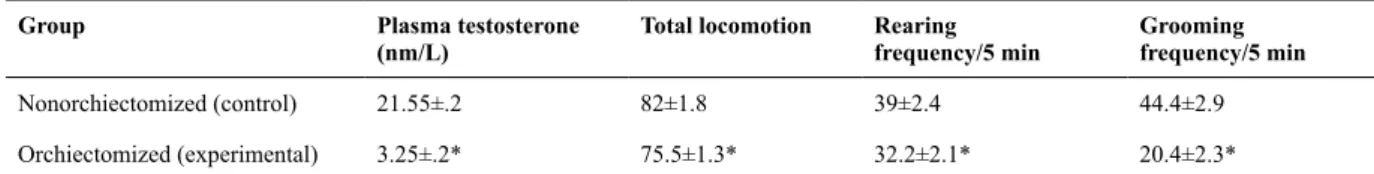
Table1. Effect of orchiectomy on plasma testosterone, total locomotion, rearing, and grooming
Group Plasma testosterone
(nm/L)
Total locomotion Rearing frequency/5 min
Grooming frequency/5 min
Nonorchiectomized (control) 21.55±.2 82±1.8 39±2.4 44.4±2.9
Orchiectomized (experimental) 3.25±.2* 75.5±1.3* 32.2±2.1* 20.4±2.3*
Data are expressed as mean± SEM (n=9 per group).
Oyekunle, Ibironke and Opabunmi 116
Choate, J. V., & Resko, J. A. (1996).Effects of androgen on brain
and pituitary androgen receptors and LH secretion of male guinea
pigs. Journal of Steroid Biochemistry and Molecular Biology,59,
315-322.
Clancy, A. N., Bonsall, R. W., &Michael, R. P. (1992).
Immunohistochemical labeling of androgen receptors in the brain of
rat and monkey. Life Sciences, 50, 409-417.
Coates, J. M. (2010). From molecule to market: Steroid hormones and inancial risk-taking. Philosophical Transactions of the Royal Society of London: B. Biological Sciences, 365, 331-343.
Coplan, J. D., & Lydiard, R. B. (1998). Brain circuits in panic disorder.
Biological Psychiatry, 44, 1264-1276.
Corodimas, K. P., LeDoux, J. E., Gold, P. W., & Schulkin, J. (1994). Corticosterone potentiation of conditioned fear in rats. Annals of the New York Academy of Sciences, 746, 392-393.
Eisenegger, C., Haushofer, J., & Fehr, E. (2011). The role of testosterone in social interaction. Trends in Cognitive Sciences, 15, 263-271.
Fernandez-Guasti, A., & Martinez-Mota, L. (2005). Anxiolytic-like actions of testosterone in the burying behavior test: Role of androgen and GABA-benzodiazepine receptors. Psychoneuroendocrinology, 30, 762-770.
Frye, C. A., & Seliga, A. M. (2001). Testosterone increases analgesia, anxiolysis, and cognitive performance of male rats. Cognive, Affective and Behavioral Neuroscience, 1, 371-381.
Gahr, M. (1990). Localization of androgen receptors and estrogen receptors in the same cells of the songbird brain. Proceedings of the National Academy of Sciences of the United States of America, 87, 9445-9448.
Gallagher, M., & Holland, P. C. (1994). The amygdala complex: multiple roles in associative learning and attention. Proceedings of the National Academy of Sciences of the United States of America, 91, 11771-11776.
Greco, B., Edwards, D. A., Michael, R. P., & Clancy, A. N. (1998). Androgen receptors and estrogen receptors are colocalized in male rat
hypothalamic and limbic neurons that express Fos immunoreactivity
induced by mating. Neuroendocrinology, 67, 18-28.
Gross, C., & Hen, R. (2004). The developmental origins of anxiety.
Nature Reviews Neuroscience, 5, 545-552.
Hall, C. S. (1934). Emotional behavior in the rat: I. Defecation and urination as measures of individual differences in emotionality.
Journal of Comparative and Physiological Psychology, 18,
385-403.
Hall, C. S., & Bellenchey, E. L. (1932). A study of rat’s behaviour in a ield: A contribution to method in comparative psychology.
University of California Publications in Psychology, 6, 1-12.
Hayden-Hixson, D. M., & Ferris, C. F. (1991).Steroid-speciic
regulation of agonistic responding in the anterior hypothalamus of
male hamsters. Physiology and Behavior, 50, 793-799.
Hermans, E. J., Putman, P., Baas, J. M., Koppeschaar, H. P., & Honk, J. V. (2006). A single administration of testosterone reduces fear-potentiated startle in humans. Biological Psychiatry, 59, 872-874.
Hermans, E. J., Ramsey, N., Tuiten, A., & van Honk, J. (2004). The amygdala and anger: responses to angry facial expressions after administration of a single dose of testosterone. Human Brain Mapping, 23, S188.
Ivinskis, A. (1968). The reliability of behavioural measures obtained in the open ield. Australian Journal of Psychology, 20, 173-177.
Krishnan, V., & Nestler, E. J. (2010). Linking molecules to mood: New insight into the biology of depression. American Journal of Psychiatry, 167, 1305-1320.
Kritzer, M. F., & Creutz, L. M. (2008). Region and sex differences
in constituent dopamine neurons and immunoreactivity for intracellular estrogen and androgen receptors in mesocortical
projections in rats. Journal of Neuroscience, 28, 9525-9535.
Lang, P. J., Davis, M., & Öhman, A. (2000). Fear and anxiety: Animal models and human cognitive psychophysiology. Journal of Affective Disorders, 61, 137-159.
LeDoux, J. E. (1996). The emotional brain: The mysterious underpinnings of emotional life. New York: Simon & Schuster.
Mazur, A., & Booth, A. (1998). Testosterone and dominance in men.
Behavioral and Brain Sciences, 21, 353-363, discussion 363-397.
Prescott, R. (1970). Some behavioural effects of variables which inluence “general level of activity” of rats.Animal Behaviour, 18,
791-796.
Resko, J. A., Connolly, P. B., Roselli, C. E., Abdelgadir, S. E., & Choate, J.V. (1993). Selective activation of androgen receptors
in the subcortical brain of male cynomologus macaques by physiological hormone levels and its relationshipto
androgen-dependent aromatase activity. Journal of Clinical Endocrinology and Metabolism, 76, 1588-1593.
Schulkin, J., Gold, P. W., & McEwen, B. S. (1998). Induction
of corticotropin-releasing hormone gene expression by
glucocorticoids: Implication for understanding the states of fear and anxiety and allostatic load. Psychoneuroendocrinology, 23,
219-243.
Svensson, A. I., Berntsson, A., Engel, J. A., & Soderpalm, B. (2000). Disinhibitory behavior and GABAA receptor function in serotonin-depleted adult male rats are reduced by gonadectomy.
Pharmacology Biochemistry and Behavior, 67, 613-620.
Tamminga, C. A. (2010). The hippocampal formation in schizophrenia.American Journal of Psychiatry, 167, 1178-1193.
Tietz, N. W. (1995). Clinical guide to laboratory tests, 3rd edition (p.
578-580). Philadelphia: W. B. Saunders.
Tillfors, M., Furmark, T., Marteinsdottir, I., Fischer, H., Pissiota, A., Langstrom, B., & Fredrikson, M. (2001). Cerebral blood low in subjects with social phobia during stressful speaking tasks: a PET study. American Journal of Psychiatry, 158, 1220-1226.
van Honk, J., Peper, J. S., &Schutter, D. J. (2005). Testosterone reduces unconscious fear but not consciously experienced anxiety: Implications for the disorders of fear and anxiety. Biological Psychiatry, 58, 218-225.
van Honk, J., Schutter, D. J. L. G., Hermans, E. J., Putman, P., Tuiten, A., & Koppeschaar, H. (2004). Testosterone shifts the balance
between sensitivity for punishment and reward in healthy young
women. Psychoneuroendocrinology, 29, 937-943.
van Honk, J., Tuiten, A., Hermans, E., Putman, P., Koppeschaar, H., Thijssen, J., Verbaten, R., & van Doornen, L. (2001). A
single administration of testosterone induces cardiac accelerative
responses to angry faces in healthy young women. Behavioral Neuroscience, 115, 238-242.
Verma, A., & Moghaddam, B. (1996). NMDA receptor antagonists
impair prefrontal cortex function as assessed via spatial delayed
alternation performance in rats: Modulation by dopamine. Journal of Neuroscience, 16, 373-379.
Walsh, R. N., & Cummins, R. A. (1976). The open-ield test: A critical review. Psychological Bulletin, 83, 482-504.
Zahrt, J., Taylor, J. R., Mathew, R. G., & Arnsten, A. F. (1997).
Supranormal stimulation of D1 dopamine receptors in the rodent
prefrontal cortex impairs spatial working memory performance.