Testosterone concentration in a bovine Bos
indicus with bilateral varicocele. Case report
Concentração de testosterona em um bovino Bos
Indicus com varicocele bilateral. Relato de caso
Marion Burkhardt KOIVISTO1; Maria Cristina Rui LUVIZOTTO1; Guilherme Padua NOGUEIRA2; Wilter Ricardo Russiano VICENTE3; Marcelo Tadeu Alvarenga COSTA4
CORRESPONDÊNCIA PARA: MARION BURKHARDT DE KOIVISTO Curso de Medicina Veterinária Faculdade de Odontologia UNESP - Campus de Araçatuba Rua Clóvis Pestana 793 Caixa Postal 341 16050-680 Araçatuba – SP e-mail: [email protected]
1- Departamento de Clínica, Cirurgia e Reprodução Animal da Faculdade de Odontologia da UNESP, Araçatuba – SP 2- Departamento de Apoio, Produção e Saúde Animal da Faculdade de Odontologia da UNESP, Araçatuba – SP
3- Departamento de Medicina Preventva e Reprodição Animal da Faculadade de Ciências Agrárias e Veterinárias da UNESP, Jaboticabal – SP
4 - Central VR, Araçatuba – SP
SUMMARY
A bovine, Bos indicus, with bilateral varicocele diagnosticed by palpation and ultrasound observation was observed during 24 months measuring scrotal circumference, semen quality, testosterone concentrations comparing with seasonal variation and other animals of the same species. The abnormal morphology of major defects and minor defects didn’t change between the bull and the other animals, however, the total defects were higher during the summer in the bull with varicocele (49,9%±6,9) when compared with the other bulls (27,9%±2,9). The animal showed higher percentage of major defects during the summer, comparing with the other seasons of the year. For the animal with varicocele testosterone levels were significantly higher during the different season’s of the year, whereas decreasing levels in the summer were seen in all Bos indicus. The clinical diagnostic was confirmed by necropsy. This pathology, characterized by a bilateral varicocele with thrombosis of the spermatic cord vessels, showed that the thermoregulation suffered establishing severe testicular degeneration. As seric testosterone increased suggesting lack of steroid retention at the testicle by the pampiniform plexus, the sperm production was abnormal.
KEY-WORDS: Varicocele. Bovine. Semen. Testosterone. Bos indicus.
INTRODUCTION
V
aricocele is a dilation and tortuosity of the veins of the pampiniform plexus and the cremasteric veins9. Jensen 8 defined it to be a local disturbance characterized by sacular dilatation and thrombosis in the internal spermatic vein. It is observed occasionally in stallions, rarely in the bull, and in about 1-2% of rams, in which occurrence increases with age. In Brazil, Fêo et al.6 reported a case of varicocele in bovine.The etiology of varicocele is unknown, but in humans deficiency of valves in veins draining the testis is considered likely. Deficiency of elastic and fibrous tissue in surrounding fascia is another possibility. Varicoceles appear as dark red nodules, 1-3 cm or more in diameter, enclosed in fascia of the spermatic cord proximal to the testis. Dissection of varicoceles may reveal large organizing laminated thrombi9. All varicoceles are thrombosed; changes associated with larger varicoceles include testicular mineralization suggesting a deficiency of thermoregulatory mechanism of the testis5, 9, 12.
In adolescent varicocele in humans, testicular degeneration is evidenced by premature germ cells sloughing, decreased number of large-state forms of germ
cells (secondary spermatocytes, spermatids, and spermatozoa) and some degree of tubular sclerosis 9.
Detailed histologic studies have revealed an apparently independent sclerosis involving almost 50% of both arteries and veins in the spermatic cords of rams. The arterial changes, which tend to be bilateral, consist of fibroplasia of the tunica intima. A variable degree of intimal and or medial mineralization may also be seen in branches of the testicular artery in the spermatic cords of rams and bucks. A review of literature showed few data referring to varicocele in bovine. The present study was carried out in order to observe seminal characteristics, hormonal findings of testosterone levels (nmol/L) in the systemic blood, and an anatomy-pathological study of the spermatic cords and testes in a Bos indicus bull with bilateral varicocele.
MATERIAL AND METHOD
RESULTS
The physical examination of the animal revealed a nodular enlargement of the spermatic cords. The scrotum had a marked clinical enlargement and pendulous supplementation. Semen was collected in an artificial vagina
twice a week in intervals of three days during two years completing 184 collections. Immediately following collection, the semen was evaluated according to standard procedures for volume, mass-activity, motility, concentration and morphology.
Monthly scrotal circumference was measured with a scrotal tape and blood samples were taken in the jugular vein. The blood was centrifuged immediately and the plasma harvested and stored at –20°C. Plasma concentrations of testosterone were determined by solid-phase I125 radioimmunoassay (Coat-A-Count Diagnostic Products Cooperation, USA). All samples were processed in duplicate. During the experiment ultrasound images (Scanner 480 Vet – Pie Medical) with a linear transducer were performed. The frequency used was 5,0MHz. Changes were observed at the spermatic cords of both testes. Following culling the reproductive tracts were examined macroscopically. Pieces of testis, epididymides and
Figure 1. Monthly variation in testosterone concentration (nmol/L) between the bull with varicocele and other’s Bos indicus during a 2
years period. Araçatuba, 2000.
spermatic cords were fixed in Bouin’s fluid for histopathological studies. The pieces were embedded in paraffin wax and sections cut at 5 microns were stained with hematoxylin and eosin11.
Other nine bulls Bos indicus also were evaluated in order to compare with the animal which showed the pathological change.
For statistical analysis the nonparametric Friedman test were performed comparing the variation of abnormal spermatozoa during the different seasons of the year. The Mann-Whitney test was used in order to compare the average value of the bulls with the other Bos indicus. The values are expressed in average ± SD.
Figure 3
Spermatic cord with firm nodules at palpation (a= right testicle; b= left testicle). Araçatuba, 2000.
Figure 4
Varicocele with vascular thrombosis (left testicle). Araçatuba, 2000.
Figure 5
Microscopic aspects of vascular thrombosis. t (thrombosis), v (vessel), h (hemosiderin). HE. Obj. 40x. Araçatuba, 2000.x
conformation and, on palpation, painful sensibility of the testes. The presence of a right scrotal frenulum results in moderate lateral rotation of 90° of the testes.
During the period of observation, the average of scrotal circumference was 46,4 ± 1,1 cm and the seminal values were: volume 3,8 ± 2,8 ml, motility 59,3 ± 5,7 %, mass-activity 3,6 ± 0,8, concentration 1660,6 ± 353,7 spermatozoa/mm3.
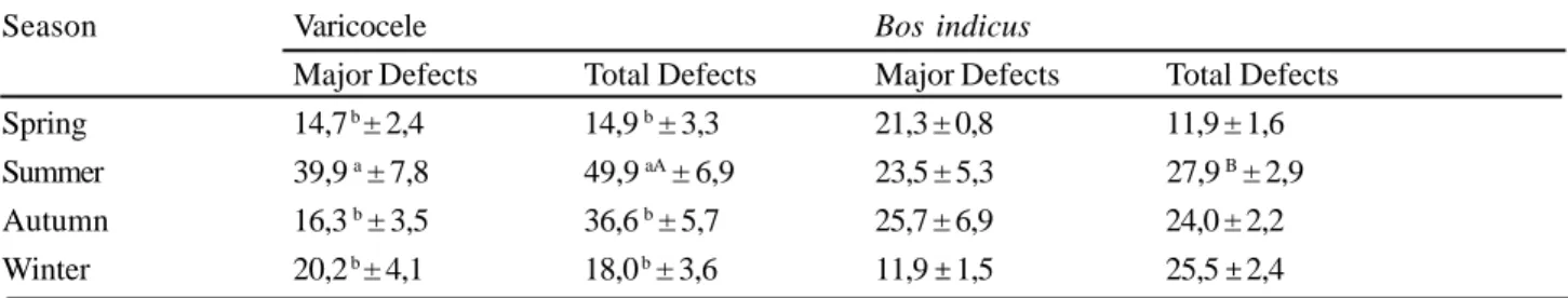
Considering the seasonal changes there were significant difference (p < 0,005) of major defects and total defects in the summer comparing with the other seasons (Tab. 1). During the two year period the bull showed average values of major and total defects (25,4 ± 16,1 %; 11,1 ± 8,2 % and 35,3 ± 3,1 %; 23,0 ± 1,5 %, respectively), higher than other bulls of the same species. The average percentage of minor defects in the bull didn’t show significant difference comparing with the other animals (9,9 ± 2,9%; 12,2 ± 7,6%, respectively).
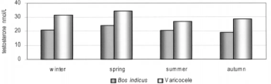
Referring to the plasma concentrations of testosterone, the monthly mean values were higher in the bull with varicocele when compared with the other Bos indicus bulls (Fig. 1).
Figure 2 shows a seasonal analysis of the plasma concentration of testosterone found in the bull with varicocele and the bulls Bos indicus.
Palpating the spermatic cords two nodules of firm
Table 1
Seasonal variation of abnormal spermatozoa morphology determined during 24 months in a bull with varicocele compared with the seasonal average morphological abnormalities of spermatozoa in nine bulls of the same breed. Araçatuba, 2000.
Different minuscule letters denote difference between the seasons Different capital letters denote difference between the animals
Season Varicocele Bos indicus
Major Defects Total Defects Major Defects Total Defects Spring 14,7 b ± 2,4 14,9 b ± 3,3 21,3 ± 0,8 11,9 ± 1,6 Summer 39,9 a ± 7,8 49,9 aA ± 6,9 23,5 ± 5,3 27,9 B ± 2,9 Autumn 16,3 b ± 3,5 36,6 b ± 5,7 25,7 ± 6,9 24,0 ± 2,2 Winter 20,2 b ± 4,1 18,0 b ± 3,6 11,9
RESUMO
Um reprodutor bovino, Bos indicus, com varicocele bilateral detectado por palpação e ultra-sonografia foi acompanhado por um período de 24 meses quanto à biometria testicular, valores espermáticos e concentração de testosterona comparados entre as estações do ano e outros animais da mesma espécie. As alterações morfológicas dos defeitos maiores e menores não variaram entre o touro com a patologia e os demais touros, no entanto, durante o verão o touro com varicocele apresentou maior percentual de defeitos totais se comparado aos demais touros da mesma espécie (49,86%±6,9 e 27,91%±2,9). O animal apresentou maior percentual de defeitos maiores no verão se comparado às outras estações do ano. Os achados de necrópsia confirmaram o diagnóstico clínico. Pode-se concluir que esta patologia, caracterizada por trombose nos vasos do cordão espermático, comprometeu a termoregulação determinando degeneração testicular severa. O aumento das concentrações de testosterona sérica sugerem a diminuição da retenção de esteroides nos testículos pelo plexo pampiniforme, a produção espermática estava anormal.
UNITERMOS: Varicocele. Bovino. Sêmen. Testosterona. Bos Indicus.
CONCLUSION
According to the histological findings, seminal evaluation and hormonal quantification, this case refers to a severe testicular degeneration caused by bilaterla varicocele followed by an increase of proximal droplets and increase in the plasma concentration of testosterone.
ACKNOWLEGEMENT
The authors gratefully acknowledge Laine Margareth Gabas for her assistance. This work was supported in part by FUNDUNESP.
DISCUSSION
Scrotal circumference was higher in the bull with varicocele comparing with the other animals of the same group (42,6 to 41,0 cm) indicating a patologic condition confirmed through palpation, ultrasound and through histologic features. Through microscopic examination the testes showed moderate to severe degeneration evidenced by partial or total lack of spermatogenesis, as well as multinuclear cells in the lumen of the seminiferous tubules that were calcified similar to the description of Desphande et al.4, Ezzi et al.5; Machado et al.12 and Jubb et al.9.
During 24 month’s seminal values like volume, motility, massactivity and concentration were not affected by the bilateral varicocele. So the average values for seminal characteristics were similar for the Zebu breed7.
The average values of morphologic abnormalities were higher than in the other bulls of the same species. The consistency were evident at the left, one of them measuring about 4,0 cm of diameter proximal to the testes and the other with 1,5 cm more distant. At the right spermatic cord there was a nodule of 2,0 cm with the same characteristics described before (Fig. 3 and 4). The testicular consistency or tone was decreased loosing the normal resilience.
Through ultrasound examination at the left testicular parenchym it was possible to detect small focal disseminated hyperecoic areas. At the spermatic cords the areas corresponding to the nodules showed compact hypoecoic image of low density.
The macroscopic evaluation of the spermatic cords revealed dilatation of the veins and arteries and large organizing laminated thrombi, with focus of calcification at the center. The testes with altered consistency, showed in the middle several hardened points, suggesting mineralization.
Microscopic examination revealed sclerosis and fibroplasia of the tunica intima and media of the testicular arteries in the spermatic cords occurring pigment deposition of hemosiderin close to the mural thrombosis (Fig. 5).
percentage of spermatozoa with abnormal morphology considering the major defects 25,4 ± 16,0 % and total defects 35,3 ± 15,2 %, showed a higher incidence than the standard values for the species. The average of minor defects 9,9 ± 2,9 % were within reference values2, 3, 7.
Considering the seasonal changes there were significant difference (p < 0,005) of major defects and total defects in the summer comparing with the other seasons. These results are in agreement with the observations of Koivisto et al.10. The prevailing major defect was the proximal droplet and according to Ott15 and Barth and Oko1 would reveal a defect at the level of the seminferous tubules confirmed through the findings of testicular degeneration by histological studies.
Considering the plasma concentrations of testosterone, the monthly mean values were higher in the bull with varicocele when compared with the other Bos indicus bulls (Fig. 1), suggesting that the secretion rate of this steroid was modified due to the present pathology 14.
Received: 26/03/2001 Accepted: 27/02/2002
REFERENCES
1. BARTH, A. D.; OKO, R. J. Abnormal morphology of bovine spermatozoa. 1. ed. Iowa State: University Press, 1989. p. 285
2. BLOM, E. The ultrastructure of some characteristic sperm defects and a proposal for a new classification of the bull spermiogram.
Nord. Vet. Med., v.25, n.7, p.383-391, 1973.
3. COLÉGIO BRASILEIRO DE REPRODUÇÃO ANIMAL. Manual
para exame andrológico e avaliação de sêmen animal. Belo
Horizonte: Colégio Brasileiro de Reprodução Animal, 1998. p. 49.
4. DESHPANDE, K. S.; DIWAN, J. G.; WAKANKAR, C. C.; DESHPANDE, B. R. Varicocele in a Merino ram. Indian Veterinary
Journal, v. 56, n. 3, p. 236-239, 1979.
5. EZZI, A.; LADDS, P. W.; HOFFMANN, D.; FOSTER, R. A.; BRIGGS, G. D. Pathology of varicocele in the ram. Australian
Veterinary Journal, v. 65, n. 1, p. 11-15, 1988.
6. FÊO, J. C. S. A.; BERNABE, C. R.; VISINTIN, J. A.; GUERRA, J. A case of varicocele in a bull. In: INTERNATIONAL CONGRESS ON ANIMAL REPRODUCTION AND ARTIFICIAL INSEMINATION, 9. 1980, Madrid, Espanha. Anais... Madrid, 1980. v. 3, p. 252.
7. FONSECA, V. O. et al. Procedimentos para avaliação
andrológica e do sêmen congelado. Belo Horizonte: Colégio
Brasileiro de Reprodução Animal, 1992, p. 79.
8. JENSEN, R. Diseases of sheep. Philadelphia: Lea and Febinger, 1974. p. 6-8.
9. JUBB, K. V. F.; KENNEDY, P. C.; PALMER, N. Pathology of
domestic animals. 4. ed., San Diego: Academic Press, 1992, vol.
3, p. 633.
10. KOIVISTO, M. B.; NOGUEIRA, G. P.; COSTA, M. T. A Bos taurus taurus x Bos taurus indicus. Fluctuations of the sperm abnormalities (previous note). In: CONGRESSO PANAMERICANO DE CIÊNCIAS VETERINÁRIAS, 15., 1996, Campo Grande, MS, Brasil. Anais... Campo Grande, 1996. p. 385.
11. LUNA, L. G. Manual of histologic staining methods of
the armed forced institute of pathology. 3. ed. New York:
Mac-Graw-Hill, 1968. p. 258.
12. MACHADO, R.; SIMPLÍCIO, A. A.; SANTA ROSA, J. Varicocele bilateral em ovino Ile de France (bilateral varicocele in Ile de France). In: CONGRESSO BRASILEIRO DE REPRODUÇÃO ANIMAL, 9., 1996, Belo Horizonte, MG, Brasil. Anais... Belo Horizonte: Colégio Brasileiro de Reprodução Animal, 1991. p.407
13. MALMGREM, L. Experimental induced testicular alterations in boars: Hormonal changes in mature and peripubertal boars. Acta Vet. Scand., v. 31, n. 1, p. 97-107, 1990.
14. McDONALD, L. E.; PINEDA, M. H. Veterinary
endocrinology and reproduction. 4. ed., Philadelphia: Lea &
Febinger, 1989. p. 571.
15. OTT, R. S. Breeding soundness examination of bulls. In: MORROW, D. A. Current therapy in theriogenology. London: W.B. Saunders, 1986. p. 1143.
16. PRABHAKAR, J.; CHIMBONI, M.; MALMGREN, L.;FREDRIKSSON, G.; MADJEY, A. Effects on Testosterone and LH Concentrations of induced Testicular Degeneration in Bulls.
Acta Vet. Scand., v. 31, n. 4, p. 505-507, 1990.
17. RHYNES, W. E.; EWING, L. L. Testicular endocrine function in Hereford bulls exposed to high ambient temperature. Endocr., v. 92, n.2, p. 509-515, 1973.