Copyright
© ABE&M todos os dir
eitos r
eser
vados.
Influence of estradiol and
triiodothyronine on breast
cancer cell lines proliferation
and expression of estrogen and
thyroid hormone receptors
Efeito do estradiol e da triiodotironina na proliferação de linhagens celulares de câncer de mama e na expressão de receptores de estrógeno e hormônio tireoidiano
Sílvia Helena Cestari1, Nancy Bueno Figueiredo1,
Sandro José Conde1, Sueli Clara1, Maria Lucia Hirata Katayama2, Carlos Roberto Padovani3, Maria Mitzi Brentani2, Célia Regina Nogueira1
ABSTRACT
Objective: To better understand the estrogen (E2) agonist action of triiodothyronine (T3) the effects of these hormones on ER negative MDA-MB-231 breast cancer cells were compared with
those on S30, a clone of MDA-MB-231 stably transfected with ERα cDNA, in terms of proliferation
and modulation of hormone receptors. Results: Growth experiments showed that MDA-MB-231
was not modulated by any hormone or tamoxifen (TAM). Treatment with E2, 10-8M or 10-9M had
little effect on S30 proliferation. T3 at 10-8M significantly inhibited proliferation.This effect was not
reverted by TAM. Treatments with 10-8M concentration of E2 or T3reduced ERα gene expression
in S30, an effect partially blocked by association with TAM, with no effect on TR expression. These
results suggest that, in S30, 10-8M T3 has a similar action to E2 relative to ERα gene modulation.
Conclusions: Such results emphasize the need of determining T3 levels, before the introduction
of antiestrogenic forms of treatment in breast cancer patients. Arq Bras Endocrinol Metab. 2009;53(7):859-64.
Keywords
Triiodothyronine; estradiol; breast neoplasms; cell line
RESUMO
Objetivo: Para compreender melhor a ação da triiodotironina (T3) agonista de estrógeno (E2), foram comparados os efeitos destes hormônios em células de câncer de mama MDA-MB-231
ER negativas com um clone de MDA-MB-231, transfectado estavelmente com o cDNA de ERα
(S30), em termos de proliferação e modulação dos receptores hormonais. Resultados:
Experi-mentos de crescimento mostraram que MDA-MB-231 não foi modulada por qualquer hormônio ou pelo tamoxifeno (TAM). O crescimento de S30 foi essencialmente inalterado por tratamento
com E2 10-9M ou 10-8M, mas T3 10-8M inibiu significativamente a proliferação quando
compara-da a ambas concentrações de E2. Esse efeito não foi revertido pelo TAM, sugerindo um
resul-tado não genômico, independente de ERE. Tratamentos com 10-8M de E2 ou de T3 reduziram a
expressão do gene ERα em S30, efeito parcialmente impedido pela associação com TAM, sem
efeito na expressão de TR. Os resultados sugerem que, em S30, T3 10-8M tem ação semelhante
ao E2 com relação à modulação do gene ERα. Conclusões: Esses resultados enfatizam a
ne-cessidade de dosagem de T3 circulante antes da introdução do tratamento antiestrogênico no
câncer de mama. Arq Bras Endocrinol Metab. 2009;53(7):859-64.
Descritores
Triiodotironina; estradiol; neoplasias da mama; linhagem celular
1 Departamento de Clínica Médica, Universidade Estadual Paulista “Júlio de Mesquista Filho” (Unesp), Botucatu, SP, Brasil 2 Departamento de Radiologia, Faculdade de Medicina, Universidade de São Paulo (USP), São Paulo, SP, Brasil 3 Departamento de Bioestatística, Instituto de Ciências Biológicas, Unesp, Botucatu, SP, Brasil
Correspondence to: Célia Regina Nogueira
Departamento de Clínica Médica, Unesp
Distrito de Rubião Júnior, s/n 18618-970 – Botucatu, SP, Brasil [email protected]
Copyright
© ABE&M todos os dir
eitos r
eser
vados.
INTRODUCTION
E
strogens are pivotal in the growth of both nor-mal and neoplastic mammary tissues, and mediate most of their actions via ligand-dependent transcription factors, known as estrogen receptors (ER) (1,2). The involvement of thyroid hormone (TH) in the develo-pment and differentiation of normal breast tissue has been established (3-5). Although epidemiologic and experimental studies have associated TH pathologies with an increase in the risk of breast cancer, the role of these hormones remains controversial. Kapdy and Wolfe (6) and Mustacchi and Greenspan (7) found an association between TH ingestion and increase in the risk of mammary cancer. On the other hand, Vorherr (8) described an increase in the survival of hyperthyroid patients with breast cancer. Saraiva and cols. (9) suggest the existence of a biological link between breast can-cer in post-menopausal woman and subclinical hyper-thyroidism. Turken and cols. (10) and Gogas and cols. (11) reported an increase in the prevalence of thyroid autoimmune disease in patients with breast cancer. Cristofanilli and cols. (12) showed that spontaneous clinical hypothyroidism may decrease the aggressive-ness of breast cancer and reduce tumor incidence.Most, if not all, major triiodothyronine (T3) actions are mediated by specific high affinity nuclear receptors (TR), which are encoded by two genes, TRα and TRβ
(13). Recent results reveal substantial changes in the expression profile of TH receptors, suggesting a possi-ble deregulation that could trigger breast cancer deve-lopment (14).
In a previous work (15), we demonstrated in MCF-7 cells, an ER-positive breast cancer cell line, that T3 mimicked the effects of estradiol (E2), stimulating growth, modulating mRNA transcription of growth factors and inducing the expression and activity of E2-inducible proteins. In addition, these T3 effects were antagonized by the simultaneous addition of tamoxifen (TAM), which is a competitive inhibitor of E2 binding to ER. Similar effects, however, were not observed in the ER-negative MDA-MB-231 breast cancer cells, in spite of the presence of high TR amounts, suggesting that, in MCF-7 cells, both ligands share a common sig-naling pathway via ER. In addition, Dinda and cols. (16) indicated that in T47D breast cancer cells also ER positive, T3 regulates T47D cell cycle progression and proliferation raising the p53 level and causing hyper-phosphorylation of pRb by a common mechanism
in-volving ER and T3 receptor (T3R)-mediated pathways. In line with these results, a physiological concentration of L-thyroxine (T4) has been recently reported to in-duce MCF-7 cells proliferation measured by tritiated thymidine or appearance of proliferating cell nuclear antigen (PCNA) (17).
Jeng and cols. (18) demonstrated previously that after the stable transfection of ERα in MDA-MB-231 cells (devoid of ERα and ERβ) with an ERα vector (S30), these ER negative cells regained hormonal res-ponsiveness.
With the objective of clarifying the E2 agonist ac-tion of T3, we herein compared the effects of E2 and T3 in terms of proliferation and modulation of ER and TR in two breast cancer cell lines: MDA-MB-231, whi-ch is estrogen-insensitive, but possesses TRα and TRβ, and S30 cell, that is a MDA-MB-231 transfected with wild ERα (19).
METHODS
Cell culture and growth experiments
Cell growth was assessed by using two methods: WST-1 (Roche Diagnostics, Mannheim, Germany) – indirect method, a colorimetric assay based on the cleavage of the tetrazolium salt WST-1 by mitochondrial deshydro-genases in viable cells; and cell count – direct method, using a Neubauer chamber and trypan blue staining (Sigma, St Louis, MO, USA). Both experiments were performed in parallel and made in quadruplicates.
Copyright
© ABE&M todos os dir
eitos r
eser
vados.
media by radioimmunoassay and the expected hormo-ne concentrations were detected.
WST-1
The media were changed and hormones added dissol-ved in absolute ethanol after 24 hours (day 0), with daily media changes. Two hours before the readings of absorbance Spectra Max 190 (Molecular Devices) at a wavelength of 450 nm, 10 µL WST-1 was added to each well to determine proliferation and cell viability. The same procedure was followed for seven days.
Cell count
The media were changed and hormones added dissol-ved in absolute ethanol after 24 hours (day 0), with daily media changes. Cells were harvested in triplicate at the indicated times and cell numbers were counted. Cell numbers were plotted as a logarithmic function against time and cell population doubling time (DT) was estimated at the exponential phase.
Semi-quantitative reverse transcription-polymerase
chain reaction analyses of mRNA for ERα, PR, TRα
and TRβ
Total RNA was extracted from cells by TRIzol Reagent (Invitrogen Life Technologies, Carlsbad, CA, USA) which is based on the guanidine thiocyanate method. Frozen cells were mechanically homogenized on ice in 1 mL of ice-cold TRIzol. Total RNA was solubilized in Rnase-free H2O and quantified by measuring the opti-cal density (OD) at 260 nm. RNA purity was ensured by obtaining a 260/280 nm OD ratio of approximately 2.0. Two micrograms of RNA was reverse-transcribed with oligo dT primers and Superscript reverse trans-criptase (RT) in a total volume of 21 µL, according to standard methods (Invitrogen Life Technologies, Carl-sbad, CA, USA). Control “No RT” reactions were
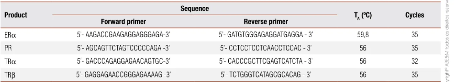
per-formed by omitting the RT enzyme. These reactions were then polymerase chain reaction (PCR)-amplified to ensure that DNA did not contaminate RNA. Primer pairs for ERα, TRα, TRβ and PR were designed from a sequence published in GenBank. PCR conditions were as follows: cycles of 94 °C for 30 seconds, annealing of specific primers (Table 1) 1 minute to ERα; 30 seconds to PR, TRα and TRβ, 30 seconds at 72 °C for exten-sion (except ERα, 1 minute). The bands corresponding to each gene were quantified by densitometry as inte-grated optical density. PCR products were run in dupli-cate on a different gel for each gene, and results were averaged. The size (then number of base pairs) of each band corresponded to the size of processed mRNA. The PCR signals were normalized by co-amplification (multiplex) of a human cyclophilin transcript with 0.5 µL of each primer, except for ERα PCR, which was performed in simplex. Final numeric values are expres-sed as a ratio between each and the internal (housekee-ping) gene, human cyclophilin.
Statistical analysis
Data are expressed as mean ± sd. Two-way ANOVA was used to analyze cellular proliferation and receptor amplification. Multiple comparisons were performed using the Tukey's test (20). The level of significance was p < 0.05.
RESULTS
T3, E2 and steroidal antagonist (TAM) action on cell proliferation
MDA-MB-231 growth was not modulated by the addi-tion of T3, E2 or TAM. The concentraaddi-tions of E2 (10 -7M and 10-8M) were equally effective in stimulating
MCF-7 cell proliferation. The simultaneous addition of TAM (10-6M) inhibited the proliferative effect of E2
(data not shown).
Table 1. Oligonucleotide primers used for polymerase chain reaction (PCR) ampliication of reverse transcribed RNA
Product Sequence TA (ºC) Cycles
Forward primer Reverse primer
ERα 5’- AAGACCGAAGAGGAGGGAGA-3’ 5’- GATGTGGGAGAGGATGAGGA - 3’ 59,8 35 PR 5’- AGCAGTTCTAGTCCCCCAGA -3’ 5’- CCTCCTCCTCAACCTCCAC - 3’ 56 35 TRα 5’- GACCCAGAGGAGAACAGTGC-3’ 5’- CACCCGCTTCGAGTCATCTA - 3’ 56 32 TRβ 5’- GAGGAGAACCGGGAGAAAAG -3’ 5’- TCTGGGTCATAGCGCACAG - 3’ 56 35
Copyright
© ABE&M todos os dir
eitos r
eser
vados.
S30 growth was not modulated by E2 or TAM. Nei-ther the treatment with two T3 concentrations, 10-10M
and 10-9M nor the concomitant addition of TAM
affec-ted DT of S30 cells in comparison with controls. The concentration of T3 (10-8M) significantly increased DT
and, therefore, significantly reduced cell proliferation when compared to the concentrations of E2, butthis effect was not reversed by TAM (Figure 1).
The results of the cell proliferation experiments per-formed in parallel were similar. Figures 1 and 2 show the data obtained by the cell count method.
Figure 1. Growth experiments in S30 breast cancer cells.
S30 breast cancer cells were cultivated with MEM without phenol red, with Earle’s Salts and 25 mM Hepes, supplemented with 1% antibiotic antimycotic solution, 200 nM L-glutamine, 10 nM non-essential amino acids (NEAA), 5% FBS, 6 ng/mL bovine insulin, and 500 μg/mL G418. Cells were seeded (2.5 x 10³ cells per well) and treated for three days with estradiol 10-9M (E2 1), 10-8M (E2 2), 10-7M (E2 3), triiodothyronine 10-10M (T3 1), 10-9M (T3 2), 10-8M (T3 3), and tamoxifen 10-6M (TAM). Results are mean of 4 simultaneous experiments ± sd with * p < 0.01 (p values determined in relation to E21 and E2 2).
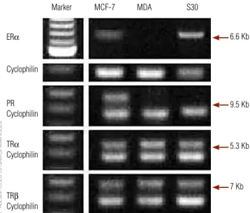
Figure 2. Expression of ERα, PR, TRα, TRβ with co-amplification of a human cyclophilin transcript in MCF-7, MDA-MB-231 (MDA) and S30 breast cancer cell lines.
Marker MCF-7 MDA S30
6.6 Kb
9.5 Kb
5.3 Kb
7 Kb ERα
Cyclophilin
PR Cyclophilin
TRα
Cyclophilin
TRβ
Cyclophilin
ER, progesterone and thyroid receptors determination in S30 cell
RT-PCR was used to detect ER, progesterone (PR) and thyroid receptors (TR) transcripts in order to better characterize our model. Both MDA-MB-231 and S30 expressed TRα and TRβ, but not PR. Only S30 cell ex-pressed ERα. MCF-7 cells, which express ER, PR and TR, were used as a positive control (Figure 2).
Effects of E2 and T3 in TRα, TRβ and ERα expression
Treatment of either MDA-MB-231 (data not shown) or S30 cells with T3 (10-8M) or E2 (10-8M), both in
the absence or presence of TAM (10-6M), did not affect
TRα and TRβ levels (data not shown). In S30 cells, the presence of E2or T3 significantly decreased ERα
expression. Association of TAM (10-6M) with either E2
(10-8M) or T3 (10-8M) reverse partially the reduction
of ERα expression induced by the presence of the hor-mones alone (Figure 3).
* p < 0.05.
Figure 3. Expression of ERα (A) and TRα (B) transcript in S30 breast cancer cell line estimated by reverse transcription-polymerase chain reaction (RT-PCR) in each treated groups.
Data were run in duplicate on different gels for each gene, and the results were averaged. Quantiication of the PCR signal was obtained by densitometric analysis of the product in integrated optical density (IOD). Gene expressions were normalized to the cyclophilin signal from the same RT product. Normalized data are expressed as means ± sd.
Doubling time (hours)
*
*
CTTAM E21 E22 E23
E21 + TAME22 + TAME23 + TAM
T31 T32 T33
T31 + TAMT32 + TAMT33 + TAM
20 16 12 8 4 0
mRNA (relative IOD)
Control TAM E2
* *
*
*
E2 + TAM T3 T3 + TAM 2,0
1,5
1,0
0,5
0,0
mRNA (relative IOD)
Control TAM E2 E2 + TAM T3 T3 + TAM 2,0
1,5
1,0
0,5
Copyright
© ABE&M todos os dir
eitos r
eser
vados.
DISCUSSION
The aim of this paper was to expand our previous in-vestigations (Nogueira and Brentani, 1996) suggesting that TH elicits estrogenic effects via ER. The effects of E2 and T3 on the proliferation and the modulation of ER and TR were examined in MDA-MB-231 stably transfected with the wild type ERα (S30) and compa-red to the parental ER-negative MDA-MB-231 breast cancer cells.
In agreement with our previous results, MDA-MB-231 growth was independent from the addition of E2, T3, and TAM (15). In S30 transfected cells, ER-mRNA levels were similar to those shown by ER res-ponsive MCF-7 cells (21). However, ER expression in MDA-MB-231 did not lead to significant effects on cell proliferation upon E2 treatment. Contrariwise several authors have reported that restoration of ERα expres-sion in MDAMB-231 leads to a ligand dependent inhi-bition of growth (22-24) an effect blocked by anties-trogens. Moreover, Moggs and cols. (25) demonstrated that this antiproliferative effect of E2 in MDAMB-231 transduced with ER is mediated by down regulation of many genes involved in cell cycle progression as revea-led by a microarray approach.
We have no clear explanation for our divergent re-sults. This abnormal behavior may be explained by the specific conditions of our S30 cells. According to Vig-non and cols. (26) quiescent cells maintained in low serum concentrations (2%) were insensitive or slightly stimulated by E2 treatment. As late passages of S30 cells were used here, it is possible that ER had lost efficiency, even though its concentrations remained high, as in ER positive hormone-independent tumors. According to Garcia and cols. (22), E2 inhibitory effects are small and reduced the total cell content by only 30-40% after four to six days of treatment. They also observed that the maximal inhibitory effect of E2 (2 nM) was compatible with the occupancy of ER sites and did not increase fur-ther with concentrations up to 1 µM. One explanation of our results may be related to the evident reduction of ERα mRNA levels after E2 treatment of S30 cells, leading to no significant effects on cell growth. T3 at 10-8M concentration also reduced ERα gene expression
in those cells as compared to controls, suggesting that E2 and T3 actions were similar. As the down regulation is partly inhibited by TAM, it seems that T3 may inte-ract with ERE or influence pathways controlled by ERE. Down regulation ERα protein by E2 was previously re-ported in MCF-7 and T47D (27), and it has been
sug-gested to be due to the degradation of ER induced by E2. However, unlike E2, 10-8M T3 treatment inhibited
S30 proliferation which is the classical effect of E2 in the same cells. As TAM could not reverse the proliferation rate of S30 cells in combination with T3, these data that suggested that this inhibitory effect of T3 is probably not dependent on ERE, pointing to a non-genomic pa-thway or resulting from different interactions with other promoter elements, including AP1 or SP1 (28,29). Ac-cording to Zhou-Li and cols. (30), antiestrogens are non competitive inhibitors of T3 only in the cells whose growth is responsive to E2.
In conclusion, our results showed that the effects of T3 in S30 growth differ from those of E2, but both hormones down regulated ERα gene expression. Thus, in absence of E2, clinically important changes in TH levels could influence pathways controlled by ER.
Acknowledgements: the authors are grateful to Dr. V. Craig Jor-dan (Robert H. Lurie Cancer Center, Chicago, IL) for having kindly provided the S30 cell line used in this work, and to Funda-ção de Amparo à Pesquisa do Estado de São Paulo (Fapesp), São Paulo (SP), Brazil.
Disclosure: no potential conflict of interest relevant to this article was reported.
REFERENCES
1. Migliaccio A, Di Domenico M, Castoria G, de Falco A, Bontempo P, Nola E, et al. Tyrosine kinase/p21ras/MAO-kinase pathway ac-tivation by estradiol-receptor complex in MCF-7 cells. EMBO J. 1996;15(6):1292-300.
2. Razandi M, Pedram A, Levin ER. Plasma membrane estrogen re-ceptors signal to anti-apoptosis in breast cancer. Mol Endocrinol. 2000;14(9):1434-47.
3. Topper YJ, Freeman CS. Multiple hormone interactions in the developmental biology of the mammary gland. Physiol Rev. 1980;60(4):1049-106.
4. Vonderhaar BK, Tang E, Lyster RR, Nascimento MCS. Thyroid hormone regulation of epidermal growth factor levels in mouse mammary glands. Endocrinology. 1986;119(2):580-5.
5. Hovey RC, Trott JF, Vonderhaar BK. Establishing a framework for the functional mammary gland: from endocrinology to morpho-logy. J Mammary Gland Biol Neoplasia. 2002;7(1):17-38. 6. Kapdi CC, Wolfe JN. Breast cancer. Relationship to thyroid
sup-plements for hypothyroidism. JAMA. 1976;236(10):1124-7. 7. Mustacchi P, Greenspan F. Thyroid supplementation for
hy-pothyroidism. An iatrogenic cause of breast cancer? JAMA. 1977;237(14):1446-7.
8. Vorherr H. Thyroid function in benign and malignant breast dise-ase. Eur J Cancer Clin Oncol. 1987;23(3):255-7.
9. Saraiva PP, Figueiredo NB, Padovani CR, Brentani MM, Nogueira CR. Profile of thyroid hormones in breast cancer patients. Brazil J Med Biol Res. 2005;38(5):761-5.
Copyright
© ABE&M todos os dir
eitos r
eser
vados.
11. Gogas J, Kouskos E, Tseleni-Balafouta S, Markopoulos C, Reve-nas K, Gogas G, et al. Autoimmune thyroid disease in women with breast carcinoma. Eur J Surg Oncol. 2001;27(7):626-30. 12. Cristofanilli M, Yamamura Y, Kau SW, Bevers T, Strom S, Patangan
M, et al. Thyroid hormone and breast carcinoma. Primary hypo-thyroidism is associated with a reduced incidence of primary bre-ast carcinoma. Cancer. 2005;103(6):1122-8.
13. González-Sancho JM, García V, Bonilla F, Muñoz A. Thyroid hormone receptors/THR genes in human cancer. Cancer Lett. 2003;192(2):121-32.
14. Conde I, Paniagua R, Zamora J, Blánquez MJ, Fraile B, Ruiz A, et al. Influence of thyroid hormone receptors on breast cancer cell proliferation. Ann Oncol. 2006;17(1):60-4.
15. Nogueira CR, Brentani MM J. Triiodothyronine mimics the effects of estrogen in breast cancer cell lines. Steroid Biochem Molec Biol. 1996;59(3-4):271-9.
16. Dinda S, Sanchez A, Moudgil V. Estrogen-like effects of thyroid hor-mone on the regulation of tumor suppressor proteins, p53 and re-tinoblastoma, in breast cancer cells. Oncogene. 2002;21(5):761-8. 17. Tang H-Y, Lin H-Y, Zhang S, Davis FB, Davis PJ. Thyroid hormone
cau-ses mitogen-activated protein kinase-dependent phosphorylation of the nuclear estrogen receptor. Endocrinology. 2004;145(7):3265-72. 18. Jeng M-H, Jiang S-Y, Jordan VC. Paradoxical regulation of estro-gen-dependent growth factor gene expression in estrogen recep-tor (ER)-negative human breast cancer cells stably expressing ER. Cancer Lett. 1994;82(2):123-8.
19. Jiang SY, Jordan VC. Growth regulation of estrogen receptor-ne-gative breast cancer cells transfected with complementary DNAs for estrogen receptor. J Natl Cancer Inst. 1992;84(8):580-91. 20. Zar JH. Biostatistical analysis. New Jersey: Prentice Hall; 1999. 21. Touitou I, Vignon F, Cavailles V, Rochefort H. Hormonal regulation
of cathepsin D following transfection of the estrogen or
progeste-rone receptor into three sex steroid hormone resistant cancer cell lines. J Steroid Biochem Mol Biol. 1991;40(1-3):231-7.
22. Garcia M, Derocq D, Freiss G, Rochefort H. Activation of estrogen receptor transfected into a receptor-negative breast cancer cell line decreases the metastatic and invasive potential of the cells. Proc Natl Acad Sci USA. 1992;89(23):11538-42.
23. Lazennec G, Bresson D, Lucas A, Chauveau C, Vignon F. ER beta inhibits proliferation and invasion of breast cancer cells. Endocri-nology. 2001;142(9):4120-30.
24. Tonetti DA, Rubenstein R, DeLeon M, Zhao H, Pappas SG, Ben-trem DJ, et al. Stable transfection of an estrogen receptor beta cDNA isoform into MDA-MB-231 breast cancer cells. J Steroid Biochem Mol Biol. 2003;87(1):47-55.
25. Moggs JG, Murphy TC, Lim FL, Moore DJ, Stuckey R, Antrobus K, et al. Anti-proliferative effect of estrogen in breast cancer cells that re-express ERalpha is mediated by aberrant regulation of cell cycle genes. J Mol Endocrinol. 1995;34(2):535-51.
26. Vignon F, Prebois C, Rochefort H. Inhibition of breast cancer gro-wth by suramin. J Natl Cancer Inst. 1992;84(1):38-42.
27. Hall LC, Salazar EP, Kane SR, Liu N. Effects of thyroid hormones on breast cancer cell proliferation. J Steroid Biochem Mol Biol. 2008;109(1-2):55-66.
28. Sharma D, Saxena NK, Davidson NE, Vertino PM. Restoration of tamoxifen sensitivity in estrogen receptor-negative breast cancer cells: tamoxifen-bound reactivated ER recruits distinctive core-pressor complexes. Cancer Res. 2006;66(12):6370-8.
29. Paech K, Webb P, Kuiper GG, Nilsson S, Gustafsson J, Kushner PJ, et al. Differential ligand activation of estrogen receptors ER alpha and ER beta at AP1 sites. Science. 1997;277(5331):1508-10. 30. Zhou-Li F, Albaladejo V, Joly-Pharaboz MO, Nicolas B, Andre J.