Copyright
© ABE&M todos os dir
eitos r
eser
vados.
Radioiodine concentration by the
thymus in differentiated thyroid
carcinoma: report of five cases
Captação do radioiodo pelo timo no carcinoma diferenciado de tireoide: relato de cinco casos
Maria Eduarda Mello1, Rodrigo C. Flamini1, Rossana Corbo1, Marcelo Mamede1,2
ABSTRACT
The radioactive iodine has been used with great value as a diagnostic and therapeutic method in patients with differentiated thyroid carcinoma previously submitted to total thyroidectomy. False-positive whole-body scans may occur due to misinterpretation of the physiologic distri-bution of the radioisotope or lack of knowledge on the existence of other pathologies that could eventually present radioiodine uptake. Thymic uptake is an uncommon cause of false-positive whole-body scan, and the mechanism through which it occurs is not completely understood. The present paper reports five cases of patients with differentiated thyroid cancer who presented a mediastinum uptake of radioiodine in a whole-body scan during follow-up. The patients had either histological or radiological confirmation of the presence of residual thymus gland. It is very important to know about the possibility of iodine uptake by the thymus in order to avoid un-necessary treatment, such as surgery or radioiodine therapy. Arq Bras Endocrinol Metab. 2009;53(7):874-9.
Keywords
Thyroid neoplasms; thyroid gland; thymus gland; iodine isotopes; radionuclide imaging
RESUMO
O iodo radioativo tem sido utilizado com grande valia como método diagnóstico e terapêutico em pacientes com carcinoma diferenciado de tireoide previamente submetidos à tireoidecto-mia total. Resultados falso-positivos na pesquisa de corpo inteiro (PCI) podem ocorrer por má interpretação da distribuição fisiológica do radioisótopo ou por não conhecimento da existên-cia de outras patologias que podem eventualmente captar o radioiodo. Captação pelo timo é uma causa incomum de resultado falso-positivo e o mecanismo pelo qual ocorre não é total-mente esclarecido. O presente trabalho relata cinco casos que apresentaram PCI positiva em mediastino durante o seguimento, com comprovação histológica ou tomográfica sugestiva de timo. Ressalta-se a importância do conhecimento dessa possível causa de falso-positivo a fim de se evitar tratamentos desnecessários. Arq Bras Endocrinol Metab. 2009;53(7):874-9.
Descritores
Neoplasias da glândula tireoide; glândula tireoide; timo; isótopos de iodo; cintilografia
1 Serviço de Medicina Nuclear,
Instituto Nacional de Câncer (Inca), Rio de Janeiro, RJ, Brasil
2 Serviço de Pesquisa Clínica,
Inca, Rio de Janeiro, RJ, Brasil
Correspondence to: Marcelo Mamede
Rua André Cavalcanti, 37, 2º andar – Fátima
22230-050 – Rio de Janeiro, RJ, Brasil
Received in Mar/15/2009 Accepted in Aug/3/2009
INTRODUCTION
D
ifferentiated thyroid cancer (DTC) responds for about 90% to 95% of all cases of thyroid malignan-cies and has a good prognosis when adequately treated. The mainstay of treatment is total thyroidectomy and debulking of secondary malignancies, if possible, such as cervical lymph nodes (1,2). Radioiodine therapy may be used as an adjunct to surgery, for ablation of remnant thyroid tissue or for the treatment of distant metastases,since it has been associated with decreased recurrence and increased survival rate (3).
false-po-Copyright
© ABE&M todos os dir
eitos r
eser
vados.
sitives WBSs, being sometimes misinterpreted as me-tastatic disease. Hence, this could lead to inappropriate treatment, such as the administration of radioiodine or surgery. The normal tissues that commonly concentra-te iodine include salivary glands, breast tissue and or-gans of the gastrointestinal and urinary systems (4-6), whereas pathological uptake has been reported in pul-monary inflammatory or neoplastic diseases, sinusitis, inflammatory diseases of the salivary glands, pericar-dial effusions, esophagus pathologies, ovarian cysts or tumours, urinary tract diseases and traumatic lesions, among others (5-17).
An uncommon cause of false-positive WBS is iodine uptake by the normal thymus, which may be misinter-preted as thyroid metastases to mediastinal nodes or to the lung (18-25). It is very important to recognize this condition and its characteristics in order to facilitate the differentiation between this normal variant and the pre-sence of metastases.
The present paper reports five cases of DTC patients submitted to total thyroidectomy and radioiodine thera-py that had iodine uptake in the mediastinum during the follow-up, and presents a review of the latest insights.
CASE 1
A 57-year-old female was histopathologically diagno-sed with papillary carcinoma of the thyroid invading the capsule of the organ after total thyroidectomy. Sub-sequently, she underwent an iodine-123 thyroid scin-tigraphy (123I-TS) and a 123I-WBS, which revealed an uptake of 1.6% and an uptake in the anterior cervical region, respectively. She received an ablative dose of 5,550 MBq (150mCi) and the 131I-WBS post-therapy was also positive in the cervical region. After nine mon-ths, the patient underwent a new 131I-WBS, which was negative, but with high levels of serum thyroglobulin
(Tg) (Table 1). Despite the negative WBS, the patient received a new treatment with I-131 (7,400 MBq; 200mCi) (26,27). The 131I-WBS post-therapy was posi-tive in the anterior mediastinum (Figure 1A) and a tho-racic computed tomography (CT) revealed soft tissue attenuation in the superior mediastinum, suggesting lymph nodes in the adipose tissue or residual thymus (Figure 1F). Eight months after the second radioiodine therapy, a 131I-WBS was still positive in the mediasti-num, however, with less intensity, and the Tg after hor-mone withdrawal was still high (728 ng/mL – Table 1). She received another dose of 7,400 MBq of I-131 and 131I-WBS obtained after the therapy was positive in the
mediastinum once again. At the moment, the patient is in ambulatory control, waiting for reevaluation.
CASO 2
A 69-year-old woman was submitted to total thyroi-dectomy with histopathological diagnosis of multifocal papillary carcinoma invading the organ capsule and sur-rounding adipose tissues. After surgery, she underwent 131I-TS and a 131I-WBS, which revealed 1% of uptake
and showed restrict uptake in the cervical region, res-pectively. She received a 5,550 MBq (150 mCi) ablative dose and the post-therapy WBS confirmed the uptake primarily found in the cervical region. The patient’s la-boratory profile is shown on Table 1. Seven months after the radioiodine therapy, a new 131I-WBS was still positive in the cervical region, but with lower intensity. She received another dose of 5,550 MBq (150 mCi) of I-131 and the post-therapy 131I-WBS was positive at the same site. Seventeen months later, a 131I-WBS was positive in the superior mediastinum (Figure 1B), and a thoracic CT showed a homogeneous mass with soft tis-sue attenuation localized in the superior mediastinum, resembling lymph node metastases (Figure 1G).
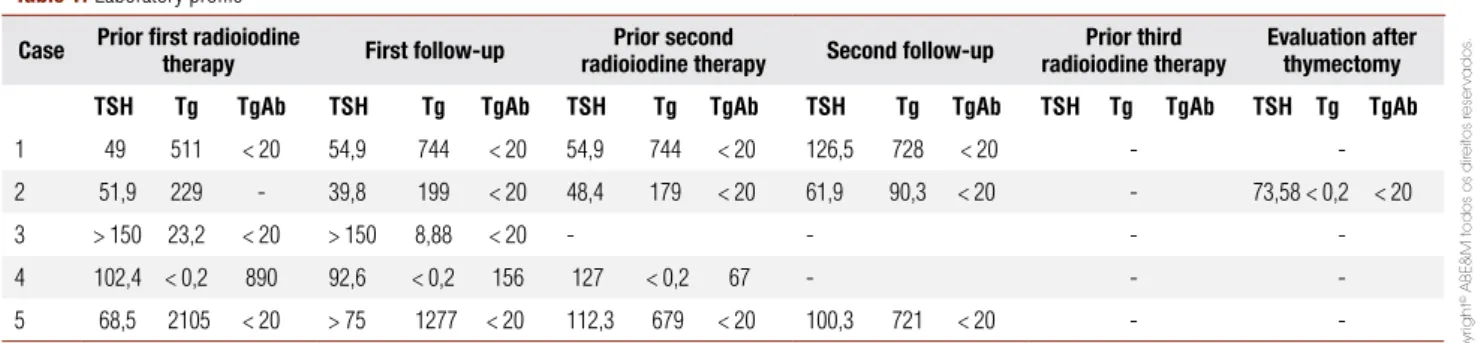
Consi-Table 1.Laboratory profile
Case Prior first radioiodine
therapy First follow-up
Prior second
radioiodine therapy Second follow-up
Prior third radioiodine therapy
Evaluation after thymectomy
TSH Tg TgAb TSH Tg TgAb TSH Tg TgAb TSH Tg TgAb TSH Tg TgAb TSH Tg TgAb
1 49 511 < 20 54,9 744 < 20 54,9 744 < 20 126,5 728 < 20 -
-2 51,9 229 - 39,8 199 < 20 48,4 179 < 20 61,9 90,3 < 20 - 73,58 < 0,2 < 20
3 > 150 23,2 < 20 > 150 8,88 < 20 - - -
-4 102,4 < 0,2 890 92,6 < 0,2 156 127 < 0,2 67 - -
-5 68,5 2105 < 20 > 75 1277 < 20 112,3 679 < 20 100,3 721 < 20 -
Copyright
© ABE&M todos os dir
eitos r
eser
vados.
dering the imaging findings and the levels of Tg (90.3 ng/mL), the mediastinum mass was resected and the histopathologic analysis was compatible with thymic tissue free of neoplastic disease and no signs of thyroid tissue. The evaluation performed nine months after the surgery revealed a negative 131I-WBS and a Tg after hormone withdrawal less than 0.2 ng/mL (Table 1).
CASE 3
A 28-year-old female underwent total thyroidectomy due to a cervical nodule and was diagnosed with pa-pillary thyroid carcinoma, follicular variant, without capsular or angiolymphatic infiltration. 123I-TS revealed an uptake of 2%, compatible with thyroid remnants, and the patient received 5,550 MBq (150 mCi) of I-131 for thyroid ablation. 131I-WBS post-therapy showed inten-se activity in the thyroid bed as well as accumulation in the anterior mediastinum (Figure 1C). CT scan showed a small amount of soft tissue attenuation in the medias-tinum that was suggestive of the thymus gland (Figure 1J). Six months after ablation, the 131I-WBS was ne-gative and Tg, after thyrotropin (TSH) estimulation, decreased (Table 1).
CASE 4
A 42-year-old woman with a cervical nodule found du-ring routine examination was submitted to total thyroi-dectomy with later diagnosis of papillary carcinoma with small solid areas poorly differentiated. The 131I-TS revea-led uptake of 3% and the 131I-WBS was positive only in the cervical, probably corresponding to thyroid remnants. She received 5,550 MBq (150 mCi) of I-131 for abla-tion and the post-therapy 131I-WBS showed uptake in the thyroid bed. During four years, the examinations, inclu-ding cervical ultrasonography (USG), 131I-WBS and CT scan, were all negative. During the follow-up period, the patient had positive anti-thyroglobulin antibodies (Table 1). The WBS obtained afterwards demonstrated radioiodine accumulation in the anterior mediastinum (Figure 1D). A CT scan showed a mediastinum mass su-ggestive of lymph node metastases (Figure 1I) and the pa-tient received 7,400 MBq (200 mCi) of I-131. Six mon-ths later, a CT scan post-therapy showed no significant changes and the mass was resected. The histopathologic examination was compatible with thymic tissue with no signs of malignancy, associated with necrotic areas, pro-bably caused by the radioiodine previously administered. Nowadays, the patient remains in ambulatory control.
Figure 1. Whole-body scans (WBS) and thoracic CT scans.
Note: 1(a,f) – Case 1 - post-therapy 131I-WBS shows cervical and upper mediastinum uptake, and CT scan shows soft tissue attenuation mass in the upper mediastinum, respectively; 1(b,g) – Case 2 - 131I-WBS
Copyright
© ABE&M todos os dir
eitos r
eser
vados.
CASE 5
A 56-year-old female was diagnosed with papillary car-cinoma of the thyroid and cervical lymph node involve-ment after total thyroidectomy and bilateral neck dissec-tion. 123I-TS showed uptake of 1.4% and 123I-WBS was positive in the cervical region. An ablative dose of 5,550 MBq (150 mCi) of I-131 was delivered and the post-therapy 131I-WBS confirmed the cervical region uptake, with no evidence of metastases. During follow-up, the patient developed enlarged cervical lymph nodes and recurrence of the disease was confirmed after cervical node dissection. Three months after surgery, she had a negative 131I-WBS and a Tg level in the face of TSH stimulation of 1277 ng/mL (Table 1). Considering the high level of Tg, despite the surgery and the negative WBS, the patient received new radioiodine treatment with 7,400 MBq (200 mCi) of I-131 and the post-therapy 131I-WBS was positive in the superior mediasti-num (Figure 1E), finding not observed during standard pre-therapy WBS using a diagnostic dose. A CT scan performed after the treatment showed the presence of a heterogeneous mass, bell shaped in the mediastinum, su-ggestive of thymus (Figure 1J), with no evidence of me-tastatic disease. Eleven months after the last radioiodine treatment, the patient had a negative 131I-WBS with Tg of 721 ng/mL with the patient off L-thyroxine therapy.
DISCUSSION
The radioactive iodine has been routinely used for the diagnosis and treatment of patients submitted to total thyroidectomy because of DTC. False-positive WBS occurs mainly due to misinterpretation of the physio-logic distribution of the radioiodine (4-6). Moreover, there are several pathologic conditions that could up-take iodine, being eventually confounded with metasta-tic disease from thyroid cancer (5-17).
Radioiodine uptake by the thymus gland is one of the possible causes of a false-positive WBS and might happen because of an intrathymic ectopic thyroid tis-sue (12,28) or thyroidal metastases to the thymus (29). However, in most of the cases, the uptake of radioiodi-ne occurs because of the presence of a residual normal thymus (20-23,25) or thymic hyperplasia (8,18,19,24).
The mechanism of radioiodine uptake by the thymus is not well understood yet. Vermiglio and cols. (23) conside-red the possibility of iodine concentration by the Hassal’s bodies present in the thymic tissue, as they resemble the follicular cells of the thyroid. More recently, Spitzweg and
cols. (30) demonstrated, for the first time at molecular level, the presence of the human Na+/I- symporter (hNIS)
by an extra thyroidal tissue, including the thymus, but with a capability of transport and concentration of iodine smaller than that presented in the thyroid gland. These findings could explain why the majority of cases found in the literature of thymic iodine uptake occur more com-monly on the post-therapy scans, when higher activities are administered, or during an evaluation some time after the ablation of the thyroid remnant, when there is more radioiodine to be taken up by the thymus (2,18,19,31).
Copyright
© ABE&M todos os dir
eitos r
eser
vados.
From the five cases reported in the present study, excluding the patient with positive thyroglobulin auto-antibodies (TgAb), the other four patients presented Tg levels higher than the normal value, without clini-cal, radiological or scintigraphic findings suggestive of metastatic disease. In an interesting way, one of the pa-tients submitted to thymectomy had an important de-crease in the levels of Tg after the surgery, without any other treatment being done between the surgery and the laboratory evaluation (Table 1). This finding arises the possibility of the thymic tissue as a source of benign production of Tg. Heath and cols. (39) demonstrated the thymic expression of Tg in adult rats and embryo-nic mice with the technique of reverse transcription and polymerase chain reaction from analyses of mRNA. Sospedra and cols. (40) found transcription of Tg in four of twelve samples of human thymus and Gotter and cols. (41) reported the existence of Tg expression in medullary epithelial cells of human thymus. In the present study, the two resected thymus were not evalu-ated by immunohistochemistry for the presence of Tg expression. Thus, randomized studies are needed to eva-luate whether the Tg production by the thymus is really effective and whether it could be stimulated by TSH.
Acknowledgement: This study was approved by the Ethics Com-mittee of Instituto Nacional de Câncer (Inca). The enclosed ma-nuscript has been read and approved by all authors, who attest that there are no financial disclosures to make.
Disclosure: no potential conflict of interest relevant to this article was reported.
REFERENCES
1. Mazzaferri EL. Management of low-risk differentiated thyroid can-cer. Endocr Pract. 2007;13(5):498-512.
2. Wilson LM, Barrington SF, Morrison ID, Kettle AG, O’Doherty MJ, Coakley AJ. Therapeutic implications of thymic uptake of radio-iodine in thyroid carcinoma. Eur J Nucl Med. 1998;25(6):622-8. 3. Mazzaferri EL, Kloos RT. Current Aproaches to primary
thera-py for papillary and follicular thyroid cancer. J Clin Endocrinol. 2001;86:1447-63.
4. Carlisle MR, Lu C, McDougall IR. The interpretation of 131I scans in the evaluation of thyroid cancer, with an emphasis on false positive findings. Nucl Med Commun. 2003;24(6):715-35. 5. McDougall IR. Whole-body scintigraphy with radioiodine-131. A
comprehensive list of false-positives with some examples. Clin Nucl Med. 1995;20(10):869-75.
6. Rudoni S, Toubeau M, Mansuy S, Vaillant G, Verges B, Brun JM, et al. False positive scintigraphic images in the surveillance of differen-tiated thyroid cancers. Ann Endocrinol (Paris). 1997;58(5):399-407. 7. Bakheet SM, Hammami MM. False-positive radioiodine
whole-body scan in thyroid cancer patients due to unrelated pathology. Clin Nucl Med. 1994;19(4):325-9.
8. Salvatori M, Salctnich I, Rufine V, Troncone L. Unusual false-posi-tive radioiodine whole-body scans in patients with differentiated thyroid carcinoma. Clin Nucl Med. 1997;22(6):380-4.
9. Hoschl R, Choy DHL, Gandevia B. Iodine-131 uptake in inflam-matory lung disease: a potential pitfall in treatment of thyroid carcinoma. J Nucl Med. 1988;29(5):701-6.
10. Misaki T, Takeuchi R, Miyamoto S, Kasagi K, Matsui Y, Konishi J. Radioiodine uptake by squamous-cell carcinoma of the lung. J Nucl Med. 1994;35(3):474-5.
11. Maslack MM. Iodine-131 accumulation in a pericardial effusion [letter]. J Nucl Med. 1987;28(1):133.
12. Greenler DP, Klein HA. The scope of false-positive iodine-131 ima-ges for thyroid carcinoma. Clin Nucl Med. 1989;14(2):111-7. 13. Ong SC, Eng DN, Sundram FX, Chan LL. A novel case of
false-positive I-131 whole-body scan in thyroid carcinoma caused by subdural hematoma. Clin Nucl Med. 2004;29(3):164-6.
14. Schneider JA, Divgi CR, Scott AM, Macapinlac HA, Sonenberg M, Goldsmith SJ, et al. Hiatal hernia on whole-body radioiodi-ne survey mimicking metastatic thyroid cancer. Clin Nucl Med. 1993;18(9):751-3.
15. Schuster DM, Alazraki N. Esophageal scarring causing false-positive uptake on I-131 whole-body imaging. Clin Nucl Med. 1998;23(5):334.
16. Lungo M, Tenembaum F, Chaumerliac P, Vons C, Mirat A, Beuzen F, et al. Ovarian endometriosis cyst with iodine 131 uptake: first case of false positive in the follow up for differentiated thyroid carcinoma. Ann Endocrinol (Paris). 2000;61(2):147-50.
17. Bakheet SM, Hammami MM, Powe J. False-positive radio-iodine uptake in the abdomen and pelvis: radioradio-iodine reten-tion in the kidneys and review of the literature. Clin Nucl Med. 1996;21(12):932-7.
18. Michigishi T, Mizukami Y, Shuke N, Yokoyama K, Noguchi M, Watanabe Y. Visualization of the thymus with therapeutic doses of radioiodine in patients with thyroid cancer. Eur J Nucl Med. 1993;20(1):75-9.
19. Connolly LP, Connolly SA. Thymic uptake of radiopharmaceuti-cals. Clin Nucl Med. 2003;28(8):648-51.
20. Bestagno M, Pagliaine R, Maira G, Pizzocaro C. Mediastinal up-take of 131-I in patients with thyroid cancer: may it be referred to normal thymus? Eur J Nucl Med. 1993;20(7):75-9.
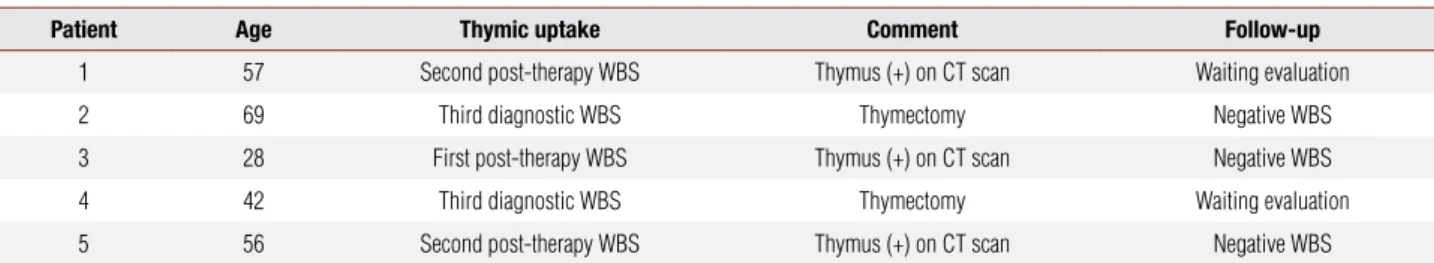
Table 2. Thymic uptake and follow-up
Patient Age Thymic uptake Comment Follow-up
1 57 Second post-therapy WBS Thymus (+) on CT scan Waiting evaluation
2 69 Third diagnostic WBS Thymectomy Negative WBS
3 28 First post-therapy WBS Thymus (+) on CT scan Negative WBS
4 42 Third diagnostic WBS Thymectomy Waiting evaluation
5 56 Second post-therapy WBS Thymus (+) on CT scan Negative WBS
Copyright
© ABE&M todos os dir
eitos r
eser
vados.
21. Davidson J, McDougall R. How frequently is the thymus seen on whole-body iodine-131 diagnostic and post-treatment scans? Eur J Nucl Med. 2000;27(4):425-30.
22. Veronikis IE, Simkin P, Braverman LE. Thymic uptake of Iodine-131 in the anterior mediastinum. J Nucl Med. 1996;37(6):991-2. 23. Vermiglio F, Baudin E, Travagli JP, Caillou B, Fragu P, Ricard M, et
al. Iodine Concentration by the thymus in thyroid carcinoma. J Nucl Med. 1996;37(11):1830-1.
24. Lee SH, Kwon HS, Yoo IR, Park HJ, Yoon KH, Cha BY, et al. Fal-se-positive iodine uptake in thymic hyperplasia on diagnostic I-123 whole body scan after total thyroidectomy. Clin Nucl Med. 2007;32(2):154-5.
25. Montella L, Caraglia M, Abbruzzese A, Soricelli A, Caputi M, Squame G, et al. Mediastinal images resembling thymus follo-wing 131-I treatment for thyroid cancer. Monaldi Arch Chest Dis. 2005;63(2):114-7.
26. Koh JM, Kim ES, Ryu JS, Hong SJ, Kim WB, Shong YK. Effects of therapeutic doses of 131I in thyroid papillary carcinoma patients with elevated thyroglobulin levels and negative 131I whole-body scan: comparative study. Clin Endocrinol. 2003;58(4):421-7. 27. Pacini F, Agate L, Elisei R, Capezzone M, Ceccarelli C, Lippi F, et al.
Outcome of differentiated thyroid cancer with detectable serum Tg and negative diagnostic (131)I whole body scan: comparison of patients treated with high (131)I activities versus untreated pa-tients. J Clin Endocrinol Metab. 2001;86(9):4092-7.
28. Spinner RJ, Moore KL, Gottfried MR, Lowe JE, Sabiston DC Jr. Thoracic Intrathymic Thyroid. Ann Surg. 1994;220(1):91-6. 29. Nam M, Chu YC, Choe W, Kim SJ, Hong SB, Kim YJ, et al.
Metas-tatic follicular thyroid carcinoma to the thymus in a 35-year-old woman. Yonsei Med J. 2002;43(5):665-9.
30. Spitzweg C, Joba W, Eisenmenger W, Heufelder AE. Analysis of human sodium iodide symporter gene expression in extra-thyroi-dal tissues and cloning of its complementary deoxyribonucleic acids from salivary gland, mammary gland, and gastric mucosa. J Clin Endocrinol Metab. 1998;83(5):1746-51.
31. Meller J, Becker W. The human sodium-iodine symporter (NIS) as a key for specific thymic iodine-131 uptake. Eur J Nucl Med. 2000;27(5):473-4.
32. Murakami M, Hosoi Y, Negishi T, Kamiya Y, Miyashita K, Yamada M, et al. Thymic hyperplasia in Graves’ Disease. J Clin Invest. 1996;98(10):2228-34.
33. Kirkeby KM, Pont A. Thymic hyperplasia in a patient with Graves` disease. J Clin Endocrinol Metab. 2006;91(1):1.
34. Brinkane A, Ounadi-Corbille W, Bellamy J, Leroy-Terquem E. Hyperplasia of the thymus in Graves` disease. A case report. Rev Pneumol Clin. 2004;60(4):239-41.
35. Nakamura T, Murakami M, Horiguchi H, Nagasaka S, Ishibashi S, Mori M, et al. A case of thymic enlargement in hyperthyroidism in a young woman. Thyroid. 2004;14(4):307-10.
36. Niendorf E, Parker J, Yechoor V, Garber JR, Boiselle PM. Thy-mic hyperplasia in thyroid cancer patients. J Thorac Imaging. 2005;20(1):1-4.
37. Kissin CM, Husband JE, Nicholas D, Eversman W. Benign thymic enlargement in adults after chemotherapy: CT demonstration. Radiology. 1987;163(1):67-70.
38. Sehbai AS, Tallaksen RJ, Bennett J, Abraham J. Thymic hyperpla-sia after adjuvant chemotherapy in breast cancer. J Thorac Ima-ging. 2006;21(1):43-6.
39. Heath VL, Moore NC, Parnell SM, Mason DW. Intrathymic expres-sion of genes involved in organ specific atoimmune disease. J Autoimmun. 1998;11(4):309-18.
40. Sospedra M, Ferrer-Francesch X, Dominguez O, Juan M, Foz-Sala M, Pujol-Borrell R. Transcription of a broad range of self-antigens in human thymus suggests a role for central mechanisms in toleran-ce toward peripheral antigens. J Immunol. 1998;161(11):5918-29. 41. Gotter J, Brors B, Hergenhahn M, Kyewski B. Medullary epithelial