The increase of cardiac output during pregnancy is the cause of heart failure in women with severe aortic valvular stenosis. Percutaneous aortic valvuloplasty has been associated with severe complications and short-term valvar restenosis. This case showed that percutaneous aortic valvuloplasty allowed both mother and fetus to survive after childbirth, and that postpartum treatment interruption resulted in maternal death in late postpartum care.
Mailing address: Walkiria Samuel Avila •
Rua Dr. Enéas Carvalho Aguiar, 44 - Cerqueira César - 05403-000 – São Paulo, SP – Brazil
E-mail: [email protected], [email protected]
Manuscript received November 25, 2008; revised manuscript received Janu-ary 23, 2009; accepted June 08, 2009
Aortic Valvuloplasty with Balloon Catheter in Maternal-fetal
Emergency in Adolescence
Walkiria Samuel Avila
1, Ludhmila Abrahao Hajjar
1, Tatiana da Rocha e Souza
1, Manuel Pereira M Gomes Junior
1,
Max Grinberg
1, Marcelo Zugaib
2Instituto do Coração do Hospital das Clínicas da Faculdade de Medicina da Universidade de São Paulo1; Departamento de Obstetricia e Ginecologia
do Hospital das Clínicas da Faculdade de Medicina da Universidade de São Paulo, SP2, Brasil
Key Words
Aortic Valve Stenosis; Balloon Dilatation; Pregnancy in Adolescence; Heart Failure.
Introduction
Severe aortic stenosis is considered to bring high risks to childbirth accounting for at least 1% of cardiopathies during pregnancy1. In general, severe aortic stenosis has
congenital etiology and maternal prognosis depends on the stenosis degree.
The increase of cardiac output in the peripartum period may cause heart failure (congestive heart failure), which must be treated with mechanical relief of valvular obstruction.
Percutaneous aortic valvuloplasty (PAVP) has been used as an alternative during pregnancy, once the main advantage it provides is avoiding the harmful effects of anesthesia and extracorporeal circulation (ECC) in fetus.
However, volemia and blood coagulation modifications during pregnancy and in the postpartum period poses additional risk of complications, even if the procedure is immediately proven successful.
This study reports the case of a pregnant adolescent with severe aortic stenosis. During her 37th week of pregnancy,
such condition evolved to a cardiogenic shock. The patient was successfully submitted to PAVP and later to a cesarean section. Nevertheless, it eventually led to death in the 6th
postpartum week.
Case Report
JASA, 18 years old, pregnant for the first time, nullipara, who has had heart murmur since childhood, presented no symptoms until the 28th week of pregnancy, when a progressive
dyspnea evolved to orthopnea. The patient had not been under prepartum care. Upon admission, she was conscious, dyspneic, cyanotic, no fever, blood pressure 80/40 mmHg, heart rate 146 bpm and blood saturation of 80% under ambient oxygen. Lung auscultation revealed crackling rales in the middle and inferior thirds of lungs and heart auscultation presented normophonetic sounds and ejective systolic murmur on left sternal edge.
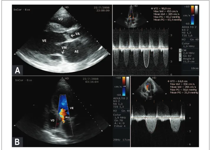
Electrocardiogram revealed sinus tachycardia and left ventricular overload. Chest radiography revealed increased cardiac area and signs of lung congestion. The measures obtained through transthoracic echocardiogram, shown on Table 1, presented an increased diameter of left cavities and greater myocardial thickness, relevant ventricular systolic impairment and bicuspid aortic valve, thickened with reduced mobility (Figure 1A).
Obstetric tests revealed: womb height 33 cm, impervious colon, no uterine contraction, single fetus in longitudinal position and cephalic presentation with 128 bpm. Ultrasound and cardiotocography showed restricted growth and well-preserved fetus vitality.
Oxygenation through nebulizer mask 8 L/min and administration of 80 mg of intravenous furesemide provided a slight improvement of such clinical condition. The medical team opted for percutaneous aortic valvuloplasty (PAVP) trying to allow a hemodynamic stabilization and later interruption of gestation through cesarean section, considering the picture of cardiogenic shock and imminent maternal death.
The procedure performed under sedation with fentanyl, propofol and etomidate started by applying a 9F introducer into the right femoral artery and femoral vein puncture to introduce a temporary pacemaker electrode impacted on the right ventricle. A left amplatzer 7F guide catheter, curve 1, and stiff 0.035”/ 2.60 m guide wire were used to transpose the aortic valve stenosis. Initial measurement evidenced a peak-to-peak gradient of 100 mmHg (Figure 2A). A Boston peripheral dilatation balloon of 4 cm diameter and 18 mm length was inflated under direct fluoroscopic control until its full expansion could be assured, with immediate deflation. A new measurement of the aortic transvalvar gradient revealed a peak-to-peak 50 mmHg (Figure 2B).
During the percutaneous aortic valvuloplasty, there was a prolonged fetal bradycardia, triggering an acute fetal suffering
e76
condition indicating an emergency cesarean section after the valvuloplasty, performed with no complications, under general anesthesia. After delivery, the patient remained stable, with no bleeding, regular uterine contractility and loquia flow.
Control Doppler ecochardiogram data are on Table 1 and in Figure 1B.
The male infant, healthy, weighing 2,170 g, gestational age of 37 weeks, Apgar indexes at 9 on the fifth and 10 on the tenth minute of life, presented good interatrial communication of 4 mm and evolved with no complications. After three days, the infant was discharged from the nursery unit.
The patient requested discharge on the third day of puerperium alleging personal problems. She did not show up for follow-up medical appointments for obstetric and cardiologic controls. According to the Maternal Mortality Committee, she was admitted to the Municipal Emergency Service on the 42nd
day after delivery, with a clinical picture of low cardiac output and heart failure, evolving to death 48 hours after.
Discussion
During pregnancy, congestive heart failure in patients with aortic stenosis is directly dependent on the valvular injury
Figure 1A – Doppler echocardiogram before and 1B after percutaneous aortic valvuloplasty.
Valve with important thickening, bivalve opening and reduced mobility. Maximum and medium aortic transvalvular gradient were 82 and 61 mmHg, respectively. The other structures (valves, ascending and descending aorta, and aortic arch) did not present anatomic alterations.
A
B
Before percutaneous
aortic valvuloplasty
After percutaneous aortic valvuloplasty
LA (mm) 45 45
LV (mm) 56 56
Mass Index (g/m2) 144 144
LF (%) 30 30
AVG (mmHg) 82/61 51/32
Aortic valvar area (cm2)
0,48 0,8
V1/V2 0,24 0,31
PAVP - percutaneous aortic valvuloplasty; LA – left atrium; LV – left ventricle; LF – left ventricular ejection fraction; AVG - aortic valve gradient; V1/V2 –
ratio between the maximum speeds on left ventricle outlow tract and on
aortic valve.
Table 1 – Measurements obtained in Transtoracic Doppler
Echocardiography before and after Aortic Percutaneous Valvuloplasty
Arq Bras Cardiol 2009; 93(6) : e76-e79
Ávila et al Emergency Aortic Valvuloplasty and Pregnancy
e77
degree, usually verified upon area measurements lower than 1 cm2. This condition gets worseon the third quarter,
when the maternal cardiac output is 40% higher than before childbirth2.
The analysis of 24 gestations in patients with aortic stenosis with average valvular area of 1cm2 revealed that 11 (45.8%)
patients presented angine, pre-syncope and heart failure symptoms, including a sudden death, upon reaching an average gestational age of 30 weeks, requiring surgery valvular change in two cases (8.3%)3.
Conventional drug therapies applied in congestive heart failure cases, which includes diuretic, vasodilator and beta-blocker drugs are relatively counter-indicated during gestation of patients with aortic stenosis, due to the disputable maternal effectiveness and the actual worsening of the placental flow.
Such restrictions determine that surgery intervention be the first choice before clinical manifestations of congestive heart failure. However, cardiac surgery during pregnancy is associated with maternal and fetus mortalities of 8.6% to 18.6%, related to the severity of maternal clinical condition and to the emergency character of the surgery4.
On the other hand, interrupting the gestation by performing cesarean or vaginal section in unstable hemodynamic conditions increases maternal risk due to blood loss and anesthesia effects. In this situation the percutaneous aortic valvuloplasty (PAVP) is intended to reduce the aortic transvalvular gradient, increase heart
rate and placental blood flow, besides improving maternal symptoms until childbirth.
Worldwide experience concerning percutaneous aortic valvuloplasty (PAVP) is still restricted to special cases, once the procedure is associated to high morbidity and its effectiveness is transitory. Nevertheless, in adolescence, a study with 148 children and adolescents showed that percutaneous aortic valvuloplasty could significantly reduce the transvalvular gradient, increase the valvular area and lower mortality levels. Then, it was concluded that the absence of valvular calcification was the most important predictor of free development of immediate and late events5.
In this case, the indication of percutaneous aortic valvuloplasty was based on the best survival indexes, both for the mother and the fetus, according to the gestational age. We believe that in the absence of fetus suffering signs, the treatment is intended to save the mother’s life, who is under a severe clinical condition. The childbirth must be performed later, based on the fetus conditions.
The good clinical conditions presented by the infant upon delivery allow us assume that intrauterine suffering during percutaneous aortic valvuloplasty was transitory and that subsequent gestation interruption allowed a favorable evolution of the newborn.
Maternal death on the 46th day after childbirth stresses the
need to continue cardiologic treatment in puerperium after percutaneous aortic valvuloplasty which, in this specific case, was palliative. Reabsorption of extravascular fluids on
uterus-Figures 2A – Pressure curve of left ventricle and aorta before and 2B after aortic valvuloplasty.
Peak-to-peak gradient measurement was 100 mmHg (Fig. 2A) before inlation. After delation, the gradient was down to 50 mmHg (Fig. 2B).
A
B
Arq Bras Cardiol 2009; 93(6) : e76-e79
Ávila et al
Emergency Aortic Valvuloplasty and Pregnancy
e78
References
placental and peripheral beds for maternal circulation and an active coagulation state make the postpartum period an equally risky one for maternal death, just as pregnancy itself.
Consider that the routine of maternal care and breastfeeding do not allow resting and obedience to restrictions made during pregnancy. Postpartum is a subject of multidisciplinary responsibility, but what we actually see is that patients can be hardly be controlled after hospital discharge. This case attests this reality, showing that the availability of the entire medical team that assisted her was not enough to avoid a fatal outcome and evidences that social problems, lack of familial support, the impact of maternal responsibility and immaturity of adolescence did not allow accurately understanding the severity of this case and disrupted further treatment.
Conclusion
We concluded that percutaneous aortic valvuloplasty may be performed in symptomatic patients in order to
allow reaching the optimal childbirth date in stable maternal hemodynamic conditions and with greater fetal survival. Nevertheless, a strict treatment must be kept in puerperium and it is equally important for maternal survival, just as during pregnancy.
Potential Conflict of Interest
No potential conflict of interest relevant to this article was reported.
Sources of Funding
There were no external funding sources for this study.
Study Association
This study is not associated with any post-graduation program.
1. Drenthen, W, Pieper, PG, Roos-Hesselink, JW, van Lottum WA, Voors AA, Mulder BJ, et al. Outcome of pregnancy in women with congenital heart disease: a literature review. J Am Coll Cardiol. 2007; 49: 2303-11
2. Avila WS, Rossi EG, Ramires JAF, Grinberg M, Bortolotto MRL, Zugaib M, et al. Pregnancy in patients with heart disease: experience with 1,000 cases. Clin Cardiol. 2003; 26 (3): 135-42.
3. Avila WS, Grinberg M, Rossi EG, Tarasoutchi F, Cardoso LF, da Luz PL, et al.
Impact of pregnancy on the natural history of severe aortic stenosis. Eur Heart J. 1996; 17 (Suppl): 444.
4. Arnoni RT, Arnoni AS, Bonini RAC, Almeida AFS, Neto CAN, Dinkuysen JJ, et al. Risk factors associated with cardiac surgery during pregnancy. Ann Thorac Surg. 2003; 76: 1605-8.
5. Moore P, Egito E, Mowrey H, Perry SB, Lock JE, Keane JF. Midterm results of balloon dilation of congenital aortic stenosis: predictors of success. J Am Coll Cardiol. 1996; 27: 1257-63.
Arq Bras Cardiol 2009; 93(6) : e76-e79
Ávila et al Emergency Aortic Valvuloplasty and Pregnancy