Clinicoradiological Session
Case 4/2009 – Fourteen-Year-Old Boy with Atrial Septal Defect and
Left Pulmonary Artery Stenosis
Edmar Atik
Hospital Sírio Libanês, São Paulo, SP - Brazil
Mailing address: Edmar Atik •
Rua Dona Adma Jafet, 74 cj 73 - Bela Vista - 01308-050 - São Paulo, SP, Brazil E-mail: [email protected]
Key words
Heart defects, Congenital pulmonary subvalvular stenosis; atrial septum / abnormalities; atrial septum / surgery.
Clinical data
The heart murmur had been identified at auscultation for the first time at four months of age, during a bronchopneumonia. Since then, the patient remained asymptomatic, physically active and did not use any specific medication.
At physical examination, the patient presented good general status, eupneic, normal skin color, acyanotic and presented normal pulses. His weight was 61.6 Kg, height was 171 cm, HR was 80 bpm and blood pressure (BP) was 110/75 mm Hg. The aorta could not be palpated at the furcula and the precordium presented slight impulses on the left sternal border and the ictus cordis could not be palpated. The second heart sound was constantly split, with the two components presenting equal intensity. The systolic murmur of +/++ intensity, harsh, of ejection, was audible on the high left sternal border. In the mitral area and throughout the dorsal area to the left, another systolic murmur, with a softer sound, was audible. The protosystolic click was evident throughout the sternal border and the liver could not be palpated.
The electrocardiogram (ECG) showed signs of right ventricular overload with an R wave of 13 mm in V1. ÂQRS was at +90o, ÂP at +70o and ÂT at +60o.
Radiographic image
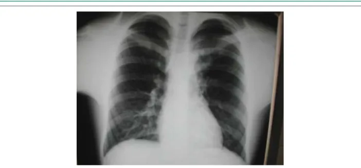
It shows the normal cardiac area with evident contrast with the pulmonary vascular net, which is decreased to the left and increased to the right. The middle arch is rectified (Figure 1).
Diagnostic impression
The radiographic image is compatible with left pulmonary artery stenosis due to the decreased arterial vascularity on this side. The increased pulmonary vascularization to the right might suggest arteriovenous blood shunt, at atrial or even ventricular level.
Differential diagnosis
Acyanogenic cardiopathies with blood shunt from left to right, such as IAC/IVC/PDA, when associated to the obstruction of the pulmonary flow, can present in a similar way. The close to normal cardiac area suggests the predominance of obstructive lesion, even in the presence of large-diameter intracavitary communications.
Diagnostic confirmation
The clinical data directed to the diagnosis of acyanogenic cardiopathy with left pulmonary artery stenosis (intense systolic murmur on the dorsal area, decreased pulmonary net to the left and right ventricular systolic overload at the ECG), associated to slight interatrial communication (mild systolic impulses on the sternal border and constantly split second sound). The echocardiogram showed interatrial communication of ostium secundum type with a 10-mm diameter, in addition to the stenosis in the beginning of the left pulmonary artery, with a 5-mm diameter and pressure gradient of 31 mmHg. The pulmonary perfusion with technetium confirmed the diagnosis through the total pulmonary flow of 94%, with 65% being directed to the right lung and 29% to the left lung.
Conduct
During surgery, ostium secundum interatrial communication with 12 mm of diameter was repaired and the stenotic left pulmonary artery was enlarged with autologous pericardium. The patient presented good evolution, with resolution of the anatomofunctional picture.
Comment
In congenital cardiopathies, radiographic details can be very useful, as in the present case, in which the decreased vascular net to the left became an unquestionable element of the presence of left pulmonary artery stenosis.
Clinico Radiological Session
Arq Bras Cardiol 2009;92(5):392-393
Figure 1 - The radiographic image shows the cardiac area close to normal, with slight ventricular arch prominence and decreased pulmonary artery net to the left, due to presumable stenosis of the left pulmonary artery.
Figure 2 - The echocardiogram shows a small atrial septum discontinuity and low, at the color Doppler, from left to right, in subcostal view, in A. Additionally, the transversal
parasternal view shows evident stenosis in the beginning of the left pulmonary artery (arrow), in B.