Arq Bras Cardiol volume 74, (nº 4), 2000
Amaral et al Congenital atresia of the left coronary artery
339
Hospital do Coração de Ribeirão Preto/Fundação Waldemar B. Pessoa, Hospital das Clínicas da Faculdade de Medicina de Ribeirão Preto, USP and Instituto do Coração do Hospital das Clínicas da Faculdade de Medicina de São Paulo, USP Mailing address: Fernando Amaral – AV. Independência, 1379 – 14025-390, Ribeirão Preto, SP, Brazil
Fernando Amaral, Carla Tanamati, João A Granzotti, Jorge L Haddad, João Ronaldo A Leite, Miguel Barbero-Marcial
Ribeirão Preto, SP - Brazil
Congenital Atresia of the Ostium of the Left Coronary Artery.
Diagnostic Difficulty and Successful Surgical
Revascularization in Two Patients
Brief communication
We report two cases of congenital atresia of the os-tium of the left coronary artery. Case 1: a six-month-old infant presenting with serious cardiac insufficiency. A noninvasive diagnosis of dilated myocardiopathy was established and the clinical picture was pharmacologi-cally compensated. When the patient was nine months of age, a hemodynamic study was performed that revealed congenital atresia of the ostium of the left coronary artery; the infant immediately underwent a successful anastomosis of the internal mammary artery with the left coronary artery. Case 2: an eleven-year-old asympto-matic boy with a history of heart murmur from the age of six months on, was refered for surgery with a diagnosis of anomalous origin of the left coronary artery from pulmo-nary trunk. A definitive diagnosis of atresia of the left co-ronary ostium was only established during surgery. Successful surgical revascularization with the left inter-nal mammary artery, and left ventricular aneurysmecto-my were performed.
Congenital anomalies of the coronary arteries,
occur-ring in 1-2% of the population 1, are classified as anomalies
at the origin (abnormal exit from the pulmonary trunk of the left coronary artery), terminal (coronary fistulae) and distri-butive (one coronary artery only) 2. Congenital atresia of
the ostium of the left coronary artery is a rather rare occur-rence that is not even mentioned in the above-cited litera-ture. Classically, it consists of total ostial atresia of the left coronary artery, possibly in association with atresia of the trunk of this vessel. Clinically, during the first months of life such patients may present with cardiac insufficiency (infantile form) or a delayed (including adults) picture of co-ronary insufficiency (adult form). We report below two pa-tients of different ages and clinical presentations, having
congenital atresia of the ostium of the left coronary artery of difficult diagnosis, who underwent successful surgical myocardial revascularization.
Case reports
Case 1 – A nine-month-old boy from Divinolândia, SP, with a clinical history of tiredness on sucking, accompanied by discrete diaphoresis from the first month of life on. At six months of age, after a case of pneumonia, a sudden worsening of his condition occurred accompanied by cardiac insufficiency. A noninvasive investigation diagno-sed dilated myocardiopathy. The patient showed signifi-cant improvement following medication, receiving a dis-charge from the hospital with a prescription of digoxin, furo-semide and captopril. At nine months of age, he was referred to clinical examination at the Heart Hospital of Ribeirão Pre-to and he was found Pre-to be in excellent general condition (weight: 10 kg). On physical examination, he had a normal pulse and upon cardiac auscultation a ++/6 systolic murmur on the 2nd – 3rd left intercostal space with normal heart
340
Amaral et al
Congenital atresia of the left coronary artery
Arq Bras Cardiol volume 74, (nº 4), 2000
(fig. 1, lower side) and an improvement of electrocardiogra-phic pattern (fig. 2 B). A recent echocardiograelectrocardiogra-phic study still shows left ventricular dilatation and hypokinesia of the late-ral wall, but also shows global improvement in the ejection fraction (=0.72).
Case 2 – An eleven-year-old male from Franca, SP with a clinical history of cardiac murmur detected on a
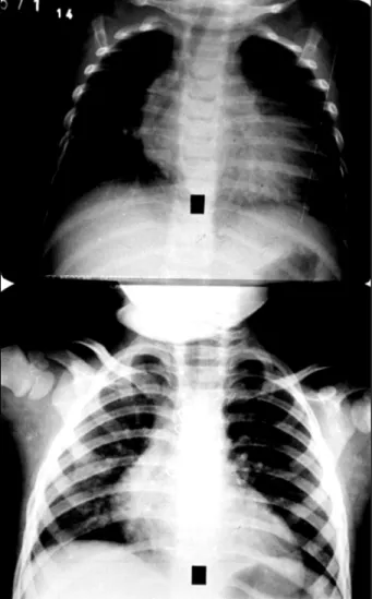
routine examination when six months old was referred to the Heart Institute of São Paulo for diagnostic investigation and treatment. When the boy was 2 ½ years old, a hemodynamic study was performed that led to a diagnosis of anomalous origin of the left coronary artery from pulmonary trunk. Surgical treatment proposed at the time was not accepted by the family; the child developed asthmatic bronchitis, no physical limitations and a good weight/structural growth pattern. Upon admittance to the hospital, he was observed to be in good general condition, had a palpable pulse and ++/6+ ejection systolic murmur in the mitral area. Thoracic radiography showed a mild increase in the cardiac area; the electrocardiogram showed signs of left ventricular strain and an inactive high antero-lateral area (fig. 4 A). An effort test according to Bruce protocol detected upslope of the ST segment in V4-V6, compatible with antero-apical ischemia. The echocardiogram revealed an anomalous origin of the left coronary artery from the pulmonary trunk, slight mitral insufficiency and an apical aneurysm of the left ventricle with an ejection fraction of 0.69. Thallium 201 scintigraphy during effort showed a persistent hypocaptation antero-septal and apical regions; technetium-99m scintigraphy Fig. 1 - Antero-posterior thoracic radiography of case 1, showing preoperative
cardiomegaly and signs of pulmonary congestion (upper); radiological improvement 11 months after surgery (lower).
Fig. 2 - Electrocardiogram of case 1, showing preoperative signs of antero-lateral infarct (A) and improved ventricular repolarization of the anterior wall 11 months following surgery (B).
Fig. 3 - A) Preoperation hemodynamic study of case 1., showing left ventri-culography in systole with marked diffuse hypokinesia: B) right coronariography in OLA showing dilated RCA with collateral circulation to LCA in the initial and final phase C) ; D) aortography of OLA showing normal opacity of RCA and contiguity but no communication of the LCA with the Valsalva sinus. OLA – ostium of the left coronary artery; RCA – right coronary artery; LCA – left coronary artery.
Arq Bras Cardiol volume 74, (nº 4), 2000
Amaral et al Congenital atresia of the left coronary artery
341
revealed diffuse and moderate hypocontractibility of the left ventricle with an ejection fraction of 0.31. A new hemo-dynamic study showed severe hypokinesia of the left ven-tricle with an antero-apical aneurysm and anomalous origin of the left coronary artery from the pulmonary trunk. Surgi-cal myocardial revascularization and aneurysmectomy were indicated. Surgery revealed a large left ventricular aneurysm with marked diffuse hypokinesia. Under extracor-poreal circulation, the pulmonary trunk was opened longi-tudinally, the left coronary ostium not becoming visible. The aorta was opened transversally; the impossibility of fin-ding the left ostium suggested a diagnosis of atresia of the left coronary ostium. Using the left internal thoracic artery, myocardial revascularization of the left coronary artery fol-lowed by aneurysmectomy were performed. At present, 3 ½ years following surgery, the youngster is asymptomatic and normal on physical examination. Thoracic radiography and the electrocardiogram denote signs of an anterolateral in-farct with significant alterations of left ventricular repolariza-tion (fig. 4 B).
Discussion
Left coronary ostium congenital atresia is an extremely rare disease; an incidence between 0.01-0.04% of “sole co-ronary” cases is admitted on the basis of results of necros-copy studies 3. The rarity of this condition can be realized
from its absence from a relevant, recently published study on congenital anomalies of coronary arteries 2, and also from
the fact that only 23 cases had been described before 1989 4.
In the last nine years, reports in the literature remain quite scarce, calling attention to the fact that only five cases were treated in a European center of reference, only one of a breast-feeding infant 5; this experience has been recently
amplified to include nine cases (Alain Serraf, personal com-munication).
The anatomical alteration consists of atresia of the os-tium of the left coronary artery, which may be associated with hypoplasia or atresia of the trunk of this vessel. Occa-sionally, serious isolated stenosis of the ostium can be met with and it may have some importance concerning the surgical technique to be employed 5.
From the clinical point of view, this anomaly may be classified as “infantile” (a precocious manifestation with cardiac insufficiency) or “adult” (manifestation of angina or sudden death); the role of the collateral circulation between the right and the left coronary arteries is considered vital at the moment of the clinical presentation of these patients. The diagnostic suspicion based on purely clinical data is difficult, because symptoms are nonspecific. On the other hand, complementary examinations play a fundamental role in the preliminary phase. Thoracic radiography may be nor-mal in the “adult” type of the anonor-maly or exhibit cardiome-galy and signs of pulmonary congestion in infants with cardiac failure. The clinical picture may be easily mistaken for dilated myocardiopathy, as occurred in the first case here presented. The electrocardiogram may become a valuable
diagnostic tool when a suspicion of this anomaly exists, as happened in the case of the first patient, where the elec-trocardiogram showed a pattern of antero-lateral infarction. An interesting phenomenon to remember is the resemblance of the clinical picture found in the patients with the anomalous origin of the left coronary artery of the pulmo-nary trunk 6. This was our first diagnostic hypothesis in both
cases, largely because of the electrocardiographic findings, as we have recently had the opportunity to demonstrate 7.
Theoretically, the diagnosis of atresia of the left coronary ostium may be obtained from the echocardiogram, in spite of difficulties due to individual anatomy. In this regard, the precise echocardiographic delimitation of the atresic portion may be difficult even if aided by Doppler. We believe this to have occurred in case 1. Diagnostic confirmation has been routinely made through the hemodynamic study, differential diagnosis of anomalous origin of the left coro-nary artery from the pulmocoro-nary trunk being of importance. In this regard, interruption of the retrograde flow in the trunk of the left coronary artery and the absence of opacity are worth noting.
In order to avoid the progress of the myocardial lesion and to control cardiac insufficiency in infants, surgery should be performed immediately after diagnosis. Despite the record of two unsuccessful cases in breast-feeding infants 6,8, present experience shows that revascularization
with the patient’s own mammary artery is a viable procedure in these cases, some reports of operations of patients in the 3 to 11 year age range being available 4,5. A technique of
surgical angioplasty in children with obstructive anomalies of the coronary arteries, including left ostium atresia, has recently been described 9. Although new, this procedure
seems to be of interest for the preservation of material for future revascularization, because in some cases saphenous vein grafts were used. Another consideration to be taken into account at the moment of surgical indication is the prevention of sudden death. Older children, adolescents and even adults presenting with few symptoms should be operated on shortly after diagnosis in order to prevent such an outcome, as already reported 10. Our patients reflect in a
342
Amaral et al
Congenital atresia of the left coronary artery
Arq Bras Cardiol volume 74, (nº 4), 2000
1. Engel HJ, Torres C, Page L Jr. Major variations in anatomical origin of the corona-ry arteries. Angiographic observations in 4.250 patients without associated congenital heart disease. Cathet Cardiovasc Diagn 1975; 1: 157-69. 2. Fernandes ED, Kadivar H, Hallman GL, Reul GJ, Ott DA, Cooley DA.
Congeni-tal malformations of the coronary arteries: the Texas Heart Institute experience. Ann Thorac Surg 1992; 54: 732-40.
3. Sharbaugh AH, White RS. Single coronary artery. JAMA 1974; 230: 243-6. 4. Koh E, Nakagawa M, Hamaoka K, Sawada T, Oga K. Congenital atresia of the left
co-ronary ostium: diagnosis and surgical treatment. Pediatr Cardiol 1989; 10: 159-62. 5. Serraf A, Baron O, Nottin E, at al. Atrésie ou sténose congénitale de l’ostium co-ronaire gauche. Revascularisation myocardique chez 5 enfants. Arch Mal Coeur 1993; 86: 587-91.
References
6. Byrum CJ, Blackman MS, Schneider B, Sondheimer HM, Kavey REW. Congenital atresia of the left coronary ostium and hypoplasia of the left main coronary artery. Am Heart J 1980; 99: 354-8.
7. Amaral F, Carvalho JS, Granzotti JA, Shinebourne EA. Origem anômala da artéria coronária esquerda do tronco pulmonar. Perfil clínico e resultados a médio prazo do tratamento cirúrgico. Arq Bras Cardiol 1999; 72: 307-13.
8. Harada K, Ito T, Suzuki Y, Shiota T, Shimada K, Takada G. Congenital atresia of the left coronary ostium. Letter. Eur J Ped 1993; 152: 539-40.
9. Bonnet D, Bonhoeffer P, Sidi D, et al. Surgical angioplasty of the main coronary arteries in children. J Thorac Cardiovasc Surg 1999; 117: 352-7.
10. Hauwaert LGV, Dumoulin M, Moerman P. Congenital atresia of left coronary os-tium. Br Heart J 1982; 48: 298-300.
test and confirmed by radioisotope evidence. Another inte-resting occurrence worth mentioning because of its rele-vance to this case was the difficulty of a preoperative diag-nosis in the face of the results of the hemodynamic study, which revealed an anomalous origin of the left coronary artery of the pulmonary trunk. In addition to these two fin-dings, another one in particular deserves attention in this patient. Four years following surgery, electrocardio-graphic evidence of worsening secondary to segmental ischemia of the left ventricle, confirmed by radioisotope evidence, could be detected. Yet, in spite of this ischemic alteration, the patient remains fully asymptomatic follo-wing great effort. It is likely that a compensatory mecha-nism effectively regulates the myocardial oxygen supply and demand relationship, thus avoiding manifestations of angina.
Due to the small amount of experience available in this field, long-term prognosis in these two cases remains uncer-tain. It becomes clear however that a delay in the diagnosis
and consequently of treatment of the patient must influence recovery of left ventricular function in a negative manner.