Case 4/2007 - A 21 Year-Old Woman with Hypertrophic
Cardiomyopathy who Presented Hemorrhagic Shock and
Disseminated Intravascular Coagulation Following Oophorectomy
due to Ovarian Cyst Rupture
Márcio Silva Miguel Lima, Antônio Laurinavicius, Mauro Canzian
Instituto do Coração do Hospital das Clínicas - FMUSP, São Paulo, SP - Brazil
Mailing address: Vera D. Aiello •
InCor – Av. Dr. Enéas de Carvalho Aguiar, 44 – 05403-000 – São Paulo, SP E-mail: [email protected]
Key words
Cardiomyopathy, hypertrophic; shock, hemorrhagic; disseminated intravascular coagulation; ovariectomy.
Section editor: Alfredo José Mansur ([email protected])
Associated editors: Desidério Favarato ([email protected]) Vera Demarchi Aiello ([email protected])
costal margin.
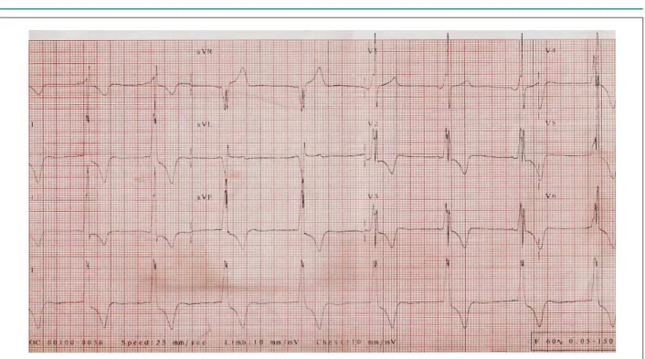
The electrocardiogram (June 2004) revealed sinus rhythm, a heart rate of 62 bpm, a short P-R interval, SÂQRS +90° tending forward, positive delta wave in V1, Q waves
in V5 e V6, and alterations in ventricular repolarization. The electrocardiographic pattern was not significantly different from the pattern the patient had shown since 2002 (Fig. 2).
Marked left ventricular hypertrophy was observed on the echocardiogram. The systolic function was interpreted as normal even though the first echocardiogram-based assessment had suggested lowered systolic function. When the test was reviewed, the interpretation of the lowered function was attributed to the asynchronous movement of the interventricular septum (table 1).
After the syncopal episodes, an in-depth electrophysiological investigation was performed. An electrophysiological study performed in June 2004 revealed the presence of two anomalous pathways of atrioventricular cardiac impulse conduction– one of left posteroseptal topography and another of left lateral topography.
The patient underwent successful radiofrequency ablation of the posteroseptal pathway. The other pathway was not accessed due to the long duration of the procedure. The patient was discharged and prescribed treatment with losartan 50 mg, carvedilol 50 mg, and acetylsalicylic acid 100 mg, which reduced the chest palpitation crises and prevented the recurrence of syncope.
The patient was readmitted in March 2005 to undergo ablation of the posterolateral pathway. After the procedure, the heart rate decreased to 28 bpm, followed by cardiac arrest in the presence of pulseless electrical activity, which was reversed. The cardiogenic shock persisted, requiring orotracheal intubation and use of vasoactive drugs.
The echocardiogram made in 2005 showed marked hypertrophy, left ventricular dilation and reduced systolic function, once again attributed to the paroxysmal movement of the septum.
Table 2 shows dynamic electrocardiogram progression data. The patient recovered progressively and was discharged from hospital.
Two months later (May 13, 2005), she sought medical care at the emergency department of a nearby hospital due to severe abdominal pain. The clinical examination showed A 21-year-old female patient was referred to the hospital
during the postoperative phase of an exploratory laparotomy for hemorrhagic acute abdomen.
The patient had a history of heart disease since birth. While still an infant, dyspnea was observed and at the medical evaluation a heart murmur was diagnosed. At the age of two, she underwent cardiac catheterization that revealed findings consistent with hypertrophic cardiomyopathy. The echocardiography showed left ventricular hypertrophy, with preserved systolic function (table 1).
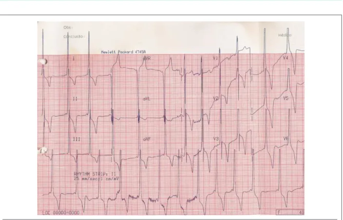
At that time (1991), the electrocardiogram showed sinus rhythm, with a PR interval of 80 msec, presence of a positive delta wave in leads I, II, aVF, V1, and V6, left
ventricular overload, and strain-type alterations in ventricular repolarization (fig. 1).
The condition progressed during childhood with the patient experiencing dyspnea during physical exertion and heart palpitations; these symptoms remained until20 years of age when she began to experience syncopal episodes preceded by precordial pain andtriggered by medium-level efforts. She took prescribedatenolol until the age of 18 (2003), when the drug was replaced by carvedilol.
Several dynamic 24-hour electrocardiograms were performed, which revealed few and isolated atrial and ventricular extrasystoles until 2004, when sustained ventricular tachycardia was recorded.
Table 1 - Echocardiographic progression
1989 1990 1991 1993 2002 2004 2005
Age (years) 6 7 8 9 18 19 20
Interventricular septum (mm) 13 14 16 15 19 26 20
Left ventricular posterior wall (mm) 11 14 16 15 19 25 20
Diastolic diameter of the left ventricle (mm) 37 31 28 42 58 54 65
Left ventricular ejection fraction (%) 82 - - - - 30* 22*
∆ D% 50 50 48 34
Aorta (mm) 19 N N N 25 26 23
Left atrium (mm) 22 N N N 33 41 38
Valve ModMI ModMI
Left intraventricular gradient (mmHg) 15
-Right intraventricular gradient (mmHg) 25
-* considered as non-reliable due to the presence of asynchronous movement of the interventricular septum. N - normal; ModMI - moderate mitral insufficiency
Fig. 1 - ECG – presence of preexcitation (delta wave), left ventricular overload and changes in strain ventricular repolarization.
signs suggestive of peritoneal irritation and anemia. The diagnosis was acute abdomen due to a ruptured ovarian cyst. The patient was readmitted, underwent left oophorectomy, and was discharged from hospital.
One week later, the patient still had abdominal pain and her dyspnea worsened. She was seen at the emergency room of the Hospital das Clínicas and, after medical evaluation, underwent a new exploratory laparotomy.
On physical examination (May 20, 2005), the patient’s blood pressure was 90/60 mmHg, her heart rate was 80 bpm, the lungs showed no abnormalities, and the heart sounds were rhythmic with no auscultatory changes. Mild hepatomegaly and pain were evident at the site of the operative wound, but no edema was observed.
The electrocardiogram (May 20, 2005) showed sinus rhythm, a heart rate of 83 bpm, a short P-R interval, SÂQRS +90 tending forward, a positive delta wave in V1, Q waves in V5 and V6, as well as alterations in ventricular
repolarization.
Laboratory test findings (May 21, 2005) were: hemoglobin 8 g/dl, hematocrit 26%, leucocytes 6300/mm³, platelets 153000/mm³, creatininemia 1.2 mg/dl, blood glucose 92 mg/dl, serum sodium 137 mEq/l and potassium 5.8 mEq/l. The patient remained in shock with diffuse bleeding refractory to treatment and died on May 22, 2005.
Fig. 2 - ECG. Presence of preexcitation (delta wave), left ventricular overload and changes in strain ventricular repolarization, similar to the previous one (Fig 1).
Table 2 - Dynamic echocardiographic progression
1990 1992 2000 2001 2004
Age (years) 7 9 16 17 20
Minimum heart rate (bpm) 43 48 39 45 37
Maximum heart rate (bpm) 166 146 122 126 113
Isolated ventricular extrasystoles 0 0 119 105 1,253
Atrial extrasystoles 17 17 61 12 33
Non-sustained ventricular tachycardia - - - - 1 (5 beats)
Clinical aspects
This is the clinical case of a young female patient diagnosed with hypertrophic cardiomiopathy (HCM) shortly after birth. By definition, HCM is a disease of genetic etiology characterized by marked myocardial hypertrophy with no clinically identifiable cause, that is, the increase in myocardial mass should not be related to potential etiologies such as systemic blood pressure or aortic stenosis, for instance1. The
prevalence of HCM in the population is approximately 1:500.2
It is the most common genetic heart disease, and is observed in 0.5% of the non-selected patients routinely referred to undergo echocardiogram. The age groups most affected are older children and young adults.
tends to be reduced in size, and at end-systole is obliterated. From the histological point of view, HCM is characterized by myocardiocyte hypertrophy, fiber disarray and fibrosis.
Clinical presentation varies a lot, but the outstanding triad for diagnosis is the association of dyspnea, thoracic pain, and syncope in young individuals, symptoms clearly observed in the patient in question. Dyspnea was the earliest symptom. Dyspnea may have a progressive development depending on the elevation of diastolic blood pressure due to reduced complacency and the well-known dynamic left ventricular outflow tract obstruction. Later, dyspnea may be caused by systolic dysfunction.
The chest pain reported by the patient was due to myocardial ischemia triggered both by increased metabolic demand and reduced coronary perfusion due to obstruction of the left ventricular outflow, or the alteration of large vessels with myocardial bridges and reduction of vessel lumen diameter. Alterations in the microcirculation with a low capillary-to-myocyte ratio are also observed.
The physical examination revealed a murmur compatible with mitral insufficiency, which may occur in hypertrophic cardiomyopathy especially when there is a large anterior systolic movement of the mitral valve. Typically, a systolic murmur is heard along the left sternal border and varies in intensity and duration during maneuvers that alter ventricular filling, such as the Valsalva maneuver and moving from squatting to standing.
The diagnosis of this patient was made during infancy through ancillary tests. There are no pathognomic findings on these tests to corroborate a diagnosis of CHS. The ECG shows classic signs of ventricular hypertrophy, such as increased QTS amplitude with left axis deviation and repolarization changes. In the clinical case we present, there was also a sign of preexcitation represented by delta waves, but this can occur also in patients with hypertrophy without preexcitation. The best test for a diagnosis and further follow-up is the echocardiogram that assesses both the structural aspect, defining the extent of myocardial mass increase, as well as the resulting functional consequences, i.e., the presence of an intracavitary gradient, anterior movement of the mitral valve with or without mitral insufficiency, and also the overall function of the left ventricle. According to the report, the asynchronous movement of the septum made it very difficult to assess the systolic function. In this case, other methods could have been used, such as radioisotope ventriculography enabling a more objective measure of the ejection fraction. Cardiac magnetic resonance could also be employed for the measurement, as the technique has exceptional structural resolution and will likely become the “gold standard” test for this diagnosis, especially for the non-obstructive forms of the disease. Although this patient had undergone coronary angiography, this test is not fundamental for a diagnosis. Its main role is to assess the intracavitary pressures more accurately and define the presence and size of the gradients.
It is extremely important to define whether there is left ventricular outflow tract obstruction or not; this obstruction is defined as a gradient greater than 30mmHg at rest or
greater than 50mmHg under provocative tests, as with the use of dobutamine. Therefore, the echocardiographic gradient of the patient (15mmHg) does not represent left ventricular outflow tract obstruction, which occurs in 30% to 50% of the patients. In this case, in the absence of a significant intraventricular gradient, provocative maneuvers can be used to identify a potential left ventricular outflow obstruction.
The annual mortality rate due to hypertrophic cardiomyopathy ranges from 1% to 6%, and most cases are of sudden death. In an epidemiological study conducted with non-selected patients, three large groups of types of death were observed: sudden death, heart failure and cerebrovascular accident associated with atrial fibrillation.3
The patient progressed with chest pain, syncope, and malignant arrhythmia detected by Holter monitoring, which meant that her clinical state could lead to a high risk of sudden death, considering that this fatal event is more frequent in young patients3. The consensus about hypertrophic
cardiomyopathy reached by the American and European Societies of Cardiology, headed by Professor Barry Maron, among others, indicates unexplained syncope and sustained spontaneous ventricular tachycardia as the major risk factors for sudden death. Other factors are prior cardiac arrest, family history of sudden cardiac death, wall thickness of more than 30 mm, hypotensive response to physical exertion, and non-sustained ventricular tachycardia (NSVT) detected by Holter monitoring4. It is important to emphasize that 5% of
the patients without any known risk factor may experience sudden death5.
Syncope associated with sustained ventricular tachycardia was the main indication for the use of an implantable cardioverter defibrillator (ICD) as the primary preventive measure against sudden death in a study conducted to validate this therapy6. In the study, it was also important to observe
that the great majority ofmalignant arrhythmias occurred in asymptomatic patients orthose with Class II heart failure, duringphysical inactivity or lightactivities. The ICD effectively protected high-risk patients by reverting sustained ventricular tachycardias and ventricular fibrillation. On the other hand, it must be remembered that ICDs are expensive and not innocuous, since inappropriate shocks may worsen quality of life and can, paradoxically, even trigger potentially lethal arrhythmias. Antiarrhythmic drugs have not been proven effective in preventingsudden death5.
An electrophysiological study (EPS) is not the best alternative for risk stratification since it renders inconclusive results5. Berh et al7 assessed the accuracy of EPS to determine
The concomitant findings characteristic of HCM and ventricular preexcitation by an accessory pathway in the same patient lead us to question a possible connection, beyond mere coincidence, between both conditions. Such an association, although uncommon, has already been noted by several authors and has been the subject of etiological and physiopathological speculation. Braunwald et al8 have
hypothesized that anomalous ventricular activation could result in regional myocardial hypertrophy and that, on the other hand, localized hypertrophy could hinder the isolation effect of the atrioventricular ring. One condition could, therefore, lead to a greater incidence of the other. Nevertheless, later observations suggest that the association of both phenotypes could be restricted to some familial groups (with an autosomaldominant patternofinheritance), thus reflecting a specific genetic etiology distinct from that observed in cases of isolated HCM. A large family has been described in which a mutation in chromosome 7q3 was expressed as ventricular hypertrophy, ventricular preexcitation, or both9. However, these patients had a few
peculiar characteristics: the histological test revealed a larger proportion of fibrosis than what is commonly found in isolated HCM (although already reported in several series of cases of HCM associated with Wolff-Parkinson-White), as well as a higher incidence of total atrioventricular blockage. Progression to dilatation seems to be significantly more frequent among patients with both conditions when compared to those with isolated HCM10, and this could be
the case of the patient in question.
Treatment is heterogeneous, consistent with the clinical presentation. Clinical treatment with negative chronotropic drugs is first-line therapy, and beta-blockers are the preferred agents. The nondihydropiridinic calcium channel antagonists (verapamil and diltiazem), and disopiramide (a type IA antiarrhythmic drug) are monotherapy alternatives for patients who can not tolerate beta-blockers or as adjuvant therapy. This patient was initially treated with atenolol, but the results were not favorable, as it happens in a minority of cases.
Refractoriness to clinical treatment is an indication for invasive therapies in HCM. These include the use of a double-chamber pacemaker, alcohol septal ablation by selective catheterization of a septal branch of the anterior descending artery, and septal myotomy-myomectomy that has a larger number of cases and results published in the literature. These measures are indicated for refractory cases as mentioned above, and in cases with significant left-ventricular outflow obstruction, which is not applicable to this case. Cardiac transplantation is the treatment indicated in cases of total clinical and invasive refractoriness to treatment, as well as advanced-stage heart failure.
Adverse progression of the disease occurs in a minority of cases. Fortunately, the great majority of patients remain asymptomatic throughout life. In a recent publication, Arteaga et al11 showed that, in a follow-up period of up to 17 years
(average of 7 years), the overall progression of HCM was benign, with a 15-year accumulated survival rateof87.9%. They observed that the main causes of disease-related death correlated with the NYHA functional classification and a wall thickness greater than 30 mm, a characteristic already
established in a specific study conducted earlier12.
In 5% to 10% of the patients, HCM progresses to marked systolic dysfunction with progressive left ventricular wall thinning and cavity enlargement. Onset of CHF during childhood is a significant unfavorable prognostic factor. Nevertheless, as was the case here, CHF was not the final development. The patient experienced an acute abdominal hemorrhagic event (ruptured ovarian cyst) with later complications, a problem that may affect any young woman and has no causal relationship with the underlying heart disease.
(Dr. Márcio Silva Miguel Lima, Dr. Antônio Laurinavicius)
Diagnostic hypothesis
Hypertrophic cardiomyopathy, with marked diastolic dysfunction; final event – disseminated intravascular coagulation.
Autopsy
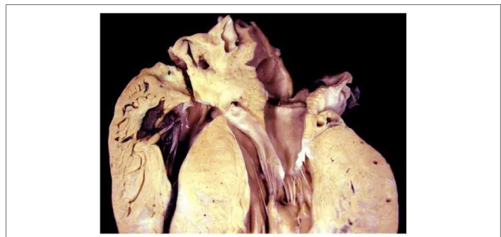
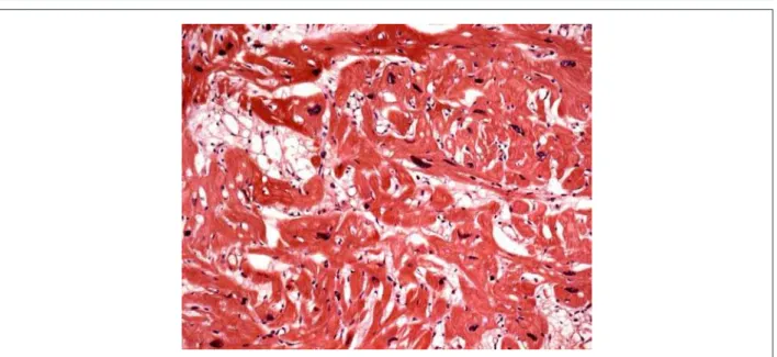
The corpse was in anasarca and, at the opening of the thoracic and abdominal cavities, approximately 280 ml of a clear yellow citrine-colored fluid were found in the pericardial sac, as well as a hemorrhagic collection of nearly 1560 ml in the abdomen. The heart was significantly enlarged, weighing 834 g. After opening of the cavities and removal of blood clots, diffuse hypertrophy of the ventricular myocardium was noteworthy, and the free left ventricular wall was slightly thicker than the interventricular septum (figure 3). The atria and the right ventricular cavity were not enlarged, contrasting with the mild increase in left ventricular cavity volume. A cross-section at the mid third of the ventricle showed large confluent areas of diffusely distributed fibrosis (figure 4). Microscopic analysis showed diffuse hypertrophy of cardiomyocytes and extensive fiber disarray (figure 5). Valve leaflets, as well as coronary arteries, showed no significant changes.
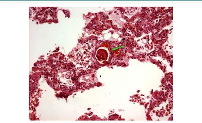
The liver and lungs presented gross- and microscopic signs of chronic passive congestion. Additionally, microthrombi were found in the hepatic sinusoids (figure 6) and in pulmonary vessels (figure 7).
Focal infarctions were observed in the kidneys.
(Dr. Mauro Canzian)
Anatomopathological diagnoses
Hypertrophic cardiomyopathy; alterations secondary to congestive heart failure; recent thrombosis in hepatic and pulmonary microvasculature consistent with disseminated intravascular coagulation; acute tubular necrosis secondary to hypovolemic shock; early focal infarction in the kidneys.
(Dr. Mauro Canzian)
Comments
Fig. 3 - Longitudinal section of the basal region of the heart showing a marked degree of ventricular myocardial hypertrophy.
Fig. 4 - Cross-section in the middle third of the ventricles showing a marked degree of ventricular myocardial hypertrophy and extensive whitish confluent areas of fibrosis.
conditions due to hemorrhagic acute abdomensecondary to the rupture of an ovarian cyst two weeks before her death. The patient underwent exploratory laparotomy that revealed intra-abdominal hemorrhage. In the early postoperative phase, a new peritoneal hemorrhagic collection was found, which progressed with refractory shock despite multiple blood transfusions and volemic replacement. The patient underwent a new exploratory laparotomy, and progressed postoperatively to a clinical condition consistent with disseminated intravascular coagulation (DIC). Microscopic analysis revealed foci of acute microcirculation thrombosis, especially in the
liver, which corroborated the hypothesis of DIC.
Hypertrophic cardiomyopathy is a relatively common genetic disease of variable penetrance (1:500 in the overall population)13. It is also known as idiopathic hypertrophic
subaortic stenosis or hypertrophic obstructive cardiomyopathy, characterized primarily by myocardial hypertrophy in the absence of other possible causes. The clinical picture varies from virtually imperceptible symptoms to significant limitations of physical activity and recurring arrhythmias14. Its classical
morphological pattern is a disproportionate and asymmetric thickening of the ventricular septum, predominantly in its subaortic region relative to the free left ventricular wall, although in 10% of the cases such asymmetry is not found15. As
evidenced in the present case, the main microscopic findings are extensive cardiomyocyte hypertrophy associated with fiber disarray and interstitial fibrosis15,16. It is the main cause
of sudden death among young individuals and an important cause of morbidity-mortality in elderly patients. More than 150 mutations in ten different genes codifying sarcomeric proteins have been identified. Most of the genetic mutations have been found in the gene encoding the beta-myosin heavy chain, cardiac troponin T, alpha-tropomyosin, and myosin-binding protein C15,17,18. The rate of complications associated
with entities such as sudden death, terminal heart failure, or fatal cerebrovascular accident is 1% to 2%14, and a significant
percentageof the patients have a life expectancy similar to that ofthe rest of the population. In general, the increase in mortality rates correlates directly with the development of complications13, as in the present case in which death was
probably due to hypovolemic shock, with a possible septic contribution, even though no infectious foci were found, whether gross- or microscopically. The early focal renal infarctions were probably due to the systemic migration of microthrombi formed in the left cardiac chambers.
Fig. 5 - Histological section of the myocardium showing hypertrophy and cardiomyocyte disarray. (Hematoxylin-eosin, Objective 20X)
Fig. 7 - Histological section of the lung, showing a focus of recent thrombosis occluding a small-diameter vessel. (Haematoxylin-eosin, Objective 20X)
References
1. Richardson P, McKenna W, Bristow M, Maisch B, Mautner B, O’Connell J, et al. Report of the 1995 World Health Organization/International Society and Federation of Cardiology Task Force on the definition and classification of the cardiomyopathies. Circulation. 1996; 93: 841-2.
2. Rick AN, David RH. Hypertrophic obstructive cardiomyopathy. N Engl J Med. 2004; 350: 1320-7.
3. Maron BJ, Olivotto I, Spirito P, Casey SA, Bellone P, Gohman TE, et al. Epidemiology of hypertrophic cadiomyopathy-related death. Circulation. 2000; 102: 858-64.
4. Maron BJ, McKenna WJ, Danielson GK, Kappenberger LJ, Kuhn HJ, Seidman CE, et al. ACC/ESC expert consensus document: a report of the American College of Cardiology Foundation Task Force on Clinical Expert Consensus Documents and the European Society of Cardiology Committee for Practice Guidelines. J Am Coll Cardiol. 2003; 42: 1687-713.
5. Maron BJ, Estes MNA, Maron MS, Almquist AK, Link MS, Udelson JE. Primary prevention of sudden death as a novel treatment strategy in hypertrophic cardiomyopathy. Circulation. 2003; 107: 2872-5.
6. Maron BJ, Shen WK, Link MS, Epstein AE, Amquist AK, Daubert JP, et al. Efficacy of implantable cardioverter-defibrillators for the prevention of sudden death in patients with hypertrophic cardiomyopathy. N Engl J Med. 2000; 342: 365-73.
7. Behr ER, Elliott P, Mckenna WJ. Role of invasive EP testing in the evaluation and management of hypertrophic cardiomyopathy. Card Electrophysiol Rev. 2002; 6: 482-6.
8. Braunwald E, Morrow A, Cornell W. Idiopathic hypertrophic subaortic stenosis. Clinical, homodynamic and angiographic manifestations. Am J Med. 1960; 29: 924-45.
9. MacRae CA, Ghaisas N, Kass S, Donnelly S, Basson CT, Watkins HC, et al. Familial hypertrophic cardiomyopathy with WPW syndrome maps to a locus on chromosome 7q3. J Clin Invest. 1995; 96: 1216-20.
10. Shibata M, Yamakado T, Imanaka-Yoshida K, Isaka N, Nakano T. Familial hypertrophic cardiomyopathy with WPW syndrome progressing to ventricular dilation. Am Heart J. 1996; 131 (6): 1223-5.
11. Arteaga E, Ianni BM, Fernandes F, Mady C. Benign outcome in a long-term follow-up of patients with hypertrophy cardiomyopathy in Brazil. Am Heart J. 2005; 149: 1099-105.
12. Spirito P, Bellone P, Harris KM. Magnitude of left ventricular hypertrophy and risk of sudden death in hypertrophy cardiomyopathy. N Engl J Med. 2000; 342: 1778-85.
13. Maron BJ. Hypertrophic cardiomyopathy: a systematic review. JAMA. 2002; 287: 1308-20.
14. Elliott P, McKenna WJ. Hypertrophic cardiomyopathy. Lancet. 2004; 363: 1881-91.
15. Schoen FJ. The Heart. In: Kumar V, Abbas AK, Fausto N. Robbins and Cotran Pathologic basis of disease, 7th ed. Philadelphia: Elsevier Saunders; 2004. p. 555-618.
16. Higuchi ML, Aiello VD, Gutierrez PS. Coração. In: Brasileiro G Fº. Bogliolo Patologia. 7a ed. Rio de Janeiro: Guanabara Koogan; 2006. p. 408-456. 17. Marian AJ, Roberts R. The molecular genetic basis for hypertrophic
cardiomyopathy. J Mol Cell Cardiol. 2001; 33: 655-70.