Editor: Alfredo José Mansur
Associate Editors: Desiderio Favarato; Vera Demarchi Aiello
Mailing address: Alfredo José Mansur – Incor – Av. Dr. Enéas C. Aguiar, 44 – 05403-000 – São Paulo, SP
Clinicopathologic Session
Case 4/99 – A 63-year-old man with heart failure 3 years after bioprosthetic replacement of mitral and aortic heart valves - Instituto do Coração do Hospital das Clínicas - FMUSP
A 63-year-old man sought medical treatment due to dyspnea during slight exertion, increased abdominal volu-me and edema, which had started two weeks prior to presen-tation at the clinic. He also complained of a cough and fever that he experienced in the week prior to presentation.
He reported his dyspnea had started 9 years before, progressing to dyspnea during slight exertion and edema of the lower limbs. He also complained of chest pain, triggered by exertion and relieved by rest. The patient was referred to INCOR for therapy (9/6/93).
The patient was diagnosed with rheumatoid arthritis in 1998 and had been previously treated for syphilis. Serolo-gical test results for Chagas’ disease were negative.
On physical examination (9/6/93), the patient showed a regular pulse rate (80 bpm), his blood pressure (BP) was 130/80 mmHg and he had an increased venous jugular pressure. Lung examination failed to show any abnormality. His heart examination revealed a first sound with diminished intensity, a systolic murmur +++/4+ in the mitral area, radiating to the axilla, and a rumbling diastolic murmur ++/ 4+ in the left sternalborder. The patient had ascites and his liver was felt 5cm below the right costal margin.
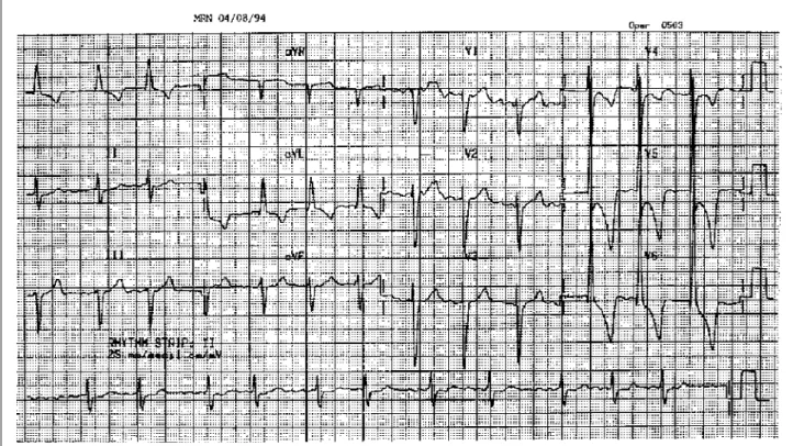
The electrocardiogram (ECG) performed in August 94 showed atrial fibrillation (AF), heart rate (HR) of 80bpm, QRS -20º backwards, left ventricular hypertrophy and secondary ventricular repolarizationabnormalities (fig. 1). Chest X-ray showed an enlarged cardiacsilhouette (+++/ 4+), a bulging medium arch, andenlargement of the hilar vessels and of the pulmonary vasculature.
The echocardiogram (1/10/94) showed marked dilation and hypokinesia of the left ventricle (LV), pulmonary hypertension, marked aortic regurgitation and slight mitral and tricuspid regurgitation (tab. I).
The hemodynamic (tab. II) and cineangiographic studies (2/94) showed normal coronary arteries, a dilated LV, with diffuse hypokinesia (+++), moderate mitral regur-gitation and marked aortic regurregur-gitation.
The patient received the diagnoses of aortic regur-gitation and mitral regurregur-gitation secondary to ventricular dilation, both of unknown origin.
He received captopril, 50mg; digoxin, 0.25mg; furose-mide, 40mg; and hydrochlorothiazide, 50mg, as well as methotrexate and chloroquine, which he had been using for rheumatoid arthritis.
Surgery for aortic and mitral regurgitation was consi-dered and performed in another clinic (6/6/94). Bioprosthetic heart valve replacement was performed in the aortic posi-tion, # 27, and in the mitral posiposi-tion, # 33. He developed AF with low ventricular response and required a pacemaker up to day 26 postoperatively (PO).
The pathological examination of the aortic valve showed thickening of the cusps as a result of fibrosis and hyalinization, as well as scattered areas of myxoid pattern. The diagnosis of degenerative aortic valve disease with fibrosis and calcification was established.
During subsequent medical visits (9/2/94), he reported dizziness and blurredvision. His heart rate was 50 bpm, and he was no longer using digoxin, beta-blockers or calcium channel blockers. A prolonged ECG was performed (5/12/ 95), which revealed periods of asymptomatic bradycardia, with ventricular escapeand a maximal pause of 2s, as well as frequent ventricular extrasystoles (tab. III).
The echocardiogram (2/14/96) showed normal func-tion and dimensions of the LV and pulmonary hyper-tension, as well as normal mitral and aortic bioprostheses (tab. I).
His dizziness and syncope became worse and the patient was admitted to the hospital.
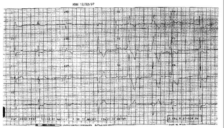
The physical examination failed to reveal any abnor-mal findings. The ECG (2/12/97) showed AF, with a HR of 50 bpm and QRS with variable morphologies, i.e., someti-mes narrower, sometisometi-mes showing a conduction abnor-mality of the left branch and anterior displacement of the loop in the horizontal plan (ventricular escape rhythm) (fig. 2). Laboratory test results (2/12/97) documented a hemo-globin level of 11.4g/dL and a hematocrit of 0.35. Serum levels were the following: creatinine, 1.2mg/dL; urea, 54mg/dL; sodium, 137mEq/L; and potassium, 4.4mEq/L. Coagulationtests were normal.
The echocardiogram (2/13/97) showed a slight hypoki-nesis of the LV, a dilated right ventricle (RV), marked pulmonary hypertension, stenosis of the mitral prosthesis and a small atrial septal defect, fossa ovalis-like (tab. I).
During the follow-up visit after hospital discharge (3/ 18/97) the patient was still complaining of dizziness; he reported dyspnea during slight exertion and the presence of dark black stools. Warfarin was discontinued and papa-verine, 200mg/d, was prescribed.
In 3/22/97, he sought medical assistance due to diffuse chest pain, diagnosed as myofascial pain.
Four months later (6/23/97), he complained of chest pain, dyspnea during medium exertion and dry cough that had appeared in the previous week. There were no signs of heart failure (HF) on physical examination. Chest X-ray revealed an enlarged cardiac silhouette (++/4+) and signs of pulmonary congestion. The cough improved after the patient received intravenous diuretics.
Five days later, he sought medical assistance due to the presence of melena. His plasma hemoglobin level was 10g/dL and the patient received 3 units of red cell concentrate.
During the medical follow-up visit (5/13/97) he did not have dyspnea, but the rotational vertigo, nausea and syncope persisted. Romberg’s sign was negative and there were signs of lack of motor coordination. He was diagnosed with vestibular or cerebellar dysfunction and referred for neurological assessment.
In the beginning of October 97, he developed dyspnea during slight exertion and orthopnea, in addition to increased abdominal volume and edema of the lower limbs. On physical examination (10/5/97), the patient was pale and afebrile, his heart rate was 86bpm and his BP was 120/80mmHg. Lung examination showed rales at both lung bases. Heart examination showed a systolic murmur (++/ 4+) in the mitral area and in the lower left sternal border. He had a bulging abdomen and there were no vis-ceromegalies. There was edema of the lower limbs (+/4+).
Fig. 1 - Electrocardiogram. Atrial fibrillation, left ventricular hypertrophy and ventricular repolarization abnormalities.
Table I - Echocardiograms
Measurements 1/10/94 2/14/96 2/13/97 12/4/97
Septum (mm) 9 13 9
-Posterior wall (mm) 9 13 8 -Left ventricle
Diastolic diameter (mm) 78 56 58 59 Systolic diameter (mm) 61 37 40 43 Ejection fraction 0.52 0.71 0.67 ∆D%= 28
Aorta (mm) 32 45 41
Left atrium (mm) 53 47 65 Right ventricle (mm) 24 14 40 LV-Ao gradient (mmHg) 24.6 29 24
LA-LV gradient - 4 13 12
Systolic pressure RV 55 77 98 82
LV- left ventricle; Ao- aorta; LA- left atrium.
Table II - Hemodynamic study *
Chamber/pressure Systolic Early- End- Mean diastolic diastolic
Right atrium 5
Right ventricle 28 0 5
Pulmonary artery 28 10 16
Pulmonary wedge 12
Left ventricle 140 0 12
Aorta 140 80 100
The patient was advised to restrict the ingestion of fluids and was given the following prescription: digoxin, 0.25mg; hydrochlorothiazide, 50mg;captopril, 37.5mg (instead of the previous 25mg dose), and 40mg daily of furosemide were added.
Two months later (12/4/97), he sought medical assis-tance due to dyspnea during slight exertion that had started 2 weeks before and fever that had started 1 week before. On physical examination (12/4/97), the patient was pale (+++/ 4+), his jugular venous pressure was increased, his limbs showed signs of decreased perfusion, his heart rate was 75bpm and his BP was 105/75mmHg. Lung examination
revealed rales at both lung bases; heart examination showed a systolic murmur (++/4+), which could be heard in all auscultatory foci. There was ascites and edema of the lower limbs.
Laboratory tests (12/4/97) revealed the following serum levels: urea of 85mg/dL, creatinine of 1.8mg/dL, glucose of 51mg/dL, sodium of 137mEq/L, and potassium of 4.2mEq/L. Blood gas analysis (room air) without O2 enrich-ment showed the following values: pH of 7.39, partial CO2 pressure of 19mmHg, partial O2 pressure of 57mmHg, saturation of O2 of 85.7%, bicarbonate concentration of 11.3mEq/L, and base excess < 11.5.
The ECG revealed paced rhythm. The echocardiogram (12/4/97) showed dilation and hypokinesiaof the RV, normal left ventricular function, an extremely thickened biopros-thesis in the mitral position, with a valvar area of 0.6cm2,
without signs of vegetation, and moderate tricuspid regurgitation (tab. I).
A few hours later, he developed hypotension (70/ 40mmHg), rales at the lower third of the lungs, abdominal distension and respiratory failure, requiring orotracheal intubation and respiratory support. There was no increase of the BP with the use of dobutamine and adrenaline, and the patient died as a result of bradycardia that progressed to asystole (12/4/97).
Discussion
Clinical findings - This patient suffered from functio-nal class III (NYHA) HF for 4 years. He was diagnosed with
Fig. 2 – Electrocardiogram. Predominance of atrial fibrillation with a high degree of atrioventricular block and periods of ventricular escape rhythm; left ventricular hypertrophy and ventricular repolarization abnormalities.
Table III – Prolonged electrocardiogram (Holter system)
Variables 5/12/95 6/19/96
Main rhythm Atrial fibrillation Atrial fibrillation Heart rate (bpm)
during placement 49 56
Minimal 35 28
(ventricular escape) (ventricularescape)
Maximal 98 139
Maximal pause (s) 2 2.3 Ventricular extrasystoles 6894 (295/h) 15953 (664/h) ventriculares
NSVT 5 1485
Idioventricular rhythm 532 Multiple (2525 beats)
Episodes of
sudden bradycardia 74
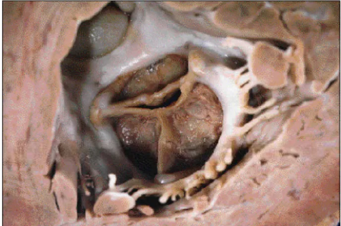
Fig. 3 – Superior view of the heart, with the atrium and great vessels removed. Note the massive thrombosis of the left atrium (arrows), with occlusion of the mitral orifice. The aortic bioprosthesis (Ao) shows a slight thickening and superficial apposition of soft, whitish material.
aortic and mitral valve regurgitation and surgery was considered, based on the disabling symptoms and on the ventricular dysfunction 1. Six months later, the patient
underwent surgery with replacement of the aortic and mitral valves by bioprostheses.
In a 63-year-old patient, degenerative disease is the most likely etiology for aortic regurgitation. However, be-cause of the past history of this patient such a diagnosis deserves caution. Rheumatoid arthritis may cause valvar lesion, mainly aortic 2,3. The pathological examination of
the patient’s valve does not rule out rheumatoid valvar disease, in which the findings of the pathological lesion are similar to those we described. One must remember that the patient had a severe form of rheumatoid disease, requiring immunosuppressivetherapy. Other possible etiologies are luetic aortitis, as the patient had suffered from syphilis in the past 4,5. However, as he was treated for syphilis and
still the mitral valve was affected, this hypothesis is improbable.
After surgery, the patient showed improvement in ventricular function and diameters. In spite of this impro-vement, he experienced dizziness and syncope, and the prolongedECG revealed maximum pauses of 2s. This conduction abnormality could be attributed to surgical manipulation, as long-term pacing was required in the postoperative period 6. Rheumatoid arthritis may affect the
conduction system of the heart, leading to atrioventricular block 7. Considering this, definite pacing with VVIR
pace-maker was indicated. Dizziness persisted in spite of the pacemaker, but the syncope disappeared. At the time the pacemaker was implanted, a dysfunction of the mitral valve prosthesis with stenosis and a 13mmHg gradient were detected on the echocardiogram. One month later, the patient developed dyspnea during exertion, which may indicate hemodynamic repercussion of the stenosis of the prosthesis.
Anemia is another condition that could account for the cardiac failure in this patient. After receiving the pacemaker, the patient was discharged with the prescription of war-farin; he had signs of melena one month later and the drug was discontinued. After this episode, he returned for medical assistance due to decompensated HF and, a few days later, he again experienced gastrointestinal bleeding, which required blood transfusion. This patient’s anemia may have multiple causes, including gastrointestinal bleedingas a result of the use of nonsteroidal anti-inflamma-tory agents.Although there is no such report by the patient, these drugs are frequently used in the treatment of rheuma-toid arthritis. In addition, he used methotrexate. This drug, which is a folate inhibitor, may cause myelotoxic effects, reducing erythropoiesis. We should also take into account the hemolysis caused by valvar prostheses as a possible factor in the genesis of anemia. The origin of the gastroin-testinal bleeding was not determined; thus, we may not con-tinue to speculate about its origin and etiology. The patient showed worsening of the HF, possibly as a result of pro-gressive prosthesis dysfunction. This was supported by
the echocardiogram, which revealed severe stenosis of the prosthesis in the mitral position, aggravated by anemia. A prosthesis dysfunction of this magnitude is infrequent within only 3 years of the pacing procedure, mainly when one takes into account the fact that this is an elderly patient. Finally, although the patient had a normal ventricular function, when he developed hypotension, he received dobutamine and noradrenaline. These drugs are usually employed in cases of ventricular dysfunction. In the present case, it is likely that the main hemodynamic abnormality occurred as a result of the stenosis of the mitral prosthesis. In this case, surgery would have been the only effective therapeutic option.
(Dr. Guilherme Spina)
Possible diagnoses - HF caused by stenosis of the
valvar prosthesis, aggravatedby anemia resulting from gastrointestinalbleeding and rheumatoid arthritis.
Autopsy
Fig. 4 – Mitral prosthesis, ventricular view, showing thickening and superficial apposition of soft, whitish material. Notice also the presence of remnants of the native mitral valve.
Fig. 5 – Histologicalsection of mitral prosthesis leaflets, showing infectious endocarditis with multiple Gram-positive cocci. BB, X 130 (original magnification).
foci of fibrosis (sclerosis of the myocardium). The histolo-gicalexamination of the mitral and aortic prostheses revealed, in both, the presence of multiple clusters of bacteria, identified in histochemistryas Gram-positive cocci (fig. 5). The lungs weighed 1,550g and showed marked passive chronic congestionand focal areas of edema. The liver showed centrolobular hemorrhagic necrosis as a result of hemodynamic shock.
(Dr. Luiz Alberto Benvenuti)
Anatomicopathological diagnoses - 1) Occlusive
thrombosis of the LA; 2) infectious endocarditis (IE) of the mitral and aortic prostheses, caused by Gram-positive cocci; 3) morphological evidence of congestive HF.
Comments
This is a case report of a patient previously diagnosed with rheumatoid arthritis, marked aortic regurgitation and slight mitral regurgitation, who underwent mitral and aortic valve replacement with bioprostheses in another clinic. We did not examine the valves removed; however, there is a report of thickened aortic cusps as a result of fibrosis and hyalinization, with areas of calcification, without reference to the presence of an inflammatory process. Although we cannot rule out definitely the rheumatic etiology in this case, the findings reported are consistent with aortic valve disease in rheumatoid arthritis.
Rheumatoid arthritis may affect the pericardium, the myocardium and the heart valves. Even though the pattern of valvar involvement is similar to that observed in rheu-matic disease, with a predominance of mitral valve lesions, aortic regurgitation is the main valvar dysfunction with clinical repercussion 8. Regurgitation is rarely significant;
however, there are reports of surgical mitral 9 or aortic 8,10
valve replacement. The classical anatomicopathological finding in the valvar lesion is the rheumatoid granuloma, but it is common to find only nonspecific lesions, such as fibro-sis, areas of neovascularization and mucoid degeneration 1.
The present case suggests that there is an involve-ment of the aortic and mitral valves by rheumatoid arthritis, or only of the aortic valve, with slight mitral regurgitation as a result of LV dilation, which is usually significant in marked aortic regurgitation.
The patient developed progressive HF in spite of the valvar replacements. This was probably related to the worsening condition of the LV, which showed marked eccentric hypertrophy and myocardial sclerosis. In spite of the absence of stenosis of the mitral prosthesis, the development of semiocclusive thrombosis of the LA, with thrombus organization next to the atrial wall, led to impair-ment of the left ventricular filling and aggravated the pas-sive pulmonary congestion. The clinical conditions were aggravated by the IE of the prostheses and by the early thrombosis of the remaining left atrial lumen, which consti-tuted the ultimate cause of death.
(Dr. Luiz Alberto Benvenuti)
1. Nishimura R, McGoon MD, Schaff HV, Giulianni ER. Chronic aortic regurgitation – indications for surgery – 1988 Mayo Clin Proc 1988; 63: 270-80.
2. Kramer PH, Imboden JB, Waldman FM. Severe aortic insufficiency in juvenile chronic arthrtits. Am J Med 1983; 74: 1088-91.
3. Lebowitz WB. The heart in rheumatoid arthritis (rheumatoid disease). A clinical and pathological study of sixty-two cases. Ann Intern Med 1963; 58 : 102-23.
References
4. Jackman JD Jr, Rudolf JD. Cardiovascular syphilis. Am J Med 1989; 87: 425-33. 5. Aizawa H, Hasegawa A, Arai M, Naganuma F, et al. Bilateral coronary ostial stenosis
and aortic regurgitation due to siphilitic aortitis Intern Med 1998; 57: 56-9. 6. Cohn LH, Collins JJ, Disesa VJ, et al. Fifteen – year experience with 1678 Huncock
Eletrocardiographic Holter monitoring in patientes with rheumatoid arthritis according acording to Steinbrocker’s criteria, functional index, value of Waaler-Rose titre and duration of disease. Clin Rheumatol 1998; 17: 369-77. 8. Kramer PH, Imboden JB Jr, Waldman FM, Turley K, Ports TA. Severe aortic
insufficiency in juvenile chronic arthritis. Am J Med 1983; 74: 1088-91.
9. Mullins PA, Grace AA, Stewart SC, Shapiro LM. Rheumatoid heart disease presenting as acute mitral regurgitation. Am Heart J 1991; 122: 242-5. 10. Bortolotti U, Gallucci V, Russo R, Glorioso S, Schivazappa L, Thiene G. Aortic