AbstrAct
Purpose: To investigate the short-term (1 week) and long-term (8 weeks) pro-tective effects of zinc administration on radioiodine (RAI)-induced lacrimal gland damage of rats.
Methods: A total of 40 rats were divided into two groups: an RAI group (n=20), which was administrated a single dose of 3 mCi of 131I and 1 mL physiologic saline for 7 days by gastric gavage, and a zinc group (n=20), which received a single dose of 3 mCi of 131I and 1 mL of physiologic saline containing zinc sulfate at a concentration of 10 mg/kg concentration for 7 days by gastric gavage. All rats underwent tear function tests before and 1 week after RAI administration. About 1 week after irradiation, half of the animals in each group were sacrificed and the extraorbital lacrimal glands were removed for histopathological exami-nation. The remaining animals of the groups underwent the same procedures at 8 weeks after irradiation.
results: In the RAI and zinc groups, the mean tear production was 3.75 ± 1.55 and 3.65 ± 1.53 mm at baseline, 2.10 ± 1.07 and 3.30 ± 1.34 mm at week 1 (p=0.004), and 3.22 ± 1.48 and 3.50 ± 1.78 mm at week 8, respectively; further, the mean corneal luorescein staining scores were 4.65 ± 2.16 and 4.80 ± 2.21 points at baseline, 7.85 ± 1.90 and 5.45 ± 2.06 points at week 1 (p=0.001), and 5.44 ± 2.13 and 4.90 ± 2.08 at week 8, respectively. The histopathological changes in rat lacrimal glands at weeks 1 and 8 were consistent with the tear function test results.
conclusions: Zinc treatment seems to be protective against RAI-induced lacrimal gland damage of rats, particularly in the acute period.
Keywords: Antioxidant; Lacrimal apparatus; Iodine radioisotopes; Zinc; Radia-tion-protective agents; Animals; Rats
RESUMO
Objetivo: Investigar se o tratamento com zinco tem efeito protetor, no curto prazo (1 semana) e longo prazo (8 semanas), sobre os danos induzidos na glândula lacrimal por iodo radiotativo (RAI) em ratos.
Métodos: Quarenta ratos foram divididos em dois grupos. No grupo RAI (n=20) foi administrada uma única dose de 3 mCi 131I e 1 cc de solução salina fisiológica duran-te 7 dias, por gavagem gástrica. O grupo zinco (n=20) recebeu uma dose única de 3 mCi 131I e 1 cc de solução salina fisiológica contendo sulfato de zinco na concentra-ção de 10 mg/kg durante 7 dias por gavagem gástrica. Os testes de funconcentra-ção lacrimal foram realizadas para todos os animais antes e após uma semana da administração da RAI. Em seguida, após 1 semana da administração, metade dos animais de cada grupo foi sacrificada e as glândulas lacrimais extraorbitais foram removidas para exame histopatológico. Os animais remanescentes dos grupos foram submetidos aos mesmos procedimentos após 8 semanas a radiação.
Resultados: As médias de produção lacrimal foram de 3,75 ± 1,55 e 3,65 ± 1,53 mm na linha de base, 2,10 ± 1,07 e 3,30 ± 1,34 mm na 1a semana (p=0,004), e 3,22 ± 1,48 e 3,50 ± 1,78 mm na 8a semana, para os grupos RAI e zinco, respectivamente. As pon-tuações médias de coloração fluoresceína foram 4,65 ± 2,16 e 4,80 ± 2,21 no início do estudo, 7,85 ± 1,90 e 5,45 ± 2,06 na primeira semana (p=0,001), 5,44 ± 2,13 e 4,90 ± 2,08 pontos na 8a semana, para os grupos RAI e zinco, respectivamente. As alterações histopatológicas das glândulas lacrimais em 1 e 8 semanas foram consistentes com os testes de função lacrimal resultados.
Conclusões: O tratamento de zinco parece ser protetor sobre os danos glândula la-crimal induzidos por RAI em ratos, especialmente no período agudo.
Descritores: Antioxidantes; Aparelho lacrimal; Radioisótopos do iodo; Zinco; Pro-tetores contra radiação; Animais; Ratos
Short- and long-term effects of zinc treatment on lacrimal gland histopathology
and tear functions tests in radioiodine-administered rats
Efeitos de curto e longo prazo do tratamento com zinco na histopatologia da glândula lacrimal
e funções lacrimais em ratos que receberam iodo radioativo
Firdevs Ornek1, damla erginturk acar2, ugur acar3, Ozdemir Ozdemir2, Hasan ikbal atilgan4, niHat Yumusak5, basak bOztOk Ozgermen6
Submitted for publication: March 22, 2016 Accepted for publication: October 7, 2016
1 Department of Ophthalmology, Ankara Training and Research Hospital, Ankara, Turkey. 2 Department of Ophthalmology, Zekai Tahir Burak Women’s Health Training and Research Hospital,
Ankara, Turkey.
3 Department of Ophthalmology, Faculty of Medicine, Hacettepe University, Ankara, Turkey. 4 Department of Nuclear Medicine, Necip Fazil City Hospital, Kahramanmaras, Turkey. 5 Department of Pathology, Faculty of Veterinary Medicine, Harran University, Sanliurfa, Turkey. 6 Department of Surgery, Faculty of Veterinary Medicine, Aksaray University, Aksaray, Turkey.
Funding: No specific financial support was available for this study.
Disclosure of potential conflicts of interest: None of the authors have any potential conflict of interest to disclose.
Corresponding author: Damla Erginturk Acar. E-mail: [email protected]
Approved by the following research ethics committee: Dışkapı Yıldırım Beyazıt Animal Laboratory
(#2014/33).
IntroductIon
Radioiodine (RAI) treatment with 131I is a viable therapeutic option for thyroid diseases, such as hyperthyroidism and diferentiated thyroid carcinoma(1). 131I is taken up by the thyroid tissue as well as other glands, especially the salivary and lacrimal glands, as each has similar sodium iodide symporters (NIS) that uptake iodide into the cells(2,3). Consequently, β-radiation from 131I exerts cytotoxic efects in these glands. Even though many studies have shown radiation dama-ge to the salivary glands(4-7), relatively few studies have investigated the side efects of RAI in the lacrimal glands(7-9).
Several studies have been conducted to investigate the efects of therapeutic agents, such as pilocarpine(4), amifostine(10,11), vitamin C(12), and vitamin E(13), to prevent RAI-induced damage to the salivary glands. It was recently demonstrated that montelukast provided long-term protection against RAI-induced damage to the rat lacrimal glands(14). Moreover, our group previously showed that vitamin E and lycopene protected the rat lacrimal glands from RAI-induced early histopatho-logical damage(15,16).
unwanted RAI-induced oxidative damage(17). Free oxygen radicals can interact and destroy cell membranes and organelles(18). Depending on the oxidative mechanism, most radioprotective agents studied so far have been antioxidants.
It is well-known that zinc is an essential antioxidant mineral. The beneicial antioxidant and/or radioprotective efects of zinc to eye diseases, such as age-related macular degeneration, have been sup-ported by many studies(19-23).Zinc plays an important role in nucleic acid metabolism, cell growth and diferentiation, and wound healing, while zinc deiciency can result in various disorders, such as growth and cognitive impairment, immune dysfunction, malabsorption syn-drome, alopecia, epidermal disorders, and chronic liver diseases(24,25). This study aimed to investigate the possible short-term (1 week) and long-term (8 weeks) protective efects of zinc administration against histopathological changes associated with RAI-induced dys-function of the rat lacrimal glands.
Methods
The study protocol was approved by the Local Ethics Committee of Animal Experiments of Dışkapı Yıldırım Beyazıt Animal Laboratory (Ankara, Turkey) and was conducted in accordance with the Associa-tion for Research in Vision and Ophthalmology Statement for the Use of Animals in Ophthalmic and Visual Research.
About 40 Wistar albino female rats aged 6-10 months and weighing 250-300 g were housed under standard laboratory condi-tions with a constant temperature of 21 ± 2°C and a relative humi-dity of 65-70% under 12-h light/dark cycle, and were fed standard rat chow and water ad libitum. The rats were acclimated for at least 1 week before the study and then were divided into two groups: an RAI group (n=20), which received 3 mCi of 131I and 1 mL of phy-siologic saline by gastric gavage(26), and a zinc group (n=20), which received 3 mCi of 131I and 1 mL of physiologic saline containing zinc sulfate (Zinco®; Berko Ilaç ve Kimya San. A.S., Istanbul, Turkey) at a concentration of 10 mg/kg body weight by gastric gavage(23). The physiologic saline and zinc were administered 2 days before the RAI therapy and were continued for 5 days posttherapy. Corneal fluorescein staining (CFS) and meniscometry testing were perfor-med for all rats. The tear function tests were repeated for all rats, 1 week after administration of 131I, and half of the rats (n=10, each group) were then sacrificed and their extraorbital lacrimal glands were removed bilaterally for histopathological examination. The remaining animals in both groups (n=19, one rat in the RAI group died due to an unknown cause) underwent tear function tests, 8
weeks after administration of 131I; moreover, the rats were sacrificed and their extraorbital lacrimal glands were removed. During these procedures, the rats were immobilized by intraperitoneal injection of 50 mg/kg of propofol (Abbott, Istanbul, Turkey).
T
EARFUNCTIONTESTINGThe tear volume of the right eyes of the rats was measured using Strip Meniscometry Tubes (SMTube®, Echo Electricity Co., Ltd., Japan), which is a recently developed, simple, swift, and noninvasive method that is well-correlated with the conventional Schirmer’s test, as des-cribed in detail elsewhere (Figure 1 A)(27). In brief, a tear-absorbing strip was applied to the conjunctival sac of the lower eyelid for 5 s. The quantity of wetness was interpreted as tear volume in millimeters (mm) (Figure 1 B).
CFS of the left eyes of the rats was performed using a cobalt blue ilter under a slit-lamp microscope, as described in detail elsewhere (Figure 1 C)(28). In brief, the degree of corneal staining was graded using a 16-point scale 90 s after application of 1 µL of 1% luorescein.
P
ATHOLOGICALANALYSISThe lacrimal glands were ixed in 10% neutral bufered formalin (pH 7.2-7.4), embedded in parain, and sliced into 4 µm-thick parain sections, which were stained with hematoxylin and eosin, and then observed under a light microscope (Olympus BX-50; Olympus Cor-poration, Tokyo, Japan) at 40- to 400-fold magniication in a masked fashion. The irst three sections and every 10th section thereafter were stained. All of the stained sections, approximately 15 slides per speci-men for the extraorbital lacrimal glands, were studied. Histopatho-logical changes were evaluated according to a previously published grading system by an experienced animal pathologist (NY)(29).
D
ATAANALYSISQuantitative variables are presented as the mean ± standard de-viation (SD) and minimum and maximum values. Nominal data are presented as frequencies and percentages. The results of the Sha-piro-Wilk test showed that the data were not normally distribu ted. Comparisons of quantitative variables among groups were asses-sed using the nonparametric Mann-Whitney U test. The Friedman test was used to assess differences among time points (baseline, week 1, and week 8). The chi-square test, Fisher’s exact test, and Fisher-Freeman-Halton test were used to compare qualitative mea-surements among groups. All statistical analyses were performed using IBM-SPSS software (ver. 21.0; IBM-SPSS, Inc., Chicago, IL, USA). A probability (p) value <0.05 was considered statistically significant.
Figure 1. A) Administration of a meniscometry tube strip on left eye of a rat. B) A representative tear volume test result. In this case, the test demonstrated a quantity of wetness equal to 4 mm. C) Evaluation of corneal luorescein staining of the rat eye with a cobalt blue ilter.
b
results
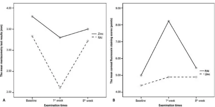
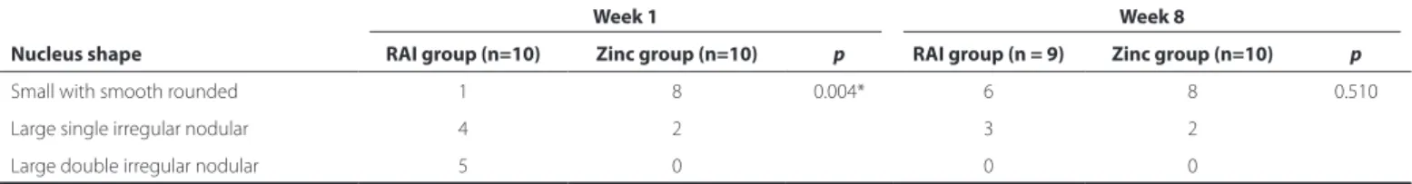
The mean meniscometry test results for the RAI and zinc groups were 3.75 ± 1.55 and 3.65 ± 1.53 mm at baseline, 2.10 ± 1.07 and 3.30 ± 1.34 mm (p=0.004) at week 1, and 3.22 ± 1.48 and 3.50 ± 1.78 mm at week 8, respectively (Table 1, Figure 2 A). The mean CFS scores of the RAI and zinc groups were 4.65 ± 2.16 and 4.80 ± 2.21 points at baseline, 7.85 ± 1.90 and 5.45 ± 2.06 points (p=0.001) at week 1, and 5.44 ± 2.13 and 4.90 ± 2.08 points (p=0.604) at week 8, respectively (Table 1, Figure 2 B).
Variation in cell shape (p=0.001), variation in cell size (p=0.023), poorly deined acidophilic cell outlines (p=0.023), abnormal lobular pattern (p=0.005), abnormal peripheral basophilia (p=0.001), the existence of periductal iniltration (p=0.023), perivascular iniltration (p=0.005), and irregular nucleus shape (p=0.004) were less frequently observed in the zinc group than in the RAI group during the pathology sections from week 1 (Tables 2 and 3, Figures 3 and 4).
The existence of periductal and/or periacinar ibrosis (p=0.023) was less frequently observed in the zinc group than in the RAI group in the pathology sections from week 8 (Table 2). Other pathological indings were similar between groups at week 8.
dIscussIon
Administration of RAI is a widely accepted treatment modality for thyroid diseases; however, besides its therapeutic efects on thyroid
tissue, ionizing radiation may cause cellular oxidative damage both directly (by disruption of DNA integrity) and indirectly (by free oxygen radical production)(30). Although the exact mechanism of RAI-induced lacrimal gland damage remains obscure, NIS function is suspected to be involved. It has been shown that NIS proteins are expressed in lacrimal glands(31), suggesting that RAI is taken up by lacrimal gland cells via NIS, which may be the main cause of lacrimal gland dysfunc-tion following RAI therapy.
Solans et al.(7) found that the subjective symptoms of lacrimal gland dysfunction were consistent with ocular dryness and both were relatively common in patients following RAI therapy. Fard-Es fahani et al.(8) reported that high-dose RAI therapy caused a reduction in tear secretion by the lacrimal glands and that symptoms of dry eye syndrome, such as redness and burning sensation, occurred signii-cantly more often in the RAI exposed group (p=0.003 and 0.001, respectively); however, foreign body sensation was similar between the exposed and unexposed groups (p=0.279). We previously inves-tigated the efects of low-dose (≤30 mCi) RAI treatment on lacrimal gland function and found that even low-dose RAI caused a decrease in Schirmer’s and break up time tests values, and an increase in ocular surface disease index and impression cytology scores(9).
Although amifostine is the only drug approved by the Food and Drug Administration for prevention of xerostomia during radiotherapy, a recently published study claimed that it is inefective(32). In this review study, the authors defended that amifostine has no signiicant
radio-table 1. tear function test results of the rAI and zinc groups
tear function tests
baseline Week 1 Week 8
rAI (n=20) Zinc (n=20) p rAI (n=20) Zinc (n=20) p rAI (n=9) Zinc (n=10) p
Meniscometry (mm) 3.75 ± 1.55 3.65 ± 1.53 0.804 2.10 ± 1.07 3.30 ± 1.34 0.004* 3.22 ± 1.48 3.50 ± 1.78 0.780 Mean ± SD
CFS (point) 4.65 ± 2.16 4.80 ± 2.21 0.848 7.85 ± 1.90 5.45 ± 2.06 0.001* 5.44 ± 2.13 4.90 ± 2.08 0.604 Mean ± SD
CFS= corneal luorescein staining; statistical analysis= Mann-Whitney U test. *= statistically signiicant.
Figure 2. A) The mean meniscometry test results and B) mean corneal luorescein staining scores at baseline, week 1, and week 8.
Figures 3. Representative histopathologic indings of the extraorbital lacrimal glands (3) at week 1 and (4) week 8. (Hematoxylin and eosin staining, igure 2 and igure 3 B: x20 magniication; bar: 50 μm, Figure 3 A: x10 magniication; bar: 100 μm). A) In the RAI group, the igure shows severe inlammation, periductal and perivascular iniltration (asterix), double irregular nucleus shape (arrowheads), atypical cell size (thin arrows), and atypical cell shape (thick arrows). B) In the zinc group, the igure shows decreased inlammatory changes (arrows). C) In the zinc group, the igure shows decreased periductal iniltration (arrow).
A b c
A b
Figure 4. Representative histopathologic indings of the extraorbital lacrimal glands (3) at week 1 and (4) week 8. A) In the RAI group, the igure shows inlammatory changes (asterics) and severe ibrosis (arrowheads). B) In the zinc group, the igure shows decreased inlammatory changes (arrow) and much less ibrosis (arrowhead).
table 2. Morphological parameters of rat extraorbital lacrimal glands in the rAI and zinc groups
Morphologic parameters
Week 1 Week 8
rAI group (n=10) Zinc group (n=10) p rAI group (n=9) Zinc group (n=10) P
Cell shape variation (+) 09 1 0.001* 2 1 0.582*
Cell size variation (+) 08 2 0.023* 2 1 0.582*
Poorly deined acidophilic cell outlines (+) 08 2 0.023* 3 2 0.628*
Abnormal lobular pattern (+) 09 2 0.005* 2 2 1.000*
Abnormal peripheral basophilia (+) 10 2 0.001* 3 2 0.628*
Periductal / periaciner ibrosis (+) 01 0 1.000* 7 2 0.023*
Periductal iniltration (+) 08 2 0.023* 3 1 0.303*
Perivascular iniltration (+) 08 1 0.005* 2 0 0.211*
Statistical analysis= Fisher’s exact test; *= statistically signiicant.
table 3. histological indings according to the nuclear characteristics of rat extraorbital lacrimal glands in the rAI and zinc groups
nucleus shape
Week 1 Week 8
rAI group (n=10) Zinc group (n=10) p rAI group (n = 9) Zinc group (n=10) p
Small with smooth rounded 1 8 0.004* 6 8 0.510
Large single irregular nodular 4 2 3 2
Large double irregular nodular 5 0 0 0
protective efect on RAI-induced salivary gland damage in patients with diferentiated thyroid cancer. Amifostine administration has also several side efects that limit its use, such as emesis, hypotension, somnolence, and sneezing, and less commonly a metallic taste, hy -po calcemia, lushing, chills, weakness, and idiosyncratic reactions. Therefore, it is necessary to develop a radioprotective agent that is efective, safe, easily available, easily administered, and cost efective. Miao et al.(19) indicated that 3-month zinc therapy entirely prevented the pathological changes associated with diabetes, such as oxidative damage, inlammation, and remodeling, in the aorta of diabetic mice. In addition, they found that zinc therapy provided an increase in the function and expression of the potent antioxidant nuclear factor like-2. Dani et al.(20) found that zinc supplementation to RAI-treated rats signiicantly attenuated the adverse efects of RAI by altering the levels of malondialdehyde, glutathione, superoxide dismutase, and catalase. They also found that signiicant oxidant/antioxidant changes occurred in the red blood cells of the rats following RAI admi nistration, while zinc has been shown to act as a radioprotector. Furthermore, Dhawan et al.(21) suggested that zinc sulfate may be a promising ra-dioprotective agent for the thyroid gland. Accordingly, the opinion that zinc can prevent RAI-induced damage due to its antioxidative properties, has gained weight.
Zinc is a well-known trace element that plays crucial roles in various pathways, especially cellular growth and diferentiation. Gong et al.(33) investigated the efects of zinc deiciency on the cornea and con-junctiva of rats by electron microscopy and mass spectrometry, and identiied growth defects of the microvilli and microplicae of the ocular surface tissues, a reduction in the number of conjunctival goblet cells, and alterations in the levels of other trace elements and vitamin C in zinc-deicient rats(33). Similarly, Kanazawa et al.(34) evaluated the efects of zinc and vitamin A separately on the ocular surface of rats by creating ive diferent groups, and found a synergistic interaction between zinc and vitamin A for the healthy maintenance of the ocu-lar surface epithelium(34).
We previously reported the results of two studies about the efects of two separate radioprotective agents (vitamin E and lycopene) on acute RAI-induced lacrimal gland damage(15,16); however, the chronic efects of antioxidants on the lacrimal glands were not evaluated and tear function testing was not conducted in these former studies. In another reported study, Koca et al.,(14) investigated whether RAI treatment causes morphological damage to rat lacrimal glands and whether montelukast sodium has protective efects against RAI-indu-ced long-term (3 months) lacrimal gland damage; however, the acute adverse efect of RAI treatment was not investigated and the tear function test was not conducted, as in our former studies. Nonetheless, these results showed that RAI application resulted in atrophy and ibrosis of the rat lacrimal glands 3 months after RAI administration, and that intraperitoneal montelukast sodium appli cation prevented the negative efects of RAI without hampering ablation of the thyroid gland. In these three studies, all of the lacrimal glands (harderien, extraorbital, and intraorbital) were removed for histopathological exa-mination; whereas, in this study, only the extraor bital lacrimal glands of rats were removed because the extraor bital lacrimal gland is the most accessible.
The study aimed to evaluate the short- and long-term radiopro-tective activity of zinc. The results demonstrated that tear function was adversely afected by RAI application and zinc treatment pro-tected against these adverse efects, especially in the acute period. Furthermore, variations in cell shape and size, poorly deined acido-philic cell outlines, abnormal lobular pattern and peripheral basophilia, the existence of periductal and perivascular iniltration, and irregular nuclear shape were less frequently observed in the pathology sec-tions at week 1 of the zinc group than in the RAI group, and periductal and/or periacinar ibrosis was observed in the extraorbital lacrimal glands at week 8. In particular, the results of the tear function test and histopathological analysis of the RAI group at week 8 showed that the
tear function tests improved to baseline levels and the lacrimal glands were histopathologically healed, as compared to week 1. Hence, long-term zinc treatment following RAI administration is not needed. As ibrosis is a result of the lacrimal gland damage in the long term, only periductal and/or periacinar ibrosis was statistically signiicant at week 8 between the two groups.
There were some limitations to this study that should be addressed. First, to maintain compliance with the “reduction principle in animal experiments,” a relatively small number of rats were tested. Second, other tear function tests (e.g., tear break up time test, impression con-junctival cytology, etc.), which are easy to administer to humans, were not conducted(9).
The results of this study showed that zinc treatment is effective in preventing RAI-induced lacrimal gland damages in rats, especially in the acute period. In the future, ophthalmologists can chose the cheapest, most accessible, and most harmless antioxidant agents for planned RAI treatment after conirming these positive results in clinical studies.
reFerences
1. Carpenter CC, Griggs RC, Loscalzo J. Cecil Essentials of Medicine. 5th ed. Philadelphia:
WB Saunders; 2001.
2. De La Vieja A, Dothan O, Levy O, Carrasco N. Molecular analysis of the sodium/iodide symporter: impact on thyroid and extrathyroid pathophysiology. Physiol Rev. 2000; 80(3):1083-105.
3. Mandel SJ, Mandel L. Radioactive iodine and the salivary glands. Thyroid. 2003;13(3): 265-71.
4. Silberstein EB. Reducing the incidence of 131I-induced sialadenitis: the role of pilo-carpine. J Nucl Med. 2008;49(4):546-9.
5. Cavalieri RR. Iodine metabolism and thyroid physiology: current concepts. Thyroid. 1997;7(2):177-81.
6. Bohuslaviziki KH, Brenner W, Lassmann S, Tinnemeyer S, Tönshof G, Sippel C, et al. Quan titative salivary gland scintigraphy in the diagnosis of parenchymal damage after treatment with radioiodine. Nucl Med Commun. 1996;17(8):681-6.
7. Solans R, Bosch JA, Galofré P, Porta F, Roselló J, Selva-O’Callagan A, et al. Salivary and lacrimal gland dysfunction (sicca syndrome) after radioiodine therapy. J Nucl Med. 2001;42(5):738-43.
8. Fard-Esfahani A, Mirshekarpour H, Fallahi B, Eftekhari M, Saghari M, Beiki D, et al. The efect of high-dose radioiodine treatment on lacrimal gland function in patients with diferentiated thyroid carcinoma. Clin Nucl Med. 2007;32(9):696-9.
9. Koca G, Acar U, Atilgan HI, Erginturk Acar D, Altiparmak UE, Demirel K, et al. Changes in conjunctival cytology and tear function tests with radioiodine treatment for hyper-thyroidism. Ann Nucl Med. 2013;27(8):694-9.
10. Capizzi RL, Oster W. Chemoprotective and radioprotective efects of amifostine: an update of clinical trials. Int J Hematol. 2000;72(4):425-35.
11. Bohuslavizki KH, Klutmann S, Jenicke L, Brenner W, Feyerabend B, Henze E, et al. Ra-dioprotection of salivary glands by S-2-(3-aminopropylamino)-ethylphosphorothioic (amifostine) obtained in a rabbit animal model. Int J Radiat Oncol Biol Phys. 1999; 45(1):181-6.
12. Liu B, Kuang A, Huang R, Zhao Z, Zeng Y, Wang J, et al. Inluence of vitamin C on salivary absorbed dose of 131I in thyroid cancer patients: a prospective, randomized, single-blind, controlled trial. J Nucl Med. 2010;51(4):618-23.
13. Bhartiya US, Raut YS, Joseph LJ, Hawaldar RW, Rao BS. Evaluation of the radioprotecti ve efect of turmeric extract and vitamin E in mice exposed to therapeutic dose of ra-dioiodine. Indian J Clin Biochem. 2008;23(4):382-6.
14. Koca G, Yalniz-Akkaya Z, Gültekin SS, Yumusak N, Demirel K, Korkmaz M, et al. Radio-protective efect of montelukast sodium in rat lacrimal glands after radioiodine treatment. Rev Esp Med Nucl Imagen Mol. 2013;32(5):294-300.
15. Acar U, Atilgan HI, Acar DE, Yalniz-Akkaya Z, Yumusak N, Korkmaz M, et al. The efect of short-term vitamin E against radioiodine-induced early lacrimal gland damage. Ann Nucl Med. 2013;27(10):886-91.
16. Acar DE, Acar U, Yumusak N, Korkmaz M, Acar M, Atilgan HI, et al. Reducing the histo-pathological changes of radioiodine to the lacrimal glands by a popular anti-oxidant: lycopene. Curr Eye Res. 2014;39(7):659-65.
17. Nagler RM. Efects of head and neck radiotherapy on major salivary glands-animal studies and human implications. In Vivo. 2003;17(4):369-75.
18. Konings AW, Coppes RP, Vissink A. On the mechanism of salivary gland radiosensiti-vity. Int J Radiat Oncol Biol Phys. 2005;62(4):1187-94. Erratum in: Int J Radiat Oncol Biol Phys. 2006;64(1):330.
19. Miao X, Wang Y, Sun J, Sun W, Tan Y, Cai L, et al. Zinc protects against diabetes-in-duced pathogenic changes in the aorta: roles of metallothionein and nuclear factor (erythroid-derived 2)-like 2. Cardiovasc Diabetol. 2013;12(1):54.
changes on the antioxidant system and on the morphology of red blood cells in rats. Hell J Nucl Med. 2006;9(1):22-6. Comment in: Hell J Nucl Med. 2007;10(1):35; author reply 35-6.
21. Dhawan D, Singh Baweja M, Dani V. Zinc sulphate following the administration of iodine-131 on the regulation of thyroid function, in rats. Hell J Nucl Med. 2007;10(3): 167-71.
22. Chew EY, Clemons TE, Agrón E, Sperduto RD, Sangiovanni JP, Kurinij N, et al. Long-Term Efects of Vitamins C and E, β-Carotene, and Zinc on Age-Related Macular Degene-ration: AREDS Report No. 35. Ophthalmology. 2013;120(8):1604-11.
23. Ertekin MV, Tekin SB, Erdogan F, Karslioglu I, Gepdiremen A, Sezen O, et al. The efect of zinc sulphate in the prevention of radiation-induced dermatitis. J Radiat Res. 2004; 45(4):543-8.
24. Murakami M, Hirano T. Intracellular zinc homeostasis and zinc signaling. Cancer Sci. 2008;99(8):1515-22.
25. Brown KH, Peerson JM, Rivera J, Allen LH. Efect of supplemental zinc on the growth and serum zinc concentrations of prepubertal children: a meta-analysis of randomized controlled trials. Am J Clin Nutr. 2002;75(6):1062-71.
26. Mitrofanova E, Unfer R, Vahanian N, Link C. Rat sodium iodide symporter allows using lower dose of 131I for cancer therapy. Gene Ther. 2006;13(13):1052-6.
27. Ibrahim OM, Dogru M, Ward SK, Matsumoto Y, Wakamatsu TH, Ishida K, et al. The
eicacy, sensitivity, and speciicity of strip meniscometry in conjunction with tear function tests in the assessment of tear meniscus. Invest Ophthalmol Vis Sci. 2011; 52(5):2194-8.
28. Lin Z, Liu X, Zhou T, Wang Y, Bai L, He H, et al. A mouse dry eye model induced by topical administration of benzalkonium chloride. Mol Vis. 2011;17:257-64. 29. Rios JD, Horikawa Y, Chen LL, Kublin CL, Hodges RR, Dartt DA, et al. Age-dependent
alterations in mouse exorbital lacrimal gland structure, innervation and secretory response. Exp Eye Res. 2005;80(4):477-91.
30. Riley PA. Free radicals in biology: oxidative stress and the efects of ionizing radiation. Int J Radiat Biol. 1994;65(1):27-33.
31. Spitzweg C, Joba W, Schriever K, Goellner JR, Morris JC, Heufelder AE. Analysis of human sodium iodide symporter immunoreactivity in human exocrine glands. J Clin Endocrinol Metab. 1999;84(11):4178-84.
32. Ma C, Xie J, Jiang Z, Wang G, Zuo S. Does amifostine have radioprotective efects on salivary glands in high-dose radioactive iodine-treated diferentiated thyroid cancer. Eur J Nucl Med Mol Imaging. 2010;37(9):1778-85.
33. Gong H, Takami Y, Amemiya T, Tozu M, Ohashi Y. Ocular surface in Zn-deicient rats. Ophthalmic Res. 2004;36(3):129-38.
34. Kanazawa S, Kitaoka T, Ueda Y, Gong H, Amemiya T. Interaction of zinc and vitamin A on the ocular surface. Graefes Arch Clin Exp Ophthalmol. 2002;240(12):1011-21.