O
r i g i n a la
rt i c l e1 7 Arq Bras Oftalmol. 2017;80(1):17-20 http://dx.doi.org/10.5935/0004-2749.20170006
ABSTRACT
Purpose: To report the visual rehabilitation outcomes and complications of patients fitted with mini-scleral rigid gas-permeable (RGP) contact lenses (mini-SCLs) after penetrating keratoplasty.
Methods: We retrospectively reviewed 27 eyes (21 patients) that were fitted with mini-SCLs between October 2013 and December 2014. We analyzed demographic data, previous corneal disorders, visual outcomes, interval from keratoplasty to contact lens fitting, topographic and specular microscope data, fitted contact lens parameters, and complications. The patients were divided into two groups according to the elapsed time since surgery: Group A, grafts with <10 years (n=14 eyes); and Group B, grafts with ≥10 years (n=13 eyes).
Results: Lens use was discontinued in four eyes, and microbial keratitis developed in one eye during follow-up. No corneal graft rejection was observed. The mean interval between grafting and initial contact lens fitting was 10.6 ± 7.3 years (range: 1-29 years). The most frequent reason for keratoplasty was keratoconus (22 eyes, 81.4%). The mean contact lens-corrected visual acuity (CLCVA) was 0.09 ± 0.12 logMar (range: 0.50-0.00 logMar). The average topographic astigmatism, mean steepest keratometry (Kmax), and average cellularity on specular microscopy were
6.19 ± 3.49 diopters (D), 58.4 ± 7.8 D, and 1,231 ± 723 cells/mm2, respectively. Conclusions: Mini-SCL use allowed successful visual rehabilitation after corneal keratoplasty, particularly in patients who required corrective lenses for low visual acuity and were unable to wear RGP contact lenses. Our results indicate that mi-ni-scleral lenses may be an option for the treatment of corneal irregularities, such as those associated with keratoplasty.
Keywords: Corneal transplantation; Keratoplasty, penetrating; Contact lenses; Ke-ratoconus; Astigmatism; Corneal diseases/rehabilitation
RESUMO
Objetivos: Avaliar a reabilitação visual e complicações com o uso de lentes de con-tato rígidas gás-permeáveis mini-esclerais em pacientes submetidos ao transplante penetrante de córnea.
Métodos: Estudo retrospectivo de 27 olhos (21 pacientes) adaptados com lentes de contato mini-esclerais entre outubro de 2013 e dezembro de 2014. Informações demo-gráficas, doença corneana prévia, acuidade visual, tempo decorrido entre transplante e adaptação da lente, dados topográficos e de microscopia especular, parâmetros da lente de contato adaptada e complicações foram analisadas. Os pacientes foram divididos em dois grupos, levando em consideração o tempo decorrido do transplante de córnea: menos de 10 anos (Grupo A, n=14 olhos) e mais de 10 anos (Grupo B, n=13 olhos). Resultados: Quatro olhos desistiram do uso da lente de contato e 1 paciente apresentou quadro de ceratite infecciosa durante o período de acompanhamento. Não ocorreu episódio de rejeição de botão corneano transplantado durante o período avaliado. O tempo médio entre o transplante e a adaptação da lente de contato foi de 10,6 ± 7,3 anos (variação de 1 a 29 anos) e a causa mais frequente de ceratoplastia foi ceratocone (22 olhos, 81,4%). A acuidade visual média corrigida com lente de contato foi de 0,09 ± 0,12 logMar (variação de 0,50 a 0.00 logMar). O astigmatismo topográfico médio foi de 6,19 ± 3,49 dioptrias (D), a ceratometria média mais curva (Kmax) foi 58,4 ± 7,8 D e a celularidade média na microscopia especular foi 1.231 ± 723 células/mm2. Conclusões: Este estudo retrospectivo mostra o sucesso da adaptação de lentes de contato mini-esclerais na reabilitação visual após o transplante de córnea, especial-mente em pacientes com baixa acuidade visual com óculos e intolerância ao uso de lentes de contato rígidas gás-permeáveis. Nossos resultados demonstram que as lentes de contato mini-esclerais são um opção para córneas com irregularidades corneanas, assim como aquelas após o transplante de córnea.
Descritores: Transplante de córnea; Ceratoplastia penetrante; Lentes de contato; Ce-ratocone; Astigmatismo; Doenças da córnea/reabilitação
INTRODUCTION
Keratoplasty remains one of the best options, and in some ca-ses, it is the only available option for visual rehabilitation in patients with various corneal pathologies. However, this procedure remains associated with poor postoperative visual outcomes, such as posto-perative astigmatism, irregularity, and anisometropia(1-3). Studies of
keratoplasty outcomes have demonstrated that acceptable postope-rative astigmatisms of <3.00 diopters (D) and 5.00 D were achieved in 27-34%(3-5) and 18-23%(6,7) of treated patients, respectively, although
these rates depended on the surgical indication.
Currently, some surgical and nonsurgical procedures are available to manage unsatisfactory results and to achieve visual improvement as well as binocularity. Approximately 10% of patients who undergo
ke ratoplasty require another surgical intervention(8), and of these
pro-cedures, elective suture removal is the easiest and often the first option during the first year of follow-up(9). Other surgical procedures may be
performed three months after the removal of all sutures, depending on evidence of stability(10). Well-tolerated surgical approa ches for the
correction of postoperative residual ametropy include the relaxa-tion of incisions on steeper axes and compressive sutures on flatter axes(8,11), excimer laser correction(12,13), intra-cor neal ring seg ment
(ICRS) implantation(14,15), and intraocular lenses(16). Nonsurgical strategies
include spectacles and contact lenses-although, as spectacles tend to offer insufficient correction of strong or irregular astigmatism, as well as anisometropia, contact lenses remain a better option for visual improvement and achievement of binocularity. However, contact
Visual rehabilitation using mini-scleral contact lenses after penetrating keratoplasty
Reabilitação visual após transplante penetrante de córnea com lentes de contato mini-esclerais
Guilherme AndrAdedo nAscimento rochA1, PAtríciA oliveirA BrAGA miziArA1, AnA clArA vieirAde cAstro2, Arthur AndrAdedo nAscimento rochA2
Submitted for publication: July 24, 2016 Accepted for publication: October 7, 2016
1 Hospital Oftalmológico de Brasília (HOB), Brasilia, DF, Brazil.
2 Centro de Referência em Oftalmologia (CEROF); Universidade Federal de Goiás (UFG), Goiânia, GO, Brazil.
Funding: No specific financial support was available for this study.
Disclosure of potential conflicts of interest: None of the authors have any potential conflicts of interest to disclose.
Corresponding author: Guilherme Andrade do Nascimento Rocha. Av. L2 Sul, SGAS 607 - Módulo “G” - Asa Sul - Brasília, DF - 70200-670 - Brazil - E-mail: [email protected] Approved by the following research ethics committee: Hospital Oftalmológico de Brasília (#CAAE:
Vi s ua lr e h a b i l i tat i o nu s i n gm i n i-s c l e r a lc o n ta c tl e n s e sa f t e rp e n e t r at i n gk e r at o p l a s t y
1 8 Arq Bras Oftalmol. 2017;80(1):17-20
lens fitting may be challenging after keratoplasty because of the grafted corneal profile, which is usually centrally flat and peripherally steep as a result of the scar between the graft and the host cornea or the tension of the sutures applied(8-10,12).
Rigid gas-permeable (RGP) contact lenses are currently conside-red the best option for patients with irregular corneas(17,18). However,
the wide variations in curvature and corneal asymmetry usually lead to decentration and intolerance. The use of hydrogel lenses is limited because of the inability of these lenses to correct irregular or highly astigmatic corneas(1,18,19). In addition, hydrogel lenses may promote
reduced gas transmission, with consequent corneal ischemia and a higher risk of graft rejection(20). In recent years, scleral contact lenses
(SCLs) have been used in patients with keratoconus or other corneal irregularities(6,21-25) and to treat or protect corneal integrity in eyes with
surface diseases(22,26). Although SCLs were used after keratoplasty in
the early 1960s(27), the lens material (PMMA) used at that time induced
high rates of corneal hypoxia, thus reducing the utility of this treat-ment. The subsequent development of gas-permeable materials, which were first described by Ezekiel(28), have led to reductions in
corneal hypoxia. As a result, SCL use has returned to daily practice. Contact lenses diameters and bearings vary widely(29), and
en-compass the following categories: (1) corneal lenses, with diameters of 8.0-12.5 mm and all lens bearing on the cornea; (2) corneo-scleral lenses, with diameters of 12.5-15.0 mm and shared lens bearing on the cornea and sclera; (3) mini-scleral contact lenses (mini-SCLs), with diameters of 15.0-18.0 mm; and (4) large-scleral lenses, with diame-ters of 18.0-25.0 mm. In the latter two types, all lens bearing is on the sclera; these lenses differ in terms of tear reservoir capacity, which is somewhat limited with mini-SCLs and unlimited with large-scleral lenses. The present study aimed to determine the visual rehabilitation outcomes and complications after corneal grafting in patients fitted with mini-SCLs.
METHODS
The Institutional Review Board approved this study, which was conducted according to the tenets of the Declaration of Helsinki. The retrospective data evaluation conducted in the present study included all patients with a history of penetrating keratoplasty who were fitted with ESCLERA™ contact lenses (Mediphacos, Buritis, MG, Brazil) at the Cornea and Contact Lenses Department of Hospital Oftalmológico de Brasília (HOB; Brasilia, DF, Brazil) between October 2013 and December 2014.
Data extracted from medical records included age, previous cor-neal disorders, interval from keratoplasty to contact lens fitting, pos-toperative uncorrected visual acuity (UVA), pospos-toperative spectacle-corrected visual acuity (SCVA), refraction in terms of the spherical equivalent (SE) and manifest cylinder (Cyl), endothelium density (CellChek XL™ specular microscope; Konan, Irvine, CA, USA), corneal mean steepest keratometry (Kmax) and astigmatism (∆K) (Pentacam™; OCULUS Optikgeräte GmbH, Wetzlar, Germany), fitted contact lens base curve, diameter, sagittal height, and contact lens-corrected visual acuity (CLCVA).
Because corneal grafts tend to manifest recurrent ectasia and in creasing astigmatism(6), we conducted an additional analysis of our
studied cohort after we divided patients into two groups according to the time elapsed after surgery: Group A, grafts with <10 years (n=14 eyes); and Group B, grafts with ≥10 years (n=13 eyes).
MINI-SClERAl lENS fEATURES
ESCLERA™ is a rigid, gas-permeable mini-SCL composed of Boston XO2™ material, with a permeability (DK) value of 141 x 10-11 (cm2 ml
O2)/(s ml mmHg) (International Organization for Standardization [ISO] - Fatt) when using surface plasma treatment. ESCLERA™ lenses are available in the following ranges in parameters: 16.0-18.7 mm in
diameter; 6.37-8.44 mm in base curve; 9.0-11.5 mm in optical zone; and 4.12-6.82 mm in sagittal height.
fITTINg PROCEDURE
A single ophthalmologist evaluated the fitting of all lenses (G.A.N.R.). Every fitting process began with two simple steps: (1) la teral visualization of the corneal shape for sagittal height predic-tion and (2) white-to white compass measurement for diameter determination. Additional aspects intended to improve fitting were subsequently evaluated to ensure the following criteria were met: the lens diameter should exceed the limbus by ≥2.0 mm; the ideal sagittal height should be approximately 100 µm; and the peripheral edge profile should not be excessively raised or, more importantly, impinge into the conjunctiva. Maintenance of a minimal sagittal height, which reduces the accumulation of tear debris, ensures a safe oxygen transmissibility index and good visual acuity.
The presence of corneal irregularity may present challenges to achieving a homogeneous sagittal height. In many cases, the eyelids compress lenses and can promote corneal contact; therefore, it is important to perform a dynamic evaluation of the cornea and con-tact lens. Once the best preliminary lens was chosen, approximately 40 minutes elapsed before the next sagittal height evaluation. Sub-sequently, after selecting the correct lens, the refractive power was determined through dynamic refraction.
RESUlTS
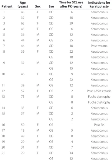
Twenty-seven eyes of 21 patients (12 females) were evaluated: 15 patients were fitted for lenses in one eye, and six patients, in both eyes. All patients had been prescribed mini-SCLs because their visual acuity was insufficiently corrected with spectacles, and they were unable to tolerate or achieve better results with other contact lens models. The mean patient age ± standard deviation (SD) was 42.3 ± 13.1 years (range: 18-75 years). The most frequent reason for keratoplasty was keratoconus (22 eyes, 81.4%), and the mean interval between grafting and SCL fitting was 10.6 ± 7.3 years (range: 1-29 years) (Table 1).
Eleven eyes (40.7%) were classified as having irregular tism, and 10 eyes (37%) were classified as having regular astigma-tism with a refractive cylinder of ≥5 D. The remaining six eyes (22.3%) had regular astigmatism with a refractive cylinder of <5 D. The overall mean SCVA and CLCVA were 0.39 ± 0.34 logMar (range: 1.8-0.0) and 0.09 ± 0.12 logMar (range: 0.50-0.00), respectively; and the CLCVA did not differ significantly between the groups (Group A: 0.10 ± 0.14 logMar versus Group B: 0.07 ± 0.09 logMar, p=0.50). However, the overall CLCVA improved relative to the SCVA (p=0.00002, paired t
test) (Figure 1). Improvements in visual acuity, defined as a gain of ≥2 decimal acuity lines, were achieved in 21 eyes (77%). Fourteen (51.8%) achieved a visual acuity of 0.00 logMar and 26 eyes (96.3%), ≥0.30 logMar. There were no statistically significant differences in en-dothelium density, manifest cylinder, corneal Kmax, and astigmatism between the groups (Table 2).
Sixteen variations of parameters were used to fit lenses to the 27 eyes. Ten eyes (37%) were fitted with 16.0 mm diameter lens; five eyes (18.6%), with 16.5 mm; 10 eyes (37%), with 17.5 mm; and two eyes (7.4%) with 18.2 mm. The most frequently fitted contact lens, which was used in five eyes (18.6%), had a 6.75-mm base curve, 16.0-mm diameter, and 4.63-mm sagittal height.
The follow-up duration for 20 eyes (74%) was greater or equal to six months. Among these eyes, six (30%) developed complications during lens use. In one patient (#10) who had undergone penetrating keratoplasty more than nine years earlier and had a bilateral corneal endothelium cell density of <1000 cells/mm3, both eyes developed
Ro c h a GaN, e t a l.
1 9
Arq Bras Oftalmol. 2017;80(1):17-20
fitting and had an endothelial cell density of <700 cells/mm3,
mini-SCL use was limited (4-6 hours per day) because of discomfort. Two other patients (#3 and #4) developed intolerance. The first decided to undergo a repeat keratoplasty, and the second selected surgical correction of the astigmatism. One patient (#9) developed microbial keratitis in the left eye during follow-up. During a slit-lamp examina-tion, this condition presented as a small paracentral 2-mm diameter stromal infiltrate associated with bulbar hyperemia. Contact lens wear was suspended, and an appropriate topical broad-spectrum antimi-crobial agent (gatifloxacin 0.5%) was administered. He exhibited a good evolution during treatment and was able to resume lens use after two weeks; no other complication or signs of infection have occur-red in the six months since this episode. Complications were not observed in the remaining seven eyes (26%) that were subjected to less than six months of follow-up. No corneal graft rejection occurred during our follow-up period.
DISCUSSION
Currently, astigmatism is most frequent cause of decreased visual acuity after keratoplasty(1-3), and this condition usually remains even
after suture removal. Many patients that have undergone pe netrating keratoplasty depend on spectacles or contact lenses to achieve the desired visual rehabilitation. Smiddy et al. reported that more than 50% of patients would need to wear contact lenses after successful penetrating keratoplasty(30). In our series, the mean UVA after corneal
transplantation was 1.4 ± 0.44 logMar, which improved to a mean SCVA with spectacles of 0.39 ± 0.34 logMar. This low level of visual rehabilitation without correction, and even with spectacles, motiva-ted us to suggest mini-SCL fitting although we consider it important to mention that all patients initially tried other types of lenses.
Because of the profile characteristics of the grafted cornea, the best contact lenses options may include hybrid lenses, small-dia meter RGP lenses, scleral lenses, reverse geometry hydrogels, and large-dia-meter inverse geometry RGP lenses(3,6,17,19). However, corneal grafts
in crease the ocular demand for oxygen(5), thus limiting the use of
hydrogels and hybrid lenses because of the increased lens thickness required to neutralize irregular or high astigmatism(19). Currently, RGP
lenses are usually considered gold standard for visual rehabilitation in patients with irregular corneas(6). However, patient adaptation may
not always be possible because of decentration, epithelial abrasions, keratitis, and discomfort, and we note that all patients in our study had a previous inability to tolerate RGP lenses after keratoplasty.
Our results from this study are similar to those reported by others. For example, 26 eyes (96.3%) in our study achieved a visual acuity of ≥0.30 logMar, and 21 eyes (77%) achieved an improvement of ≥2 visual acuity lines relative to the best SCVA. In comparison, Severinsky et al. reported that among 36 eyes fitted with SCLs, 31 eyes (94%) achieved an improvement in BCVA of ≥2 visual acuity lines, and 23 patients (82%) achieved an improvement of ≥0.5 Snellen(6). That study
also found no differences in refraction, topographic parameters, or visual acuity when the time of grafting was considered. Pullum and Bu-ckley reported a visual acuity of 20/60 or better in 77% of 530 patients referred for SCL fitting(21). Vreugdenhil reported an improvement in
decimal visual acuity from a previous mean SCVA of 0.59 to a CLCVA of 0.75 with semi-scleral lenses(22). Segal et al. reported a CLCVA of
20/40 or better in 81.8% of eyes that underwent keratoplasty(23), and
Tan et al. reported an improvement to 20/40 or better in 72% of 118 fitted eyes(24). Alipour et al. additionally described mini-SCL fitting in
56 eyes of post-keratoplasty patients and observed a visual improve-ment from 1.05 logMar (UCVA) to 0.17 logMar (CLCVA)(25).
In our series, six eyes (22%) developed complications during mini- SCL wear. Three patients (four eyes, 14.8%) failed to wear mini-SCLs. One discontinued mini-SCL use after several hours because of bilateral corneal edema, and two discontinued use because of into-lerance to the lenses. Another patient developed microbial keratitis Table 1. Demographic data of patients itted with mini-scleral contact
lenses after penetrating keratoplasty
Patient Age
(years) Sex Eye
Time for SCl use after PK (years)
Indications for keratoplasty
01 48 F OD 06 Keratoconus
02 32 F OD 10 Keratoconus
03 62 F OD 29 Keratoconus
04 43 F OS 06 Keratoconus
05 36 M OD 12 Keratoconus
06 44 M OS 25 Keratoconus
07 46 M OD 10 Post-trauma
08 39 F OD 22 Keratoconus
OS 18 Keratoconus
09 37 M OD 12 Keratoconus
OS 15 Keratoconus
10 48 F OD 09 Keratoconus
OS 22 Keratoconus
11 39 M OS 12 Keratoconus
12 52 F OS 02 Post-LASIK ectasia
13 75 M OD 08 Fuchs dystrophy
OS 08 Fuchs dystrophy
14 33 F OD 06 Keratoconus
15 37 M OD 02 Keratoconus
OS 02 Keratoconus
16 50 F OS 01 Post-RK
17 18 M OS 05 Keratoconus
18 49 F OD 08 Keratoconus
19 29 M OS 04 Keratoconus
20 31 F OD 07 Keratoconus
21 29 F OD 14 Keratoconus
OS 12 Keratoconus
CL= contact lens.
UVA= uncorrected visual acuity; SCVA= spectacle-corrected visual acuity; CLCVA= contact lens-corrected visual acuity
Vi s ua lr e h a b i l i tat i o nu s i n gm i n i-s c l e r a lc o n ta c tl e n s e sa f t e rp e n e t r at i n gk e r at o p l a s t y
2 0 Arq Bras Oftalmol. 2017;80(1):17-20
in left eye, but continued to use the lens after receiving treatment. In comparison, the complication and failure rates vary somewhat. Severinsky et al. reported usage failure in 19.4% of cases, with 10 eyes (30%) experiencing at least one episode of graft rejection and two eyes developing microbial keratitis(6). Tan et al. reported a usage failure
rate of 8% after an average follow-up of 15.3 months(24), whereas
Pullum and Buckley described a 22% of failure(21) and Segal et al., a
7.5% rate(23). In contrast, Alipour et al. reported a success rate of only
25% (i.e., failure rate of 75%)(25). Among 58 tested eyes (45 patients),
lenses were ordered for only 23 eyes (19 patients), and of these, lens usage was continued in only 14 eyes of 11 patients. Four eyes were lost after follow-up, and five others discontinued use because of contact lens intolerance in two eyes, handling difficulties in one eye, and economic reasons in one eye. In that study, the only reported complaints were conjunctival hyperemia and contact lens intolerance after three hours of use in both eyes of a single patient, and no graft related complications such as rejection or decompensation occurred.
In conclusion, this retrospective study demonstrates the successful visual rehabilitation facilitated by mini-SCL use after corneal kerato-plasty, especially in patients with low SCVA and an inability to wear RGP contact lenses. Our results indicate that mini-SCLs may be a valid option for eyes with corneal irregularities, such as those associated with keratoplasty.
REfERENCES
1. Ozkurt Y, Atakan M, Gencaga T, Akkaya S. Contact Lens Visual Rehabilitation in Kerato-conus and Corneal Keratoplasty. J Ophthalmol. 2012;2012:832070.
2. Stainer GAT, Perl T, Binder PS. Controlled reduction of postkeratoplasty astigmatism. Ophthalmology. 1982;89(6):668-75.
3. Gruenauer-Kloevekorn C, Kloevekorn-Fischer U, Duncker GI . Contact lens and special back surfasse design after penetrating keratoplasty to improve contact lens fit and visual outcome. Br J Ophthalmol . 2005;89(12):1601-8.
4. Claesson M, Armitage WJ, Fagerholm P, Stenevi U. Visual outcome in corneal grafts: a preliminary analysis of the Swedish Corneal Transplant Register. Br J Ophthalmol. 2002; 86(2):174-80.
5. Szczotka LB, Lindsay RG. Contact lens fitting following corneal graft surgery. Clin Exp Optom. 2003;86(4):244-9.
6. Severinsky B, Behrman S, Frucht-Pery J, Solomon A. Scleral contact lenses for visual rehabilitation after penetrating keratoplasty: Long term outcomes. Cont Lens Anterior Eye. 2014;37(3):196-202.
7. Kelly TL, Williams KA, Coster DJ. Corneal transplantation for keratoconus: a registry study. Arch Ophthalmol. 2011;129(6):691-7.
8. Jacobi PC, Hartmann C, Severin M, Bartz-Schmidt KU. Relaxing incisions with com-pressive sutures for control of astigmatism after penetrating keratoplasty. Graefes Arch Clin Exp Ophthalmol. 1994;232(9):527-32.
Table 2. Refractive and corneal data of eyes itted with mini-scleral contact lenses after penetrating keratoplasty
Time after surgery n SE ± SD CYl ± SD ΔK ± SD Kmax ± SD SM ± SD
<10 years 14 -5.74 ± 4.66 -3.98 ± 3.15 5.16 ± 3.02 59.09 ± 05.90 1383.6 ± 803.3
>10 years 13 -5.59 ± 4.72 -4.79 ± 2.16 7.30 ± 3.73 59.72 ± 13.02 1049.8 ± 612.8
p - 0.93 0.45 0.11 0.87 0.24
All eyes 27 -5.67 ± 4.60 -4.37 ± 2.70 6.19 ± 3.49 58.40 ± 07.80 1222.9 ± 724.5
SE= spherical equivalent (diopters; D); SD= standard deviation (D); Cyl= manifest refraction; astigmatism (D); ∆K= topographic astigmatism (D); Kmax= mean steepest keratometry (D); SM=
average endothelium cell density on specular microscopy (cells/mm2).
9. Havding G. Suture adjustment in penetrating keratoplasty. Acta Ophthalmol. 1994; 72(2):246-52.
10. Lam DS, Leung AT, Wu JT, et al. How long should one wait to perform LASIK after PKP? J Cataract Refract Surg. 1998;24(1):6-7.
11. Mandel MR, Shapiro MB, Krachmer JH. Relaxing incisions with augmentation sutures for the correction of postkeratoplasty astigmatism. Am J Ophthalmol. 1987;103(3 Pt 2): 441-7.
12. Campos M, Hertzog L, Garbus J, Lee M, McDonnell PJ. Photorefractive keratectomy for severe postkeratoplasty astigmatism. Am J Ophthalmol. 1992;114(4):429-36. 13. Forseto AS, Francesconi CM, Nose RA, Nosé W. Laser in situ keratomileusis to correct
refractive errors after keratoplasty. J Cataract Refract Surg. 1999;25(4):479-85. 14. Prazeres TM, Souza AC, Pereira NC, Ursulino F, Grupenmacher L, de Souza LB.
Intrastro-mal corneal ring segment implantation by femtosecond laser for the correction of residual astigmatism after penetrating keratoplasty. Cornea. 2011;30(12):1293-7. 15. Coscarelli S, Ferrara G, Alfonso JF, Ferrara P, Merayo-Lloves J, Araujo LP, Machado AP,
et al. Intrastromal corneal ring segment implantation to correct astigmatim after pe-ne trating keratoplasty. J Cataract Refract Surg. 2012;38(6):1006-13.
16. Alfonso JF, Lisa C, Abdelhamid A, Montés-Micó R, Poo-López A, Ferrer-Blasco T. Posterior chamber phakic intraocular lenses after penetrating keratoplasty. J Cataract Refract Surg. 2009;35(7):1166-73.
17. Wietharn BE, Driebe Jr WT. Fitting contact lenses for visual rehabilitation after pene-trating keratoplasty. Eye Contact Lens. 2004;30(1):31-3.
18. Geerards AJ, Vreugdenhil W, Khazen A. Incidence of rigid gas-permeable contact lens wear after keratoplasty for keratoconus. Eye Contact Lens. 2006;32(4):207-10. 19. Katsoulos C, Nick V, Lefferis K, Theodoro M. Fitting the post-keratoplasty cornea with
hy drogel lenses. Cont Lens Anterior Eye. 2009;32(1):22-6.
20. Mackman G, Polak FM, Sidrys L. Fluorescein angiography of soft contact lens induced vascularization in penetrating keratoplasty. Ophthalmic Surg. 1985;16(3):157-61. 21. Pullum KW, Buckley RJ. A study of 530 patients referred for RGP scleral contact lens
assessment. Cornea. 1997;16(6):612-22.
22. Vreugdenhil W, Geerards AJ, Vervaet CJ. A new rigid gas-permeable semi-scleral contact lens for treatment of corneal surface disorders. Con Lens Anterior Eye. 1998; 21(3):85-8.
23. Segal O, Barkana Y, Hourovitz D, Behrman S, Kamun Y, Avni I, et al. Scleral contact lenses may help where other modalities fail. Cornea. 2003;22(4):308-10.
24. Tan DH, Pullum KW, Buckley RJ. Medical applications of scleral contact lenses: 2. Gas permeable scleral contact lenses. Cornea. 1995;14(2):130-7.
25. Alipour F, Behrouz MJ, Samet B. Mini-scleral lenses in the visual rehabilitation of pa-tients after penetrating keratoplasty and deep anterior lamellar keratoplasty. Contact Lens Anterior Eye. 2015:38(1):54-8.
26. Siqueira AC, Santos MS, de Farias CC, Barreiro TR, Gomes JA. [Scleral contact lens for ocular rehabilitation in patients with Stevens-Johnson syndrome]. Arq Bras Oftalmol. 2010;73(5):428-32. Portuguese.
27. Ridley F. Scleral contact lenses, their clinical significance. Arch Ophthalmol. 1963;70: 740-5.
28. Ezekiel D. Gas-permeable haptic lenses. J Br Contact Lens Assoc. 1983;6:158-61. 29. Van der Worp E. A guide for scleral lens fitting [Internet] . Iowa, IA; Scleral Lens Education
Society; 2010. p. 2-3. [cited 2016 Jun 21] Available online: http://www.commons. pacificu.edu/mono/4.