INTRODUCTION
Aggressive posterior retinopathy of prematurity (AP-ROP), pre-viously known as Rush disease,” is a severe and uncommon type of retinopathy of prematurity (ROP) that demonstrates rapid progres-sion(1). Detailed criteria for the disease were defined in the Interna-tional Classification of Retinopathy of Prematurity Revisited in 2005 (ICROP)(1). It is characterized by increased dilation and tortuosity in the posterior pole vessels in all four quadrants, with flat neovasculari-zations, shunts, and hemorrhages between the vascular and avascu-lar retina. AP-ROP does not appear in any stages of classic ROP(1). The disease frequently affects zone I of the retina, but it can sometimes affect the posterior of zone II(1). It progresses without classic stages(2) and if it is not treated, it can progress rapidly to stage 5(1).
ABSTRACT
Purpose: To evaluate the retinal vascularization process after intravitreal rani-bizumab was administered to infants with aggressive posterior retinopathy of prematurity (AP-ROP).
Methods: Twenty-six eyes of 13 infants with AP-ROP who received 0.25 mg in travitreal ranibizumab were retrospectively investigated. The patients were evaluated at weekly follow-up visits, and the findings were analyzed after retinal vascularization was complete.
Results: The results showed regression in the AP-ROP of all the patients within the first 48-72 h. Average time for complete vascularization of the nasal quadrant (zone II) was postmenstrual week 45 (range 41-56), and vascularization of the temporal quadrant (zone III) was completed in the postmenstrual week 56 (range 50-65). Reactivation was observed in seven patients, on average at postmenstrual week 42; two of these patients underwent additional treatment. Two patients presented with avascular areas in the peripheral retina despite being 1 year old.
Conclusion: These results showed that retinal vascularization following intravitreal ranibizumab was completed after a delay in patients with AP-ROP. Further studies are necessary to evaluate when and how vascularization occurs after intravitreal anti-vascular endothelial growth factor treatments.
Keywords: Retinopathy of prematurity/drug therapy; Retinal neovascularization/ drug therapy; Intravitreal injections; Ranibizumab/therapeutic use; Child
RESUMO
Objetivo: Avaliar o processo de vascularização da retina após injeção intravítrea de ranibizumab aplicada em crianças com retinopatia da prematuridade posterior agressiva (AP-ROP).
Métodos: Vinte e seis olhos de 13 crianças com AP-ROP que receberam 0,25 mg de ra nibizumab intravítreo foram investigados retrospectivamente. Os resultados foram avaliados após a completa vascularização da retina, observada em acompanha-mentos semanais.
Resultados: Verificou-se que houve regressão na AP-ROP de todos os pacientes durante as primeiras 48 a 72 horas. Na média, a vascularização do quadrante nasal (zona II) foi concluída na semana 45 pós-menstrual (variação 41-56), enquanto a vascularização do quadrante temporal (zona III) foi concluída na semana 56 pós-menstrual (variação 50-65). Sete pacientes (7/13) apresentaram reativação, que aconteceram em média a 42,14 semanas pós-menstruais, dois pacientes receberam tratamento adicional. Dois pacientes apresentaram áreas avasculares na retina periférica apesar de terem um ano de idade.
Conclusões: O presente estudo mostrou que a vascularização da retina após a inje-ção intravítrea de ranibizumab foi concluída com atraso na AP-ROP. Ensaios clínicos randomizados são necessários para avaliar quando e como a vascularização acontece após tratamentos com injeções intravítreas de anti-VEGF.
Descritores: Retinopatia da prematuridade/quimioterapia; Neovascularização re-tiniana/quimioterapia; Injeções intravítreas; Ranibizumab/uso terapêutico; Criança
Several studies have reported poor outcomes in AP-ROP after laser photocoagulation(3-5), and some eyes with AP-ROP progress to retinal detachment despite early, confluent, and adequate laser treatment(6).
Vascular endothelial growth factor (VEGF) has been shown to play a role in the pathogenesis of ROP(7-9), which has led to the deve-lopment of anti-VEGF treatments as a therapeutic option. Ranibizu-mab is a humanized monoclonal antibody Fab fragment specifically designed for ocular use that acts as an anti-VEGF agent for neovas-cular disorders, including ROP(10-12). Various studies have shown that intravitreal ranibizumab is effective for the treatment of ROP(10,13-17), including case reports of its use for AP-ROP(12).
The aim of this study was to evaluate the vascularization process after intravitreal ranibizumab in a series of infants with AP-ROP.
The vascularization process after intravitreal ranibizumab injections
for aggressive posterior retinopathy of prematurity
Processo de vascularização após injeções intravítreas de ranibizumab para retinopatia
da prematuridade posterior agressiva
EminE AlyAmAç SukgEn1, yuSuf koçluk1
Submitted for publication: April 20, 2016 Accepted for publication: September 22, 2016
1 Department of Ophthalmology, Adana Numune Training and Research Hospital, Adana, Turkey.
Funding: No specific financial support was available for this study.
Disclosure of potential conflicts of interest: None of the authors have any potential conflict of interest to disclose.
Corresponding author: Emine Alyamaç Sukgen. Ege Bagatur caddesi, Adana Numune Eğitim ve Araştırma Hastanesi Yüreğir - Adana, 06520 - Turkey - E-mail: [email protected] Approved by the following research ethics committee: Adana Numune Training and Research
METHODS
This study was conducted in the Center for the Diagnosis and Treatment of Retinopathy of Prematurity in Adana Numune Training and Research Hospital, Turkey, between October 2013 and May 2015. Institutional review board approval was obtained from the center where the study was conducted, and the study was performed according to the ethical standards of the Declaration of Helsinki. The parents of all patients were informed of the effects and possible complications of intravitreal anti-VEGF injections and their written consent was obtained.
AP-ROP was diagnosed according to the ICROP criteria(1). Prior to examination, the pupils were dilated with 2.5% phenylephrine (Mydfrin®; Alcon, Fort Worth, TX, USA) and 0.5% tropicamide (Tropa-mide®; Bilim Ilac, Istanbul, Turkey). The examinations were performed using a Heine Video Omega 2 C Binocular Indirect Ophthalmoscope (Heine Optotechnik, Herrsching, Germany), operating with the Archimed Programme to obtain the images. Fundus examination with a 20 or 28 diopter lens was used to confirm the diagnosis.
Intravitreal ranibizumab was applied to 26 eyes of the 13 patients with AP-ROP. The procedure was conducted in an operating room under sterilized conditions, and the patients were monitored throughout the operation. Pupil dilation was performed by using 0.5% tropicamide and 2.5% phenylephrine drops. After achieving topical anesthesia with proparacaine HCL 0.5%, the periocular area was wiped with 10% betadine. The eyes were draped and opened using an appro-priately sized speculum. After cleaning the ocular surface with 5% betadine and waiting for 3 minutes, the procedure was performed. The area to be operated on was locally massaged with a sponge and 0.25 mg/0.025 ml ranibizumab (Lucentis®; Genentech Inc., South San Francisco, CA, USA), was injected intravitreally 1 mm into the superior temporal paralimbal area using a 30-gauge needle. Ranibizumab was administered into both of the patient’s eyes in the same operation, ensuring sterilization conditions were maintained. The same surgeon (EAS) performed all of the injections. The treatment was applied as monotherapy.
After the injections, the central retinal artery perfusion and intrao-cular pressure were checked by ophthalmic examination. Moxifloxa-cin 0.5% drops were used as postoperative prophylaxis four times a day for 1 week. Intraocular pressure was measured on the first and third days using a Tono-Pen (Tono-Pen AVIA® Applanation Tonometer; Reichert Technologies, Depew, NY, USA). The patients were moni-tored on the first day, the third day, and then on a weekly basis until zone III was vascularized; after that, they were monitored monthly.
On each visit, indirect funduscopic examinations were performed to evaluate any disease regression and reactivation, and peripheral vas cularization. Positive responses to the treatment were indicated by the disappearance of rubeosis iridis, improved pupil dilation, a de crease in retinal arterial and venous tortuosity and engorgement, regression of plus disease, and vessels continuing to vascularize toward the peripheral retina.
Reactivation was defined as the reappearance of any stage of the disease, with or without plus disease. When reactivation occurred, the patient was given supplementary treatment after consulting the parents. The patients were monitored under sophisticated laboratory parameters in terms of systemic side effect profile, particularly on the first postoperative day.
Statistical analyses were performed using SPSS software for Windows version 16.0 (SPSS Inc. Chicago, USA). The data are presented as median (range) or as mean values.
RESULTS
Table 1 summarizes the demographic data and treatment outcomes of the 13 patients included in the study (8 were female and 5 were male). The median gestational age was 28 weeks (range 24-32 weeks) and the median birth weight was 1114 g (range 550-1980) grams. In
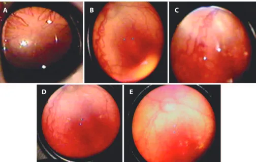
10 patients the disease affected zone I and in 3 patients it affected posterior zone II. Eight patients had rubeosis iridis. The median age at treatment was 35.4 postmenstrual, gestational weeks (range 33.5-39). The follow-up period ranged from 6 to 18 months after the primary ranibizumab therapy. Figure 1 shows a typical series of photographs (for case 13) of the progression from before treatment to six months after treatment.
Intraocular pressures were normal on the postoperative first and third days, anterior segment examinations were normal, and all the patients exhibited a significant regression of plus disease within 48-72 h after the treatment. No serious complications, such as endo-phthalmitis, retinal detachment, cataract, or intravitreal hemorrhage, or systemic side effects in the measurable parameters were observed after the injection.
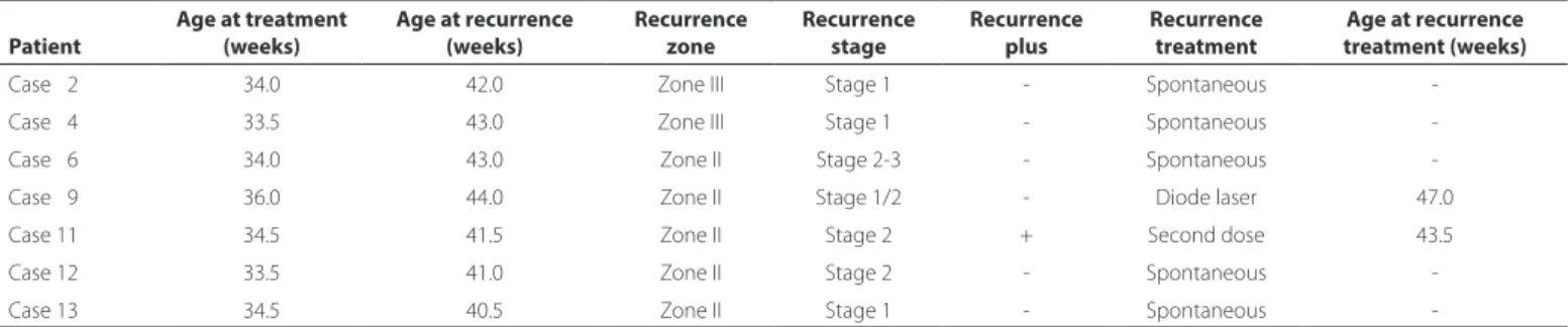
Vascularization of the nasal quadrant (zone II) was completed at a median of postmenstrual week 45 (range 41-56), and vascularization of the temporal quadrant (zone III) in postmenstrual week 56 (range 50-65) (Table 2). There were avascular areas in the peripheral retina in two patients (cases 3 and 11), even though they were one year of age. Seven patients were found to have reactivation, occurring at a median of postmenstrual week 42 (range 40.5-44) (Table 3). Reactiva-tion, which occurred as zone III stage 1 in two cases, as zone II stage 2 in two cases, and as zone II stage 1 in one case, was observed to regress spontaneously in five patients, and only two patients were given additional treatment.
Case 9 exhibited reactivation in the middle of zone II stage 2 without plus disease. As her family was unable to attend for frequent follow-ups, they requested additional treatment, and transpupillary diode laser photocoagulation was performed at postmenstrual week 47. Case 11 exhibited reactivation in zone II posterior stage 2, rapidly followed by plus disease. With her family’s consent, a second dose of ranibizumab 0.25 mg/0.025 ml was administered intravitreally at postmenstrual week 43.5.
During follow-up, it was observed that case 6 developed a fibrous band expanding to the vitreous in week 51. However, as this did not cause traction in the retina, no additional treatment was administered. There was a decrease in the volume of the fibrous band with the vascularization of the retinal periphery in the late period. Zone III vas-cularization in this patient was delayed until 65 weeks.
The mean times for the completion of zone III vascularization were 57.9 weeks for the eyes with reactivation and 54 weeks for the eyes without reactivation. Although the presence of reactivation delayed the average revascularization time, this difference was not statistically significant (p=0.171).
For zone II, the mean vascularization speed (measured as the period between the treatment week and the week when revascula-rization was complete) was 6.8 weeks in the eyes without reactivation and 12.5 weeks in the eyes with reactivation, a statistically significant difference (p=0.020). For zone III, the mean vascularization speed was 17.1 weeks in the eyes without reactivation and 23.9 weeks in the eyes with reactivation; this difference was not statistically significant (p=0.055) (Table 2).
DISCUSSION
There have been numerous studies on the use of intravitreal ra nibizumab for ROP treatment since 2012 that have demonstrated its efficacy for regression of the disease(10-12,15-17). The present study confirmed this, demonstrating that 0.25 mg intravitreal ranibizumab was effective in the regression of AP-ROP and the maintenance of retinal vascularization.
Table 1. Demographic data and pretreatment indings
Patient Sex Gestational age (weeks) Birth weight (g) Eye Zone Diagnosis Rubeosis iridis Postmenstrual age at treatment
Case 01 Male 30 1750 R Zone II post AP-ROP - 38.5
L Zone II post
-Case 02 Male 30 1400 R Zone I AP-ROP - 34.0
L Zone I
-Case 03 Male 29 1040 R Zone II post AP-ROP + 36.0
L Zone II post +
Case 04 Male 25 0550 R Zone I AP-ROP + 33.5
L Zone I +
Case 05 Female 32 1600 R Zone I AP-ROP + 37.0
L Zone I +
Case 06 Female 26 0640 R Zone I AP-ROP - 34.0
L Zone I
-Case 07 Female 29 1100 R Zone II post AP-ROP - 34.0
L Zone II post
-Case 08 Female 27 0950 R Zone I AP-ROP + 36.5
L Zone I +
Case 09 Female 28 1150 R Zone I AP-ROP + 36.0
L Zone I +
Case 10 Male 31 1980 R Zone I AP-ROP - 39.0
L Zone I
-Case 11 Female 24 0790 R Zone I AP-ROP + 34.5
L Zone I +
Case 12 Female 25 0770 R Zone I AP-ROP + 33.5
L Zone I +
Case 13 Female 28 0760 R Zone I AP-ROP + 34.5
L Zone I +
AP-ROP= aggressive posterior retinopathy of prematurity; R= right eye; L= left eye.
Figure 1. Series of photographs of a typical patient with aggressive posterior retinopathy of prematurity (case 13) treated with intravitreal ranibizumab. A) The presence of tunica vasculasa lentis before treatment. B) Plus disease in the posterior zone. C) Intraretinal neovascular vessels and shunts. D) First postoperative week. E) Six months after the operation.
A B C
Table 2. Time of vascularization
Patient
Postmenstruel age at treatment (weeks)
Age at recurrence (weeks)
Zone II completion of vascularization (weeks)
Zone III completion of vascularization (weeks)
Zone II vascularization speed (weeks)
Zone III vascularization speed (weeks)
Case 01 38.5 - 43.0 60.0 04.5 21.5
Case 02 34.0 42.0 41.0 59.0 07.0 25.0
Case 03 36.0 - 43.0 50.0 07.0 14.0
Case 04 33.5 43.0 42.0 54.0 08.5 20.5
Case 05 37.0 - 47.0 55.0 10.0 18.0
Case 06 34.0 43.0 51.0 65.0 07.0 31.0
Case 07 34.0 - 42.0 55.0 08.0 21.0
Case 08 36.5 - 42.0 54.0 05.5 17.5
Case 09 36.0 44.0 - - -
-Case 10 39.0 - 45.0 50.0 06.0 11.0
Case 11 34.5 41.5 56.0 62.0 21.5 27.5
Case 12 33.5 41.0 46.5 56.0 13.0 22.5
Case 13 34.5 40.5 42.5 51.5 08.0 17.0
Table 3. Reactivation after intravitreal ranibizumab injections
Patient
Age at treatment (weeks)
Age at recurrence (weeks)
Recurrence zone
Recurrence stage
Recurrence plus
Recurrence treatment
Age at recurrence treatment (weeks)
Case 02 34.0 42.0 Zone III Stage 1 - Spontaneous
-Case 04 33.5 43.0 Zone III Stage 1 - Spontaneous
-Case 06 34.0 43.0 Zone II Stage 2-3 - Spontaneous
-Case 09 36.0 44.0 Zone II Stage 1/2 - Diode laser 47.0
Case 11 34.5 41.5 Zone II Stage 2 + Second dose 43.5
Case 12 33.5 41.0 Zone II Stage 2 - Spontaneous
-Case 13 34.5 40.5 Zone II Stage 1 - Spontaneous
-care units, and so the age of the patient may be associated with the care protocol used in the center where the newborn was treated. A retrospective analysis of these newborn intensive care data revea-led important risk factors for ROP development. For instance, case 5 was born as one of triplets; she received oxygen treatment for 16 days, and her daily weight gain rate was 15 g. Case 10 was born as one of twins, received mechanical ventilation for 9 days and free oxygen treatment for 20 days, and suffered sepsis; his average daily weight gain was 20 g.
ICROP reported that AP-ROP usually affects zone I, but in some cases it can affect zoneposterior zone II(1). The patient in this study were in accordance with this: 10 patients had zone I and three pa-tients had posterior zone II AP-ROP. Treatment took place at a median of postmenstrual week 35.4, which is comparable with that found in previous studies(14,15,17).
The dose of ranibizumab administered has varied between pre-vious studies. For instance, Baumall et al. applied 0.2 mg ranibizumab to the 8 eyes of 4 patients with stage 1 ROP and observed reactivation in all of them(13). Zhou et al. used 0.25 mg ranibizumab in 22 eyes of 11 infants and detected reactivation in 10 infants(17). Wong et al. applied 0.25 mg ranibizumab to 6 eyes of 4 infants and detected reactivation in 5 of them(14). In contrast, Menke et al. applied 0.3 mg/0.03 ml to 6 eyes of 4 infants with zone II stage 3 ROP with plus disease and all the patients completed vascularization without recurrence(18). In addition, Chen et al.(15) and Castellanos et al.(10) reported completed vascularization in their patients without any recurrence after 0.25 mg ranibizumab.
Of the 13 infants involved in our study, reactivation was observed in 7. Two of these received additional treatment, but only one de-veloped threshold disease. Baumall et al.(13) detected reactivation between 8 and 11 weeks after the treatment and Zhou et al.(17) at a mean of 7.30 weeks after treatment. Wong(14) observed reactivation in postmenstrual week 42. These cases underwent laser ablation in avascular areas for the recurrence. The present study detected reacti-vation at a mean of 7.8 weeks after treatment, in postmenstrual week 42.6. The disease developed in more peripheral parts of the retina.
The reactivation was probably associated with the amount of reti-na vascularized prior to the elimireti-nation of the drug from the vitreous and the amount of VEGF oscillation from the remaining avascular retina. Reactivation is common after the administration of intravitreal anti-VEGF agents for AP-ROP(19). However, we believe that reactivation should be treated according to ETROP criteria because, in our study, the disease regressed spontaneously in the majority of eyes. Our approach to reactivation was to wait and follow up.
One of the most important problems regarding the use of anti -VEGF agents in ROP is when and how vascularization happens. The present study has shown that retinal vascularization was delayed com pared to physiological vascularization. This may be because anti-VEGF agents slow down the vascularization process or because revascularization speed is slower in eyes with reactivation. This is the first study to re-port the time of retinal vascularization after intravitreal ranibizumab for AP-ROP in a large series.
had avascular areas in the peripheral retina at the end of the follow-up period. Some studies have used angiography to demonstrate the presence of avascular areas in the peripheral retina after the use of anti-VEGF agents(20). One of the limitations of the present study was that we could not show complete vascularization in an angiographic way. Other limitations were that it was retrospective in nature and had a short follow-up period. Studies with wider case series and longer follow-up are needed.
In conclusion, the present study demonstrated that 0.25 mg in-travitreal ranibizumab applied inin-travitreally in AP-ROP was effective in bringing about the regression of the disease and completion of vas-cularization in majority of the cases. These effects were demonstrated anatomically with only clinical observations. Long-term studies are needed to demonstrate the effect of the anti-VEGF agents on the retinal vascularization process.
REFERENCES
1. International Committee for the Classification of Retinopathy of Prematurity. The International Classification of Retinopathy of Prematurity revisited.Arch Ophthalmol. 2005;123(7):991-9.
2. Katz X, Kychenthal A, Dorta P. Zone 1 Retinopathy of prematurity. J AAPOS. 2000;4(6): 373-6.
3. Drenser KA, Trese MT, Capone A Jr. Aggressive posterior retinopathy of prematurity. Retina. 2010;30(4):S37-40.
4. Azuma N, Ishikawa K, Hama Y, Hiraoka M, Suzuki Y, Nishina S. Early vitreous surgery for aggressive posterior retinopathy of prematurity. Am J Ophthalmol. 2006;142(4):636-43. 5. Autrata R, Krejcírová I, Šenková K, Holoušová M, Doležel Z, Borek I. Intravitreal pegapta-nib combined with diode laser therapy for stage 3+ retinopathy of prematurity in zone I and posterior zoneII.ur J Ophthalmol. 2012;22(5):687-94.
6. Gaurav S, Mangat RD, DeekshaK, et al. Aggressive Posterior Retinopathy of Prematurity: Risk factors for retinal detachment despite confluent laser photocoagulation. Am J Ophthalmol. 2013;155(1):159-64.
7. González VI, Ferrer NC, Pueyo RV. Use of anti-VEGF (anti-vascular endothelial growth factor) in retinopathy of prematurity (ROP). Arch Soc Esp Ophthalmol. 2011;86(7):207-8.
8. Romagnoli C. Risk factors and growth factors in ROP. Early Hum Dev. 2009;85(10):79-82. 9. Alon T, Hemo I, Itin A, Pe’er J, Stone J, Keshet E. Vascular endothelial growth factor acts
as a survival factor for newly formed retinal vessels and has implications for retinopathy of prematurity. Nat Med. 1995;1(10):1024-8.
10. Castellanos MA, Schwartz S, Garcia-Aguirre G, Quiroz-Mercado H. Short-term outcome after intravitreal, ranibizumab injections for the treatment of retinopathy of prematurity. Br J Ophthalmol. 2013;97(7):816-9.
11. Hoerster R, Muether P, Dahlke C, Mehler K, Oberthür A, Kirchhof B, et al. Serum concen-trations of vascular endothelial growth factor in an infant treated with ranibizumab for retinopathy of prematurity. Acta Ophthalmol. 2013;91(1):e74-5.
12. Mota A, Carneiro A, Breda J, Rosas V, Magalhães A, Silva R, et al. Combination of intra-vitreal ranibizumab and laser photocoagulation for aggressive posterior retinopathy of prematurity. Case Rep Ophthalmol. 2012;3(1):136-41.
13. Baumal CR, Goldberg RA, Fein JG. Primary intravitreal ranibizumab for high-risk reti-nopathy of prematurity. Ophthalmic Surg Lasers Imaging Retina. 2015;46(4):432-8. 14. Wong RK, Hubschman S, Tsui I. Reactivation of retinopathy of prematurity after
ranibi-zumab treatment. Retina. 2015;35(4):675-80.
15. Chen SN, Lian I, Hwang YC, Chen YH, Chang YC, Lee KH, et al. Intravitreal anti-vascular endothelial growth factor treatment for retinopathy of prematurity: comparison between ranibizumab and bevacizumab. Retina. 2015;35(4):667-74.
16. Erol MK, Deniz TC, Özdemir Ö, Tunay ZÖ, Bilgin AB, Dogan B. Spectral-Domain OCT analyses of macular changes after ranibizumab therapy for Type 1 retinopathy of prematurity. J Pediatr Ophthalmol Strabismus. 2015;52(3):152-8.
17. Zhou Y, Jiang Y, Bai Y, Wen J, Chen L.Vascular endothelial growth factor plasma levels before and after treatment of retinopathy of prematurity with ranibizumab. Graefes Arch Clin Exp Ophthalmol. 2016;254(1):31-6.
18. Menke MN, Framme C, Nelle M, Berger MR, Sturm V, Wolf S. Intravitreal ranibizumab monotherapy to treat retinopathy of prematurity zone II, stage 3 with plus disease. BMC Ophthalmol. 2015;15(1):1.
19. Mintz-Hittner HA. Intravitreal pegaptanib as adjunctive treatment for stage 3+ ROP shown to be effective in a prospective, randomized, controlled multicenter clinical trial. Eur J Ophthalmol. 2012;22(5):685-6.
20. Tahija SG, Hersetyati R, Lam GC, Kusaka S, McMenamin PG. Fluorescein angiographic observations of peripheral retinal vessel growth in infants after intravitreal injection of bevacizumab as sole therapy for zone I and posterior zone II retinopathy of prema-turity. Br J Ophthalmol. 2014;98(4):507-12.