Emotional declarative memory
assessment of patients with
mesial temporal lobe epilepsy
and patients submitted to
mesial temporal lobectomy
Lara De Vecchi Machado1, Jean Edith Frank2, Carlos Tomaz3
ABSTRACT
Epileptic seizures generate cognitive and behavioral impacts in individuals who suffer from epilepsy. Declarative memory is one of the cognitive functions that can be affected by epileptic seizures. The main objective of this work was to investigate neurocognitive function, especially the emotional working memory of patients with unilateral mesial temporal lobe epilepsy, and that of patients submitted to unilateral mesial temporal lobectomy. A face recognition test that can simultaneously recruit the frontal lobe (working memory) and mesial temporal lobe (emotional memory) was used to investigate emotional working memory. Our findings showed that the epilepsy factor significantly compromised the performance in the emotional memory test. On the other hand, surgical removal of the epileptic focus promoted an improvement in the emotional working memory of these patients, in addition to the significantly decrease in the number of seizures.
Key words: emotional memory, epilepsy, temporal lobectomy.
Avaliação da memória declarativa emocional em pacientes com epilepsia temporal mesial e pacientes submetidos à lobectomia temporal mesial
RESUMO
Crises epilépticas geram impactos comportamentais e cognitivos em indivíduos que sofrem de epilepsia. Uma das funções cognitivas que pode ser afetada pelas crises epilépticas é a memória declarativa. O objetivo do nosso estudo foi investigar funções cognitivas, especialmente a memória operacional emocional de pacientes com epilepsia temporal mesial unilateral e pacientes submetidos a lobectomia temporal mesial unilateral. Para investigar a memória operacional emocional foi utilizado um teste de reconhecimento de faces que pode recrutar simultaneamente o lobo frontal (memória operacional) e o lobo temporal mesial (memória emocional). Nossos resultados demonstram que o fator epilepsia compromete de forma significativa o desempenho no teste de memória emocional. Por outro lado, a remoção cirúrgica do foco epiléptico promoveu uma melhora na memória emocional desses pacientes, além de diminuir o número de crises.
Palavras-chave: memória emocional, epilepsia, lobectomia temporal.
Correspondence
Carlos Tomaz
Laboratory of Neurosciences and Behavior Institute of Biology, University of Brasília Campus Universitário Darcy Ribeiro Asa Norte
70910-900 Brasília DF- Brasil E-mail: [email protected]
Received 30 October 2009 Received in final form 23 March 2010 Accepted 31 March 2010
1PhD Candidate, Physiotherapist, Graduate Program in Health Sciences, Faculty of Health Sciences, University of Brasília (UnB), Brasília DF, Brazil; 2PhD, Psychologist, Laboratory of Neurosciences and Behavior, Institute of Biology, UnB; 3PhD, Full Professor, Laboratory of Neurosciences and Behavior, Institute of Biology, UnB.
Declarative memory is a long-term memory system relating to conscious rec-ollection of facts and events of an indi-vidual’s life. When emotional information combines with evoked declarative
memo-ry, a subtype of declarative memory forms: the emotional declarative memory or emo-tional memory1.
Emo-tional events are probably codiied in a way that may be-come resistant to extinction1-9. his is due to speciic
neu-ral and hormonal mechanisms that modulate memory for emotional stimuli3,10,11. hese mechanisms seem to
oper-ate in part by recruiting general cognitive mechanisms such as attention, and in part by self-mechanisms acti-vated by emotion10,11.
Many studies that assessed emotional memory have raised the possibility that the neural substrate of emotion-al memory extends to neuronemotion-al circuits within the mesiemotion-al region of the temporal lobe1,8,12-14, essentially the amygdale
and hippocampus.
Neuroimaging studies have provided important conver-gence between the indings from studies on electrical ac-tivity, drugs, lesions and neuropsychological studies, infer-ring that the amygdale and other limbic areas are involved not only in the acquisition and consolidation of emotional memory but also in its elicitation8,10,11,15. However, the role
of each hemisphere and the diferences in gender-relat-ed function of the amygdale have not been fully clariigender-relat-ed.
Epilepsy is a disorder of the nervous system that abruptly interferes in behavior, perception, movement, conscience and/or in other cortical functions. In turn, these changes lead to inability to adapt to the environ-ment, hence compromising such individuals’ quality of life. Temporal lobe epilepsy (TLE) is the most common type of epilepsy, since the temporal lobe is the most epi-leptogenic of the cerebral lobes and the most diicult one to treat pharmacologically16,17.
Studies have shown that epilepsy can impair memory acquisition and storage18. Although cognitive deicits in
epileptic patients are very frequent, the pattern and ex-tent of these deicits vary greatly. his variation is due to a number of factors, such as: [a] location and extent of the dysfunctional area or brain injury; [b] anticonvulsant medication used; [c] age at seizure onset and epilepsy du-ration and [d] seizure type and frequency18-20.
herefore, the main objective of the present work was to investigate the neurocognitive function of patients with unilateral mesial temporal lobe epilepsy, especially assess-ing the declarative memory subtype. For this purpose, the working memory test Delay-non-Matching-to-Sam-ple (DNMTS) with emotional content was used.
METHOD
Subjects
he patients, referred by physicians at the Neurolog-ical Institute of Goiânia, were properly diagnosed and presented normal intellectual capacity according to the WAIS-III test for neuropsychological assessment. he research project was approved by the Research Ethics Committee of the Federal University of Goiás. All sub-jects were informed about the research procedures and signed a free and informed consent form.
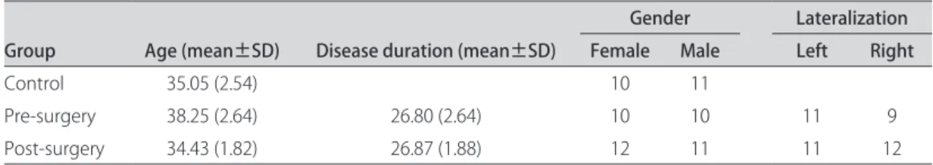
he subjects were divided into three study groups: control group, pre-surgery group and post-surgery group. he control group consisted of 21 healthy subjects (10 women and 11 men); the pre-surgery group consisted of 20 subjects with unilateral mesial temporal lobe epilep-sy. he epileptic focus was identiied by EEG-video mon-itoring and magnetic resonance (MR). his group includ-ed 10 women (ive with epileptic focus on the left and ive on the right) and 10 men (six with epileptic focus on the left and four on the right). he post-surgery group con-sisted of 23 subjects in the late postoperative period af-ter mesial temporal lobectomy, as conirmed by magnetic resonance. his group included 12 women (six with uni-lateral left lobectomy and six with uniuni-lateral right lobec-tomy) and 11 men (ive with left lobectomy and six with right lobectomy) (Table 1).
The neurocognitive test was applied to the three groups so that the results of each group could be com-pared with the other two. The computer was a Toshi-ba laptop, model Satellite M55-S135, with a 14-inch wi-descreen. he subject remained at roughly 50 cm away from the screen.
Inclusion criteria
Group 1 - Control – Individuals of both genders, aged 16 to 55 years who were not taking anti-anxiety or antidepressive drugs, without any identiied neurological disease, and who reported being right-handed.
Group 2 - Pre-surgery – Epileptic patients (both gen-ders, aged 16 to 55 years) properly diagnosed by magnet-ic resonance, electroencephalogram (EEG) and/or video monitoring with unilateral mesial temporal lobe epilepsy, without clinical signs of drug intoxication, who had not
Table 1. Demographics.
Group Age (mean±SD) Disease duration (mean±SD)
Gender Lateralization Female Male Left Right
Control 35.05 (2.54) 10 11
Pre-surgery 38.25 (2.64) 26.80 (2.64) 10 10 11 9
Post-surgery 34.43 (1.82) 26.87 (1.88) 12 11 11 12
had a seizure during the 24 hours preceding the tests, and who reported being right-handed.
Group 3 - Post-surgery – Patients (both genders, aged 16 to 55 years) with a magnetic resonance report proving that they had undergone a unilateral temporal lobectomy, including hippocampus and amygdale. he surgical tech-nique used in the Neurological Institute of Goiânia is the Spencer technique, developed in Yale University, which resects the hippocampus and amygdale as much as pos-sible. he patients did not present clinical signs of intox-ication and had not had a seizure during the 24 hours preceding the tests. hey reported being right-handed.
Neurocognitive assessment
The emotional working memory test Delay-non-Matching-to-Sample (DNMTS) is a computer-based working memory test that also assesses the emotional memory of the individual. herefore, it is believed that this test is capable of recruiting the frontal lobe (working memory) and the mesial temporal lobe (emotional mem-ory) simultaneously.
The first test was developed by Ekman & Wallace (1976) and was called Pictures of Facial Afect; it presented seven diferent emotional facial expressions and was wide-ly used in the literature on emotion. In order to increase the number of stimuli to be used in the test, the Neuro-sciences and Behavior Laboratory of UNB, in 2003, devel-oped a test with eight diferent emotional facial expres-sions (negative surprise, positive surprise, fear, anger, hap-piness, disgust, neutral and sadness) obtained by taking fa-cial pictures of actors. he test consists of a battery of stim-uli such that each battery has eight pictures of the same actor with the facial expressions listed above. his ensures that the recognition is based on the facial expression alone. he DNMTS consisted of presenting a stimulus [A] followed by an eight-second break and the presentation of two stimuli [A and B], of which one is repeated [A]. he subject has to choose the new stimulus [B].
he test consisted of a battery of two geometric stimuli, for the subject to learn, and two batteries with eight difer-ent facial expressions of two actors, one male and one fe-male. Each stimulus was presented in a 5 cm × 5 cm square. he Sysmem software (Lab. Neuroscience & Behav-ior, University of Brasília) was used for the DNMTS test. It enabled manipulation of many parameters and gener-ated automatic spreadsheets after each test, containing the response time for each attempt, the number of errors and the number of pictures.
Statistical analyses
he data were organized and analyzed using Excel® 2003 and SPSS® 13 (Statistical Package for the Social Sci-ences, Chicago, IL, USA) for Windows®.
Firstly, the mean reaction time of each attempt and the percentage of correct choices made by each individ-ual were calculated. hese data were used in the analy-sis models.
One-, two- and three-way univariate analyses of vari-ance (ANOVA) were used, covering the factors Group, Gender and Lateralization together or alone. he post hoc
analyses were performed in accordance with the Bonfer-roni method for adjusting the statistical signiicance level.
All tests were two-tailed, and statistical signiicance was set as p≤0.05.
RESULTS
Descriptive analysis of the groups
Disease durations in the pre- and post-surgery groups were not statistically diferent (p=0.983) (Table 2). Regard-ing the time elapsed since the last seizure, the post-sur-gery group presented a seizure interval signiicantly great-er than in the pre-surggreat-ery group (p<0.001) (Table 3). he mean time elapsed since surgery was 14.1 (2.09) months.
Comparison of the factor Epilepsy vs. Gender (2 × 2)
In the DNMTS test, the factor Epilepsy had a signii-cant efect on the variables of reaction time (F1,60=5.773,
p=0.019) (Fig 1A) and percentage of correct answers (F1,60=5.972, p=0.017) (Fig 1B). he pre-surgery and
post-surgery group set, representing the factor Epilepsy, had a greater reaction time (p=0.019) and a smaller percentage of correct answers (p=0.017) than did the control without epilepsy. he factor Gender and the interaction Epilepsy versus Gender did not have signiicant statistical efects on the two variables studied (Fs<0.094, ps>0.760).
Comparison between Groups and Gender (3×2)
The factor Group presented a significant effect on mean reaction time in the DNMTS test (F2,58=3.608,
p=0.033) (Fig 2A). he factor Gender and the interaction
Table 2. Mean disease duration and mean time elapsed since the last seizure.
Group Disease duration (years) Last seizure (days)
Pre-surgery 26.8 (2.64*) 6.75 (1.49*)
Post-surgery 26.87 (1.88*) 417.87 (58.76*)
*Standard deviation.
Table 3. Mean time elapsed since surgery.
Sex Time surgery (months)
Female 16.75 (1.89*)
Male 11.45 (2.28*)
Group versus Gender did not present signiicant efects on this variable (Fs<0.824, ps>0.443). he multiple com-parison procedure showed that the diference was in the comparison between the groups Control and Pre-surgery (p=0.033). Analysis of correct answer percentages in the DNMTS test did not elicit diferences that could be at-tributed to the factors Group or Gender, or to their inter-action (Fs<3.050, ps>0.054).
Comparison between Group vs. Gender vs. Lateralization (2×2×2)
Note – For this analysis model, it was not possible to use the Control group, since there were no data regard-ing its lateralization.
In the DNMTS test, neither of the factors nor the in-teraction of these factors presented a signiicant efect on mean reaction time (Fs>1.869, ps>0.179). Regarding the percentage of correct answers in the test, the analysis showed that the factors Group, Gender, Lateralization, and the interactions Group × Gender, Gender × Lateral-ization and Group × Gender × LateralLateral-ization did not have a signiicant efect on the results (Fs<1.326, ps>0.256). he interaction Group × Lateralization showed a statis-tically signiicant efect on the variable percentage of cor-rect answers (F1, 35=5.235, p=0.028) (Fig 2B). In the
pre-surgery group, subjects with epileptic focus in the left brain hemisphere had a smaller percentage of correct an-swers than did those with focus in the right. his difer-ence was not observed after surgery.
Fig 1. [A] Results from the pre-surgery and post-surgery groups, representing the factor Epilepsy. There was a greater reaction time in these groups than that of the control group (p=0.019). [B] Re-sults from the pre-surgery and post-surgery groups, representing the factor Epilepsy. There was a smaller percentage of correct an-swers in these groups than in the control group (p=0.017).
6000
5000
4000
3000
2000
1000
0
Rea
cti
on time D
NMTS (m
seg)
Epilepsy
A
Yes No
Female Male
100
80
60
40
20
0
Co
rre
ct
answe
rs D
NMTS (%)
Epilepsy
B
Yes No
Fig 2. [A] The psurgery group showed a signiicantly greater re-action time than did the control group (p=0.033). [B] In the pre-surgery group, subjects with lesions on the left side showed a sig-niicantly smaller percentage of correct answers than did those with lesions on the right side (p=0.028).
1000
800
600
400
200
0
Rea
cti
on time ref
le
x (m
s)
Group
A
Control Before surgery After surgery
Right Left Laterality 100
95
90
85
80
75
70
Co
rre
ct
answe
rs D
NMTS (%)
Group
B
Comparison of Group vs. Gender vs. Lateralization
he control group was included after two ANOVA analyses, and comparison of all the dependent variables (reaction times and percentage of correct answers) were carried out for the groups. One of the ANOVA analy-ses compared the subjects with lesions in the left hemi-sphere with the control group; the other ANOVA com-pared subjects with lesions in the right hemisphere with the control group.
he comparison between subjects with lesion in the left and control subjects showed that the factor Group had statistically signiicant efects on the variables reac-tion time in DNMTS (F2,40=4.309, p=0.020) (Fig 3A) and
percentage of correct answers in DNMTS (F2,40=5.356,
p=0.009) (Fig 3B). he multiple comparison procedure showed that the pre-surgery group had higher response
times in the DNMTS test and lower percentages of cor-rect answers than the control group (ps<0.017).
he comparison between subjects with lesion in the right and control subjects showed that the factor Group had a signiicant efect on the percentage of correct an-swers in DNMTS (F2,39=3.699, p=0.034). The multiple
comparison procedure showed statistically significant diferences between the control group and the post-sur-gery group (p=0.034) (Fig 3C): the control group had a higher percentage of correct answers than the post-sur-gery group. Statistically significant differences among the groups were not found for the other variables (Fs2,39<2.370, ps>0.106).
DISCUSSION
he main objective of the present study was to investi-gate the neurocognitive functions of patients with
unilat-8000
6000
4000
2000
0
Rea
cti
on time D
NMTS (m
s)
Group LEFT LESIONS
A
Control Before surgery After surgery
100
95
90
85
80
75
70
Co
rre
ct
answe
rs D
NMTS (%)
Group RIGHT LESIONS
C
Control Before surgery After surgery
100
95
90
85
80
75
70
Co
rre
ct
answe
rs D
NMTS (%)
Group LEFT LESIONS
B
Control Before surgery After surgery
eral mesial temporal lobe epilepsy, in particular the emo-tional working memory.
One of the results indicated that the factor epilep-sy significantly compromised the performance in the emotional memory test, since the pre- and post-surgery groups, representing the factor Epilepsy, had a signiicant efect on variable reaction time and percentage of correct answers in comparison with the control group. his ind-ing has been reported in the literature by some groups, stating that seizures can generate cognitive and behavior-al impacts in individubehavior-als with epilepsy. Furthermore, one of the cognitive functions that can be afected by seizures is the declarative memory18-21.
No signiicant diference regarding the reaction time and percentage of correct answers in the DNMTS test was found between the pre- and post-surgery groups. However, the present results show signiicantly higher reaction times in the DNMTS test for the pre-surgery group, compared with the control group. Additionally, the post-surgery group presented only an insigniicant per-formance deicit in the emotional memory test in com-parison with the control group. hese results may lead to the inference that surgical removal of the epileptic focus improved the declarative memory of these patients, es-pecially the emotional working memory.
his inding corroborates those of other studies re-ported in the literature, such as that of Aldenkamp and Bodde21. hese authors concluded from a review on
be-havior, cognition and epilepsy that the origin of cognitive or behavioral deicits was frequently related to structur-al brain damage caused by seizures. Seizures are capable of inducing progressive cellular and metabolic changes that are associated with neuronal hippocampal loss, neu-rogenesis and synaptic reorganization. he main clinical characteristic of mesial temporal lobe epilepsy is that, in most cases, the seizures become refractory to drugs, and therefore the best treatment is to remove the epileptogen-ic focus surgepileptogen-ically19,20.
Another important inding was the signiicant reduc-tion in the number of seizures in the post-surgery group in comparison with the pre-surgery group. his seems to be another good reason to remove the epileptic focus, since surgery directly inluences the control of epilepsy and may lead to improved cognition in the long run, par-ticularly in declarative memory. he literature shows that seizure frequency is one of the main factors for cognitive deicits found in patients with epilepsy18-26.
he results from the present work did not show a sig-niicant diference in performance in the emotional mem-ory tests between genders. This finding suggests that there may not be any association between lateralization and gender. Similar results have been observed in other studies, such as that of Frings et al.27 who investigated the
lateralization of hippocampal activation during memory tests related to the mesial temporal lobe, in patients with unilateral epilepsy of that same lobe with functional mag-netic resonance. hese authors did not ind any relation-ship between hippocampal activity and gender, but they found a relationship between hippocampal activity and the side of the epileptic focus.
Recently, other studies have indicated the existence of gender-associated lateralization of the amygdale that mod-ulated emotional memory, thus suggesting that the right amygdale in males and the left amygdale in females are re-sponsible for modulating emotional memory1,8,11,28.
When only lateralization and group were considered, there was a signiicant diference between the pre-surgery group and the control group regarding performance in the emotional memory test among subjects with lesions in the left hemisphere. he pre-surgery subjects with le-sion on the left side presented signiicantly worse results both for the reaction time and for the percentage of cor-rect answers in the DNMTS test, compared with the con-trol group. his poorer performance was also signiicant within the pre-surgery group when the side of the lesion was compared. Subjects with the lesion on the left side presented signiicantly higher reaction times in the DN-MTS test than did those with the lesion on the right side.
his greater reaction time in subjects with lesions on the left side is in agreement with the hypothesis proposed by Frank and Tomaz13, i.e. that the left amygdale has an
important role in the process of emotional information acquisition and therefore patients with epilepsy on the left would have a worse performance in the DNMTS test. his test investigates emotional working memory, and the acquisition phase and attention status are essential for good performance. However, other studies that attempted to elucidate the diference between the roles of the right and left amygdales in the formation of emotional memo-ries are still inconclusive1,13.
In the post-surgery group, subjects who underwent right mesial temporal lobectomy presented a lower num-ber of correct answers in the motional memory test (DN-MTS) than did the control group. his result corroborates the indings of Frank and Tomaz from a clinically similar group13, thus showing that the right hemisphere is more
specialized for processing visual stimuli of emotional nature, since the emotional stimuli used in the test are visual13,28-30.
REfERENCES
Tomaz C, Frank JE, Conde C. Integrative function of the amygdala in emo-1.
tional memory storage. Int Congr Ser 2003;1250:335-346.
Hwang DY, Golby AJ. The brain basis for episodic memory: insights from 2.
functional MRI, intracranial EEG, and patients with epilepsy. Epilepsy Behav 2006;8:115-126.
Cahill L, McGaugh JL. A novel demonstration of enhanced memory associat-3.
ed with emotional arousal. Conscious Cogn 1995;4:410-421.
Adolphs R, Cahill L, Schul R, Babinsky R. Impaired declarative memory for 4.
emotional material following bilateral amygdala damage in humans. Learn Mem 1997;4:291-300.
Phelps EA, LaBar KS, Spencer DD. Memory for emotional words following uni-5.
lateral temporal lobectomy. Brain Cogn 1997;35: 85-109. Dalgleish T. The emotional brain. Nat Rev Neurosci 2004; 5:583-589. 6.
Brierley B, Medford N, Shaw P, David AS. Emotional memory and perception 7.
in temporal lobectomy patients with amygdala damage. J Neurol Neurosurg Psychiatry 2004;75:593-599.
Dolcos F, LaBar KS, Cabeza R. Remembering one year later: role of the 8.
amygdala and the medial temporal lobe memory system in retrieving emo-tional memories. Proc Natl Acad Sci USA 2005;102:2626-2631.
Gasbarri A, Pompili A, Arnone B, et al. Declarative memory retention 9.
and emotional stimuli. A study of an Italian sample. Funct Neurol 2005;20: 157-162.
Dolan RJ, Lane R, Chua P, Fletcher P. Dissociable temporal lobe activa-10.
tions during emotional episodic memory retrieval. Neuroimage 2000;11: 203-209.
Hamann S. Cognitive and neural mechanisms of emotional memory. Trends 11.
Cogn Sci 2001;5:394-400.
Brewer JB, Moghekar A. Imaging the medial temporal lobe: exploring new 12.
dimensions. Trends Cogn Sci 2002;6:217-223.
Frank JE, Tomaz C. Lateralized impairment of the emotional enhancement of 13.
verbal memory in patients with amygdala-hippocampus lesion. Brain Cogn 2003;52:223-230.
Baxter L, Spencer B, Kerrigan J. Clinical application of functional MRI for mem-14.
ory using emotional enhancement: deicit and recovery with limbic enceph-alitis. Epilepsy Behav 2007;11:454-459.
Bell B, Giovagnoli A. Recent innovative studies of memory in temporal lobe 15.
epilepsy. Neuropsychol Rev 2007;17:455-476.
Kent GP, Schefft BK, Howe SR, Szaflarski JP, Yeh HS, Privitera MD. The ef-16.
fects of duration of intractable epilepsy on memory function. Epilepsy Behav 2006;9:469-477.
Cavazos JE, Cross DJ. The role of synaptic reorganization in mesial temporal 17.
lobe epilepsy. Epilepsy Behav 2006;8:483-493.
Lah S, Lee T, Grayson S, Miller L. Efects of temporal lobe epilepsy on retro-18.
grade memory. Epilepsia 2006;47:615-625.
Alessio A, Damasceno BP, Camargo CH, Kobayashi E, Guerreiro CA, Cendes 19.
F. Diferences in memory performance and other clinical characteristics in patients with mesial temporal lobe epilepsy with and without hippocampal atrophy. Epilepsy Behav 2004;5:22-27.
Alessio A, Kobayashi E, Damasceno BP, Lopes-Cendes I, Cendes F. Evidence 20.
of memory impairment in asymptomatic individuals with hippocampal at-rophy. Epilepsy Behav 2004;5:981-987.
Aldenkamp AP, Bodde N. Behaviour, cognition and epilepsy. Acta Neurol 21.
Scand 2005;182:19-25.
Hernandez MT, Sauerwein HC, Jambaqué I, et al. Attention, memory, and be-22.
havioral adjustment in children with frontal lobe epilepsy. Epilepsy Behav 2003;4:522-536.
Lagae L. Cognitive side efects of anti-epileptic drugs the relevance in child-23.
hood epilepsy. Seizure 2006;15: 235-241.
Chaix Y, Laguitton V, Lauwers-Cances V, et al. Reading abilities and cognitive 24.
functions of children with epilepsy: Inluence of epileptic syndrome. Brain Dev 2006;28:122-130.
Lutz MT, Helmstaedter C. EpiTrack: tracking cognitive side efects of medi-25.
cation on attention and executive functions in patients with epilepsy. Epi-lepsy Behav 2005;7:708-714.
Vingerhoets G. Cognitive efects of seizures. Seizure 2006;15: 221-226. 26.
Frings L, Wagner K, Halsband U, Schwarzwald R, Zentner J, Schulze-Bonha-27.
ge A. Lateralization of hippocampal activation difers between left and right temporal lobe epilepsy patients and correlates with postsurgical verbal learn-ing decrement. Epilepsy Res 2008;78:161-170.
Gasbarri A, Arnone B, Pompili A, et al. Sex-related lateralized efect of emo-28.
tional content on declarative memory: an event related potential study. Be-hav Brain Res 2006;168:177-184.
Campo P, Maestù F, Ortiz T, Capilla A, Fernandez S, Fernandez A. Is medial 29.
temporal lobe activation speciic for encoding long-term memories? Neu-roImage 2005;25:34-42.
Chiaravalloti N, Glosser G. Memory for faces dissociates from memory for lo-30.