ABSTRACT
http://dx.doi.org/10.1590/1678-775720160170
Fr agile X sy n dr om e: pan or am ic r adiogr aph ic
e v a l u a t i o n o f d e n t a l a n o m a l i e s , d e n t a l
m ineralizat ion st age, and m andibular angle
Aida SABBAGH-HADDAD1, Denise Sabbagh HADDAD2, Edgard MICHEL-CROSATO3, Emiko Saito ARITA2
1- Associação Paulista de Cirurgiões-Dentistas, Departamento de Odontologia para Pacientes com Necessidades Especiais, São Paulo, SP, Brasil. 2- Universidade de São Paulo, Faculdade de Odontologia, Departamento de Estomatologia, Disciplina de Radiologia, São Paulo, SP, Brasil. 3- Universidade de São Paulo, Faculdade de Odontologia, Departamento de Odontologia Social, São Paulo, SP, Brasil.
Corresponding address: Denise Sabbagh Haddad - Av. Professor Lineu Prestes, 2227 - Cidade Universitária - São Paulo - SP - 05508-000 - Phone: 55(11)3091-7831 - e-mail: [email protected]
6XEPLWWHG$SULO0RGL¿FDWLRQ$XJXVWVW$FFHSWHG$XJXVW
F
ragile X syndrom e ( FXS) is a disorder linked t o t he chrom osom e X long arm ( Xq27.3) ,ZKLFKLVLGHQWL¿HGE\DFRQVWULFWLRQQDPHGIUDJLOHVLWH,WGHWHUPLQHVYDULRXVFKDQJHV VXFKDVEHKDYLRUDORUHPRWLRQDOSUREOHPVOHDUQLQJGLI¿FXOWLHVDQGLQWHOOHFWXDOGLVDELOLWLHV
Craniofacial abnor m alit ies such as elongat ed and nar r ow face, pr om inent for ehead, br oad nose, lar ge and pr om inent ear pavilions, st rabism us, and m yopia ar e fr equent charact er ist ics. Regar ding t he oral aspect s, deep and high- ar ched palat e, m andibular prognat hism , and m alocclusion are also observed. Obj ect ive: The purpose of t his st udy was t o evaluat e t he dent al radiographic charact erist ics as described in 40 records of pat ient s w it h panoram ic radiography. Mat erial and Met hods: The pat ient s w ere in t he range of 6- 17 years old, and were divided int o t w o groups ( 20 subj ect s w ho w ere com pat ible w it h t he norm alit y st andard and 20 individuals diagnosed w it h t he FXS) , w hich w ere m at ched for gender and age. Analysis of t he panoram ic radiographic exam inat ion involved t he evaluat ion of dent al m ineralizat ion st age, m andibular angle size, and presence of dent al anom alies in bot h deciduous and perm anent dent it ions. Result s: The result s of radiographic evaluat ion dem onst rat ed t hat t he chronology of t oot h erupt ion of all t hird and second low er m olars is ant icipat ed in individuals wit h FXS ( p< 0.05) . I n t his group, supernum erary deciduous t eet h ( 2.83% ) , giroversion of perm anent t eet h ( 2.31% ) , and part ial anodont ia ( 1.82% ) were t he m ost frequent dent al anom alies. I n addit ion, an increase was observed in t he m andibular angle size in t he FXS group ( p< 0.05) . Conclusion: We conclude t hat know ledge of dent al radiographic changes is of great im port ance for dent al surgeons t o plan t he t reat m ent of t hese individuals.
Ke yw or ds: Fragile X syndrom e. I nt ellect ual disabilit y. Toot h abnorm alit ies. Panoram ic radiography. Dent ist ry.
I N TROD UCTI ON
Fragile X ( Mart in- Bell) Syndrom e ( FXS) is an inher it ed genet ic disease, w hich is lit t le k now n by m ost professionals in t he healt h area. For t his reason, it s act ual incidence in t he populat ion is st ill unknow n alt hough it s prevalence is know n t o be high. Recent st udies have show n a pre- m ut at ion prevalence in m en ( 1: 430) and wom en ( 1: 209) in t he USA16.
Th e FXS d esig n at ion is r elat ed t o a f r ag ile
VXI¿FLHQWWRLQGLFDWHPXWDWLRQLQWKHJHQHDQGWKH use of Sout hern Blot and Hybridizat ion m et hods
LVQHFHVVDU\IRU¿QDOGLDJQRVLVRIWKHV\QGURPH1.
0ROHFXODU VWXGLHV LGHQWL¿HG WKLV PXWDWLRQ LQ t he Fragile X Ment al Ret ar dat ion t y pe- 1 ( FMR1) gen e, w h ich is locat ed on t h e X ch r om osom e, and ex plains t he fragile sit e in t he subt er m inal p o r t i o n o f i t s l o n g ar m . Var i ab l e ef f ect s can b e ob ser v ed in t h e p h en ot y p ic con st it u t ion of in d iv id u als w it h t h e sy n d r om e d u e t o a g en e perm ut at ion and expansions observed in t he Fragile X Men t al Ret ar d at ion Pr ot ein ( FMRP) ( g r eat er or sm aller am ou n t of CGG n u cleot ides)6 , 9. Th e
alt erat ion in t he FMRP is repeat ed in body cells, affect ing various organic st ruct ures and funct ions, m ainly t hose linked t o t he cognit ive abilit y. Thus, int ellect ual disabilit y is t he m ost im port ant clinical m anifest at ion, which is caused because t he FMRP is absent in t he brain of t hese pat ient s18,19. The degree
of int ellect ual disabilit y is ext rem ely variable, even am ong individuals from t he sam e fam ily. How ever, VHYHUHGH¿FLHQF\VHHPVWREHWKHPRVWIUHTXHQW m an if est at ion , w h ich occu r s in 4 2 . 0 % of m en affect ed by t his m ut at ion7.
Given t he variable clinical aspect s, t he consensus in the literature is t hat t he chrom osom al or m olecular st u d y of in d iv id u als w it h in t ellect u al d isab ilit y of unk now n or igin is m andat or y t o ident ify t he individuals affect ed by t he FXS m ut at ion1,6,7,9,16,18,19.
I n t he FXS, t he typical clinical present at ion shows a classic t riad, form ed by m acroorchidism ( in m en) , large and prom inent ear pavilions, and elongat ed and narrow face7. I n t hese individuals, t he face is
longer because t heir m andibles suffer a dow nward rot at ion4. Their cephalic perim et er is increased and
t he bizygom at ic diam et er and int ernal int ercant hal dist ance are dim inished. I n addit ion, t heir height , w ingspan, and lengt h of hands, feet , and digit s are
DOVRVLJQL¿FDQWO\LQFUHDVHG2.
The behav ioral charact er ist ics pr esent in t he );6 LQFOXGH SRRU H\H FRQWDFW ÀDSSLQJ KDQGV defensive physical cont act , and im pulsivit y, as well as hyperact ivit y aggressiveness, anxiet y, and self-m ut ilat ion5,13.
The oral and facial clinical ex am inat ion is a priorit y in Dent ist ry, but few st udies were found in t he dat abases addressing t he oral m anifest at ions of t he FXS. How ever, som e aut hors cit e deep and high- arched palat e and prom inent j aw as t he m ain charact erist ics of FXS5,11. Furt herm ore, t he presence
of m acr og lossia, p ar t ial an od on t ia, st ain s an d enam el hypoplasia, shape anom alies, m acrodont ia, and unilat eral and bilat eral crossbit e have also been report ed13.
The purpose of t his st udy was t o evaluat e t he d en t al st r u ct u r e alt er at ion s, m an d ib u lar an g le m easur em ent s, and dent al m ineralizat ion st age t hrough panoram ic radiography, in individuals wit h
fragile X syndrom e.
M ATERI AL AN D M ETH OD S
This st udy was appr oved by t he I nst it ut ional Review Boar d ( CAAE 46419215.7.0000.0075) of t he School of Dent ist r y, Univer sit y of São Paulo ( FOUSP) .
Fort y clinical form s were select ed. They included panoram ic radiographic exam s of individuals aged bet w een 6- 17 years. These form s are part of t he dat abase of t he aut hor ’s privat e clinic.
The sam ple was divided int o t w o gr oups: 20 SDWLHQWVGLDJQRVHGZLWK);6FRQ¿UPHGE\PROHFXODU analysis ( m et hod of double digest ion of genom ic DNA by t he EcoRI and Eagle enzym es, followed by Sout hern blot and hybridizat ion w it h t he St B12.3 p r ob e) , n am ed FXS g r ou p an d 2 0 in d iv id u als com pat ible w it h t he norm al pat t ern, w hich w ere called cont rol group. Bot h groups w ere m at ched f or gen der an d age, in clu din g in div idu als w it h deciduous, m ixed, and perm anent dent it ion. All of t hem had anam nesis, oral clinical exam inat ion, and panoram ic radiographic exam inat ion.
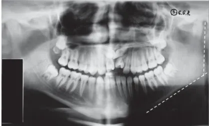
By panoram ic radiographic evaluat ion ( Figure 1 ) , t w o r ad iolog ist s an aly zed t h e m an d ib u lar angle size, chr onology of er upt ion accor ding t o Nolla’s crit eria10, and t he dent al anom alies relat ed
t o changes in shape, v olum e, posit ion such as m acr od on t ia, m icr od on t ia, f u sion , g em in at ion , co n cr escen ce, t au r o d o n t i sm , r o o t f u si o n an d lacerat ion, dens in dent e, t ransposit ion, giroversion, i m p e r f e c t a m e l o g e n e s i s , p a r t i a l a n o d o n t i a , dent al, root supernum erary, dent al num ber of all deciduous and perm anent t eet h, w het her erupt ed or not . The kappa and intraclass correlation (ICC) coeI¿cients w ere used t o t est t he int ra- and int er-rat er reliabilit y.
Nolla1 0 calcu lat ed t h e d en t al m in er alizat ion
st age dividing t he developm ent of each t oot h in 10 st ages, since t he presence of dent al crypt unt il t he com plet e form at ion of t he apex10. The aut hor
separat ed t ables for m en and w om en, w here t he DYHUDJHFDOFL¿FDWLRQVWDJHVZHUHUHFRUGHGIRUHDFK t oot h in t he age range of 6- 17 years.
Th e p an or am ic r ad iog r ap h s w er e an aly zed using a negat oscope, and t he dent al m ineralizat ion st ages w ere int erpret ed using t he Nolla’s 10- st age diagram s of dent al developm ent10.
Measur em ent s of t he m andibular angle w er e obt ained by t he int ersect ion of linear m easurem ent s t an gen t ial t o t h e m an dible r am u s an d in f er ior border.
All dat a w ere com piled int o a spreadsheet using WKH2I¿FH0LFURVRIW([FHO0LFURVRIW&RUSRUDWLRQ Redm ond, WA, USA) program . The SPSS 19® ( I BM
cor r elat ion coef f icien t s, an d St u d en t ’s t t est . Differ ences, associat ions, and cor r elat ions w er e FRQVLGHUHG VLJQL¿FDQW ZKHQ WKH WHVW GHVFULSWLYH level (p) was low er t han 0.05.
The r elat ionship bet w een m easur em ent s was
HYDOXDWHGE\WKH3HDUVRQ¶VFRUUHODWLRQFRHI¿FLHQWp) ,
ZKLFKZDVHYDOXDWHGZKHQVLJQL¿FDQWFRUUHODWLRQV w er e f ou n d . Ab solu t e p v alu es su g g est w eak
( |p_ PRGHUDWH _p| < 0.7) , and st r ong
( |p_FRUUHODWLRQV
RESULTS
The panoram ic radiographs w ere analyzed by WZRREVHUYHUV.DSSDFRHI¿FLHQWZDVXVHGWRWHVW
int ra- and int erobserver agr eem ent in all dent al anom alies. I CC was used t o t est int ra- and int er-rat er reliabilit y in t he Nolla st age and m andibular DQJOH PHDVXUHPHQWV %RWK FRHI¿FLHQWV LQGLFDWH st rong correlat ion ( > 0.8) bet w een t he param et ers analyzed.
I n m ost panoram ic radiographic exam s of our sam ple, t he age gr oup included bot h deciduous an d p er m an en t d en t it ion . Th u s, w e ev alu at ed separat ely t he fr equency of dent al anom alies in t he deciduous and perm anent dent it ions in bot h con t r ol an d FXS gr ou ps ( Table 1 ) . Th e con t r ol g r o u p d i d n o t sh o w d en t a l a n o m a l i es i n t h e d ecid u ou s d en t it ion ( n = 1 3 8 ) . How ev er, som e alt erat ions were present in t he perm anent dent it ion
Dental Anomalies Deciduous teeth Permanent teeth
Control group (n=138)
FXS group (n=106)
Control group (n=608)
FXS group (n=605)
% % % %
Macrodontia 0 0 0 0
Microdontia 0 0 0.16 0
Fusion 0 0 0 0
Gemination 0 0 0 0
Concrescence 0 0 0 0
Taurodontism 0 0 0 0
Fused roots 0 0 0 0.99
Laceration roots 0 0 0.82 1.16
Dens in dente 0 0 0 0
Transposition 0 0 0 0
Giroversion 0 0.94 0.99 2.31
Amelogenesis imperfecta 0 0 0 0
Partial anodontia 0 0 0.49 1.82
Supernumerary tooth 0 2.83 0 0
Supernumerary root 0 0 0.99 0.33
Table 1- Frequency of dental anomalies (per tooth) in the FXS and control groups
( n = 6 0 8 ) : m icr odon t ia ( 0 . 1 6 % ) , r oot lacer at ion ( 0.82% ) , gir over sion ( 0.99% ) , par t ial anodont ia ( 0.49% ) , and super num erar y r oot s ( 0.99% ) . I n t he deciduous dent it ion ( n= 106) , t he FXS group present ed giroversion ( 0.94% ) and supernum erary t oot h ( 2.83% ) . I n t he perm anent dent it ion ( n= 605) , w e observed fused root s ( 0.99% ) , root lacerat ion ( 1.16% ) , gir over sion ( 2.31% ) , par t ial anodont ia ( 1.82% ) , and supernum erary root s ( 0.33% ) .
We evaluat ed all panoram ic radiographic exam s for erupt ion chronology by t he Nolla’s approach10,
sep ar at ed t h e t w o g r ou p s, an d ob ser v ed t h at i n d i v i d u al s w i t h t h e FXS sh o w ed accel er at ed erupt ion in t he upper ( 18, p= 0.063; 28, p= 0.024) an d low er ( 3 8 , p = 0 . 0 3 3 ; 4 8 , p = 0 . 0 2 6 ) t h ir d m olars, and low er second m olars ( 37, p= 0.004; 47, p= 0.001) .
The bilat eral m andibular angle m easurem ent s w er e evalu at ed an d t h e FXS gr ou p sh ow ed an LQFUHDVHVWDWLVWLFDOO\VLJQL¿FDQWDVFRPSDUHG w it h t he cont rol group (p< 0.001; Table 2) .
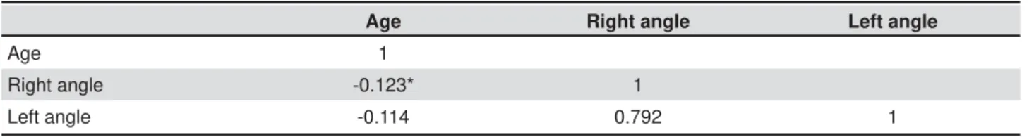
The Pearson’s t est for correlat ion bet w een age and m andibular angle indicat ed a w eak correlat ion (p= 0.44) ( Table 3) .
D I SCUSSI ON
The fragile X syndrom e is a genet ic disease w it h a great variabilit y in clinical present at ion. Unt il t he 1990s, t his syndr om e was diagnosed by clinical signs and chrom osom al st udy ( karyot ype) . However, UHFHQWVWXGLHVKDYHFRQ¿UPHGWKDWWKH3&5PHWKRG LVQRWVXI¿FLHQWWRLQGLFDWHPXWDWLRQLQWKH)05 gene in w om en affect ed by t he disease. Since t his discovery, t he m olecular exam has been included
IRU¿QDOGLDJQRVLVRIWKHV\QGURPH1,6,7,9,16,18,19.
Due t o t he FMR1 gene perm ut at ion and FMRP expansions, variable effect s have been observed in t he phenot ypic const it ut ion of syndrom ic pat ient s6,9.
Th e r adiogr aph ic ex am in at ion is on e of t h e m ost af f or d ab le com p lem en t ar y ex am in at ion s u sed in Den t ist r y f or diagn osis, plan n in g, an d im plem ent at ion of t reat m ent , being useful in all d en t al sp ecialt ies. Pan or am ic r ad iog r ap h s ar e am ong t hose exam inat ions, being a part of dent al surgeon rout ine due t o t he operat ional sim plicit y of t he equipm ent , low- dose radiat ion, low cost , yet allow ing exam inat ion of a large area of t he m axilla and m andible. Fur t her m or e, it is w idely used in epidem iological st udies in t he evaluat ion of dent al inj uries and anom alies, whose knowledge is of great value for st udies in cert ain populat ions.
Analysis of digit al panoram ic radiographs ( 1937) of individuals aged 10- 34 years showed t hat dent al absence by t oot h ex t ract ion, par t ial anodont ia, ex t r usion, m igrat ion, t ransposit ion, gir ov er sion, and carious and periapical inj uries w ere t he m ost f r eq u en t in j u r ies an d alt er at ion s, w it h h ig h er prevalence in w om en. The ones less com m on in t his gr oup w er e: changes in t he condylar head, hypercem ent osis, m andibular fract ure, odont om a, dent igerous cyst , kerat ocyst ic odont ogenic t um or, cem ent - bone per iapical dy splasia, for eign body, and cleft palat e17.
I solat ed cases of dent al radiographic evaluat ion RI);6KDYHDOVREHHQSXEOLVKHG2QHVWXG\LGHQWL¿HG t he presence of m esiodens and t aurodont ism in t he
XSSHU¿UVWDQGVHFRQGSHUPDQHQWPRODUV8. Ot her
au t h or r ep or t ed t h e p r esen ce of n on - er u p t ed supernum erary t oot h in t he apical region of t he upper right cent ral incisor, wit hout change in t he erupt ion chronology4.
,QGLYLGXDOVZLWK);6VKRZVSHFL¿FFKDUDFWHULVWLFV such as low caries prevalence, problem s of cross and open bit e, sever e occlusal w ear and dent al ch an ges in clu din g im pact ed can in e, con gen it al absen ce of pr em olar, pr em olar su per n u m erar y, and a large hypoplast ic defect in a t oot h alone, as
Groups n Right side Left side
M ± SD Min-Max M ± SD Min-Max
Control 20 122.7 ± 5.7 112-132 123.0 ± 6.3 108-136
FXS 20 131.8 ± 5.9 120-144 130.4 ± 7.6 120-142
p<0.001; M: mean values; SD: standard deviations
Table 2- Measurements of mandibular angles in patients of the control and fragile X syndrome (FXS) groups
Age Right angle Left angle
Age 1
Right angle -0.123* 1
Left angle -0.114 0.792 1
* p =0.449
com pared w it h norm al individuals15.
I n our st udy, supernum erary deciduous t eet h ( 2.83% ) , gir over sion ( 2.31% ) , par t ial anodont ia ( 1 . 8 2 % ) , l acer at ed ( 1 . 1 6 % ) an d f u sed r o o t s ( 0.99% ) , and supernum erary root ( 0.33% ) in t he perm anent t eet h w ere t he m ost frequent dent al anom alies in t he FXS group.
As obser v ed in ou r st u dy, par t ial an odon t ia is a disor der in w h ich t h er e is a failu r e in t h e dent al developm ent of deciduous or, m ore oft en, perm anent dent it ion. Part ial anodont ia is associat ed wit h cert ain disorders such as ect oderm al dysplasia, Dow n’s syndrom e, and cleft lip and palat e14. This
associat ion also occur s in super num erar y t eet h relat ed t o t he Gardner syndrom e and cleidocranial d y sp l a si a , w h i ch i s ca u sed b y a n a u t o so m a l dom inant gene. Alt hough t he ex act pr evalence of isolat ed hypodont ia or supernum erary t eet h is unknown, in m any cases t here is a fam ilial t endency for t his defect . I t r esult s fr om m ut at ions in t he polygenic syst em , which is m ost oft en t ransm it t ed in an aut osom al dom inant m anner, wit h incom plet e penet rance and variable expression. Alt hough FXS is an inherit ed genet ic disease, t hese radiographic ¿QGLQJVFDQQRWEHDWWULEXWHGWRVSHFL¿FIHDWXUHVRI t he syndrom e. Furt her st udies should be done t o FRQ¿UPWKHVH¿QGLQJV
I n all st udies involving t he fragile X syndrom e, t h e r elat ion sh ip b et w een ag e an d m an d ib u lar angle was not invest igat ed. Measurem ent of t he m an dibu lar an gle, w h ich is t h e an gle bet w een m andibular body and ram us, has been used as a t ool t o det erm ine t he age of individuals. I n t he range of 3- 13 years, age is inversely proport ional ( in degrees) t o t he angle12. According t o our st udy,
indiv iduals w it h t he fragile X sy ndr om e ex hibit an 8 - degr ee in cr ease in t h e m an dibu lar an gle w hen com pared w it h t he norm al range ( p< 0.05) . How ev er, Pear son’s cor r elat ion t est indicat ed a w eak correlat ion bet w een age and t he m easures of m andibular angle in bot h groups. This age group was chosen based on t he m andibular bone grow t h phase. How ever, t he age range, com pared w it h t he sam ple size, is far t oo wide ( 6- 17 years) t o allow a GH¿QLWLYHFRQFOXVLRQRQWKHLQFUHDVHLQPDQGLEXODU angle w it h age.
Som e classic facial charact erist ics in individuals wit h FXS, such as long face4,7, downward m andibular
rot at ion7 and skelet al open bit e14, do not m eet our
result s regarding t he increase in t he m andibular an g le. Ou r r esu lt s su g g est t h at t h e lon g f ace observed in t he pat ient s wit h FXS2,4,7,15 could explain
t he increase in m andibular angle. Furt her evaluat ion RIFHSKDORPHWULFVWXGLHVLVQHFHVVDU\WRFRQ¿UPWKH above m ent ioned hypot hesis.
Th e d e n t a l d e v e l o p m e n t a n d i t s e r u p t i o n FKURQRORJ\ PD\ EH LQÀXHQFHG E\ D QXPEHU RI fact ors such as et hnic group, gender, diet , syst em ic
d i se a se s, i n f e ct i o u s p r o ce sse s, cl i m a t e , a n d const it ut ional t ypes. Alt hough dent al er upt ion is LQÀXHQFHGE\JHQHWLFDQGHQYLURQPHQWDOIDFWRUV in m ost cases it keeps a cer t ain pat t er n, w hich can b e ap p lied in leg al m ed icin e t o est im at e t he chr onological age of indiv iduals w it hout an LGHQWL¿FDWLRQ GRFXPHQW DV ZHOO DV LQ WKH GHQWDO t reat m ent planning.
Kot ilainen and Pir inen3 ( 1999) evaluat ed t he
dent al dev elopm ent of 28 boy s ( aged 4. 9- 17. 6 years) w it h FXS and t hree girls ( aged 5.8, 10.4, and 12.7 years) w ho w ere FXS carriers. They used t he Dem ir j ian and Goldst ein ( 1976) cr it er ia for t oot h developm ent and t hose of Hagg and Taranger ( 1985) for t oot h erupt ion, and com pared t he st at ure dat a and bone m at urit y grow t h of t he individuals. They concluded t hat t he clinical dent al erupt ion of deciduous and perm anent t eet h in m en w it h FXS was precocious as com pared w it h t hat observed in FRQWUROVRIWKHVDPHDJH7KHGHQWDOFDOFL¿FDWLRQ st age was ant icipat ed in m en and het er ozygous carrier w om en, and t he height and bone m at urit y growt h did not show an ant icipat ed developm ent . Our result s w ere different from t hose of Kot ilainen and Pirinen3UHJDUGLQJGHQWDOFDOFL¿FDWLRQ
which was ant icipat ed, and dent al erupt ion, which was precocious in all t eet h, because t he evaluat ion crit eria w ere different .
CON CLUSI ON
I n d i v i d u a l s w i t h t h e f r a g i l e X s y n d r o m e show ed a higher fr equency of dent al anom alies ( s u p e r n u m e r a r y d e c i d u o u s t e e t h ( 2 . 8 3 % ) , gir over sion ( 2.31% ) , par t ial anodont ia ( 1.82% ) , lacerat ed root s ( 1.16% ) , fused root s ( 0.99% ) , and super num erar y r oot ( 0. 33% ) in t he per m anent t eet h ) w h en com par ed w it h t h e con t r ol gr ou p. Ad d it ion ally, an in cr ease w as ob ser v ed in t h e m andibular angle and accelerat ion in t he erupt ion chronology of upper and low er t hird m olars and low er second m olars.
Dent al surgeons should consider t he changes in WKHVH¿QGLQJVWRREWDLQDEHWWHUGHQWDOSODQQLQJDQG t reat m ent in individuals wit h t he fragile X syndrom e.
REFEREN CES
1- Alliende MA, Curot t o BL, Valiende G, Toro J, Sant a Maria L, González MR. Diagnost ico cit ogenet ico- m olecular del Síndrom e Xq frágil. Rev Chil Tecnol Med. 2007; 27: 1339- 46.
2 - Bu t ler MG, Pr at esi R, Wat son MS, Br eg WR, Sin g h DN. Ant hr opom et r ic and craniofacial pat t er ns in m ent ally r et ar ded m ales w it h em phasis on t he fragile X sy ndr om e. Clin Genet . 1993; 44: 129- 38.
3- Kot ilainen J, Pirinen S. Dent al m at urit y is advanced in fragile X syndrom e. Am J Med Genet . 1999; 83: 298- 301.
4- Kulkarni GV, Levine N. Fragile X ( Mart in- Bell) syndrom e. Spec Care Dent ist . 1994; 14: 21- 5.
5 - Lach i ew i cz AM, D aw so n D V, Sp i r i d i g l i o zzi GA. Ph y si cal charact erist ics of young boys w it h fragile X syndrom e: reasons
IRUGLI¿FXOWLHVLQPDNLQJDGLDJQRVLVLQ\RXQJPDOHV$P-0HG
Genet . 2000; 92: 229- 36.
6- Mazzocco MM. Advances in research on t he fragile X syndrom e. Ment Ret ard Dev Disabil Res Rev. 2000; 6: 96- 106.
7 - Min g r on i- Net t o RC, Rosen b er g C, Vian n a- Mor g an t e AM, Pavanello RC. Fragile X frequency in a m ent ally ret arded populat ion in Brazil. Am J Med Genet . 1990; 35: 22- 7.
8- Mit t al S, Rawal YB. Fragile “ X” syndrom e. A case st udy. I ndian J Dent Res. 1996; 7: 59- 62.
9 - Mu r r ay A, En n i s S, MacSw i n ey F, Web b J, Mo n t o n NE. Repr oduct ive and m enst r ual hist or y of fem ales w it h fragile X expansions. Europ J Hum Genet . 2000; 8: 247- 52.
10- Nolla CM. Developm ent of t he perm anent t eet h. J Dent Child. 1960; 27: 254- 66.
11- Pandey UB, Phadke SR, Mit t al B. Molecular diagnosis and genet ic counseling for t he fragile X m ent al ret ardat ion. Neurol I ndia. 2004; 52: 36- 42.
12- Poonacha KS, Shigli AL, I ndushekar KR. Relat ive posit ion of t he m andibular foram en in different age groups of children: a radiographic st udy. J I ndian Soc Pedod Prev Dent . 2010; 28: 173- 8. 13- Scully C. Fragile X ( Mar t in Bell) sy ndr om e. Dent Updat e. 2002; 29: 196- 8.
14- Shafer WG, Hine MK, Levy BM. A t ext book of oral pat hology. Michigan: Saunders; 1983.
15- Shellhar t WC, Casam assim o OS, Hager m an RS, Belanger GK. Or al f in d in g s in f r ag ile X sy n d r om e. Am J Med Gen et . 1986; 23: 179- 87.
16- Shelt on AL, Cor nish K, Kraan C, Geor giou- Kar ist ianis N,
0HWFDOIH6$%UDGVKDZ-/HWDO([SORULQJLQKLELWRU\GH¿FLWVLQ
fem ale prem ut at ion carriers of fragile X syndrom e: t hrough eye m ovem ent s. Brain Cogn. 2014; 85: 201- 8.
17- Varoli FP, Warm ling LV, Sant os KC, Oliveira JX. Occurrence of lesions, abnorm alit ies and dent om axillofacial changes observed i n 1 9 3 7 d i g i t al p an o r am i c r ad i o g r ap h y. J Heal t h Sci I n st . 2013; 31: 258- 61.
18- Willem sen R, Olm er R, De Diego Ot ero Y, Oost ra BA. Tw in sist er s, m onozy got ic w it h t he fragile X m ut at ion, but w it h a different phenot ype. J Med Genet . 2000; 37: 603- 4.