ABSTRACT
Occurrence and antimicrobial susceptibility of
enteric rods and pseudomonads isolated from
Sanrrangers Sales SILVA1, Maximilo de Oliveira RIBEIRO2, Francisco Isaac Fernandes GOMES1, Hellíada Vasconcelos CHAVES1, Antonio Alfredo Rodrigues e SILVA3, Iriana Carla Junqueira ZANIN1, Francisco Cesar Barroso BARBOSA1
1- Universidade Federal do Ceará, Faculdade de Odontologia, Campus Sobral, Sobral, CE, Brasil. 2- Universidade Federal do Ceará, Faculdade de Medicina, Campus Sobral,Sobral, CE, Brasil. 3- Agência Nacional de Vigilância Sanitária, Brasília, DF, Brasil.
Corresponding address: Francisco Cesar Barroso Barbosa - Faculdade de Medicina de Sobral - Universidade Federal do Ceará - Avenida Comandante Maurocélio Rocha Ponte, 100 - Derby - Sobral - CE - Brazil - 62-042-280 - Phone: +55 (88) 3611.2202 - E-mail: [email protected]
6XEPLWWHG)HEUXDU\0RGL¿FDWLRQ-XQH$FFHSWHG-XQH
A
spiration of oral bacteria leads to cardiac and respiratory infectious diseases and dentures can act as a reservoir for pathogenic microorganisms. Objective: To determine the occurrence and the in vitro antimicrobial susceptibility of enteric rods and pseudomonadsenteric/non-fermenter system. Antibiotic bacterial susceptibility was assessed by the disc diffusion method of amoxicillin, amoxicillin/clavulanic acid, doxycycline, tetracycline,
for 40 species by E-test. Results: 34 subjects (65.4%) harbored enteric rods in their prostheses. Klebsiella pneumoniae (26.5%), Escherichia coli (23.5%), and Enterobacter aerogenes (23.5%) were the most prevalent species. All organisms were susceptible to
demonstrating variable sensitivity patterns to other antimicrobials. However, the MIC
90
mL) and cefotaxime (MIC90
nosocomial diseases-related bacterial species and low susceptibility to antimicrobial drugs. Therefore, these results imply caution against the indiscriminate use of broad spectrum antibiotics in dental practice.
Ke y w or ds: Enterobacteriaceae. Pseudomonas. Drug resistance.
I N TROD UCTI ON
Variations in the oral microbiota are directly related to age and have been attributed to the use of dentures16. The oral health status declines as a
result of the aging process and individuals culminate with the need of dental prostheses, affecting their health, functional activities, and self-esteem8.
Worldwide, 810 million people are aged 60 or over, which is predicted to increase to at least two billion by 2050, 22% of the entire global population, and
challenges for oral health care delivery, to an increasingly aged population with declining oral health13,19,31.
These individuals may not be able to clean their dentures properly, which in turn could result in
complex three-dimensional architecture. One of the problems directly related to these materials is still
turn has received little attention by patients and general clinicians as its dental counterpart32.
The presence of enteric rods and pseudomonads
of their pathogenic potential and ability to adhere to solid surfaces2,17.In addition, infections caused by
of the bacterial resistance to a variety of antibiotics,
cephalosporins, aminoglycosides, carbapenems, chloramphenicol, aztreonam, trimethoprim/ sulfametaxazol, tetracycline, and doxycycline5,24.
The clinical importance of the non-fermenters gram-negative bacilli presence in the denture biofilm has significantly increased because of infections, high rates of morbidity, mortality in hospitalized subjects, and high levels of resistance. The dissemination of gram-negative bacteria with acquired antimicrobial drugs resistance is becoming a global problem. The propagation and dissemination of these microorganisms have
11,25.
Enteric microorganisms and pseudomonads have
However, there are current reports of resistant enteric rods to carbapenems30. Among bacterial
isolates resistant to ampicillin or amoxicillin, the
production of these hydrolytic enzymes seems to be the major mechanism of resistance to
resistance to tetracycline was widely disseminated in the microbial enteric rods, and many of tested microorganisms were resistant25.
Having known such information, our hypotheses are that wearing dental prostheses older people may harbor superinfecting microorganisms in their
the surface of these appliances as well as these bacterial species could possess antimicrobial resistance. Therefore, we consider relevant to determine the prevalence of Enterobacteriaceae and Pseudomonadaceae species isolated from in vitro susceptibility to antimicrobial drugs, since these opportunistic microorganisms were previously
15,28,29.
M ATERI AL AN D M ETH OD S
St u dy popu la t ion
Enteric microorganisms were isolated from the inner surface of dentures of 52 subjects within
a 2-year follow-up period (2007-2008) at the Center for Dental Specialties – Sobral, State of Ceará, Brazil. The requirements for inclusion in this study were: no history of diabetes, no use of antimicrobials in the past three months, nor other medication that could affect their systemic or local immune system. All subjects signed a written consent form that was approved by the Institutional Review Board of Federal University of Ceará (COMEPE nº 258/07).
D a t a colle ct ion
All subjects answered a questionnaire about their systemic health and a dental and soft tissue examination was performed to assess their oral
in the medical record of each patient. Initially, the surfaces of the dentures were thoroughly dried with sterilized gauze to avoid contamination by
dentures was collected with the aid of a sterile swab, and the samples were immediately placed in sterile phosphate buffered saline (PBS, pH 7.5, 0.8% NaCl) to the Department of Microbiology and Parasitology, Federal University of Ceará, in Sobral, and processed within a maximum of 2 hours after collection. Samples were dispersed by agitation (30 seconds) and serially diluted (10-1 and 10-2) in PBS.
Aliquots of 100 μL of the solutions obtained were plated on MacConkey agar (Acumedia, Lansing, Michigan, USA). Then, the plates were incubated at 36°C±1°C for 24 hours.
0LFURRUJDQLVPVDQGPLFURELDOLGHQWL¿FDWLRQ
Gram staining, colony morphology on MacConkey agar plates (Acumedia, Lansing, Michigan, USA), production of oxidase (Oxidase Newprov Strips,
Enterobacteriaceae
enteric bacilli fermenters, oxidase-negative bacilli, and non-fermenter bacilli was carried out by the system BBL Crystal Enteric/Nonfermenter (Becton Dickinson Systems, Cockeysville, Maryland, USA) according to the manufacturer’s instructions. The sample Escherichia coli ATCC 25922 was used as control.
An t im icr obia l su sce pt ibilit y t e st
hours. Thereafter, in order to perform the sensitivity test, it was used the disk diffusion method of the Clinical & Laboratory Standards Institute manual6.
The antimicrobial drugs tested were amoxicillin (10
the medium used for this test. After seeding and incubating the samples in a microbiological greenhouse at 36°C±1°C for 24 hours, the zones of inhibition were measured and the organisms were
according to CLSI references6,7. A total of 52 strains
of enteric rods and pseudomonads were submitted to susceptibility tests. For multidrug resistant species, the Minimum Inhibitory Concentration (MIC) was determined for the following drugs: cefotaxime (CT), tobramycin (TM), doxycycline
methodology used was E-test® (AB Biodisk, Solna,
Sweden) and interpretations were made according to the CLSI references6,7.
St a t ist ica l a n a ly sis
The Mann-Whitney test was used to determine differences regarding age groups for men and women, and the chi-square test was used to access differences between genders, age groups, and time of prostheses use correlated between the presence and absence of the studied microorganisms. Differences of p<0.05 were considered statistically
RESULTS
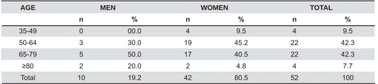
The population consisted of 52 individuals, of whom 42 (80.8%) were women, ranging from 35 to 81 years old (61.7±10.6), and 10 (19.2%) were men, ranging from 53 to 93 years old (72±13.2).
between men and women regarding age groups (p=0.245) and the average time of denture usage was around 11.09 years (±9.6) (Table 1). Regarding their systemic health status, 31 (59.6%) subjects
reported to be diagnosed with type 2 diabetes, three (5.7%) reported to have gastritis, one (1.9%) reported to be asthmatic, and one (1.9%) to have osteoporosis, while 11 (21.1%) were systemically healthy at anamnesis. Regarding their oral condition, we diagnosed three (5.7%) subjects with candidiasis, two (3.8%) with stomatitis, one (1.9%) with xerostomia, while 46 (88.4%) were found to be orally healthy at anamnesis.
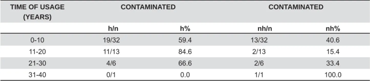
According to Tables 2 and 3, 34 (65.4%) subjects harbored enteric bacilli and/or pseudomonas species on dental prostheses. Most of these subjects (91.2%) aged over 50. Also, the studied microorganisms were found in 15 (75%) subjects in the group of subjects who wore dental prostheses over a 10-year period of time (Table 4). However,
between the time of prostheses usage and the presence or absence of microorganisms (p>0.05).
the prosthesis, since no significant difference
AGE MEN WOMEN TOTAL
n n n
35-49 0 00.0 4 9.5 4 9.5
50-64 3 30.0 19 45.2 22 42.3
65-79 5 50.0 17 40.5 22 42.3
2 20.0 2 4.8 4 7.7
Total 10 19.2 42 80.5 52 100
Table 1- Age and gender distribution of subjects in the 2007-2008 period
MICROORGANISM MEN WOMEN
n n
Only enteric bacilli 6 60.0 24 57.1
Only Pseudomonadaceae 0 00.0 1 2.4
Enteric bacilli and Pseudomonadaceae 0 00.0 1 2.4
Other species of microrganisms 0 00.0 2 4.8
Absence of microrganisms 4 40.0 14 33.3
Total of individuals 10 100.0 42 100.0
was observed in this study (p=0.74). Across the 42-woman group, 24 (57.1%) had their dentures solely contaminated with enteric bacilli, one participant (2.4%) had Pseudomonas species, and another one (2.4%) harbored enteric bacilli and pseudomonads. Furthermore, an 81-year old participant, who wore prostheses for 1 year, and a 59-year old subject, exceeding 20 years of denture use, had $cinetobacter iZoI¿ and Burkholderia pseudomallei
dental prostheses.
Regarding the species found, Klebsiella pneumoniae, Escherichia coli, and Enterobacter aerogenes were the bacteria most prevalent detected, being isolated from dentures of nine (26.5%), eight (23.5%), and eight (23.5%) individuals, respectively. It was also detected Citrobacter freundii (11.8%), Klebsiella ozaenae (11.8%), Klebsiella oxytoca (8.8%), Enterobacter cloacae (8.8%), Serratia marcescens (8.8%), Serratia liquefaciens (8.8%), Enterobacter gergoviae (5.9%), Pseudomonas aeruginosa (5.9%), Pseudomonas putida (2.9%), Enterobacter
sakasakii (2.9%), Burkholderia pseudomallei (2.9%), and $cinetobacter iZof¿ (2.9%).
Table 5 lists the antimicrobial susceptibility of all detected bacterial species. All strains were
one of E. coli. Most species were resistant to amoxicillin or amoxicillin associated with clavulanic acid. The results from the MIC test on the bacterial species showed multidrug resistance patterns (Table 6): 33.4% of the K. pneumoniae species were resistant to cefotaxime (CT) and 22.2% to doxycycline (DC), being 11.1% of these organisms resistant to the other analyzed antimicrobial drugs.
Moreover, 37.5% and 12.5% of E. coli strains were resistant to doxycycline and tobramycin (TM), respectively, while 12.5% of Enterobacter aerogenes were resistant to the same antibiotics. In addition, 25% of species of Citrobacter freundii were resistant to doxycycline and imipenem (IP) and 33.3% of E. cloacae demonstrated insensitivity to doxycycline. The Serratia marcescens strains showed resistance to cefotaxime and to tobramycin. The two (100%) Serratia liquefaciens strains and
AGE MEN WOMEN TOTAL
h/n K h/n K h/n K
35-49 0/0 0.0 3/4 75.0 3/4 75.0
50-64 2/3 66.7 11/19 57.9 13/22 59.1
65-79 3/5 60.0 12/17 70.6 15/22 68.2
1/2 50.0 2/2 100.0 3/4 75.0
All ages 6/10 60.0 28/42 66.7 34/52 65.4
Table 3- Number of analyzed subjects with dental prostheses harboring enteric rods, Pseudomonas, Acinetobacter spp., and Burkholderia spp. associated with groups of age and gender
n= subjects in each group
h= harboring subjects in each group h%= percentage of harboring subjects
TIME OF USAGE (YEARS)
CONTAMINATED CONTAMINATED
h/n K nh/n QK
0-10 19/32 59.4 13/32 40.6
11-20 11/13 84.6 2/13 15.4
21-30 4/6 66.6 2/6 33.4
31-40 0/1 0.0 1/1 100.0
Table 4- Number of subjects wearing harbored or non-harbored dental prostheses by enteric rods, Pseudomonas,
Acinetobacter spp., and Burkholderia spp. associated for time of dental prostheses usage
n= subjects in each group
the only one Pseudomonas aeruginosa (50%) showed values of MIC90 for doxycycline and respectively (Tables 6 and 7).
D I SCUSSI ON
Enteric bacilli and pseudomonads are opportunistic pathogens at different human body sites. The oral cavity functions as a reservoir for pathogenic species3,17. As a result of the aging process, the oral
health status declines, often leading to defective oral/denture hygiene in geriatric subjects, being the reasons for this fact the lack of hygiene education of these subjects or of those caring for them. Consequently, oral hygiene is often neglected, resulting in poor oral health and in an increase in the presence of local or general infections that can be related with the presence of enteric microorganisms in the oral cavity4,9.
Here we show that dental prostheses can harbor
this study; in addition, these microorganisms can be even resistant to antimicrobial drug therapy
as demonstrated here. This is really alarming because, albeit many of these subjects might not be systemically compromised by any disease at the moment they house such pathogens in their oral cavity, they could suffer from any illness that could dampen and compromise their immune system, making them, in turn, more susceptible to be infected by the oral opportunistic pathogens they are harboring.
Thus, a proper denture hygiene protocol is essential, since it could prevent these individuals from exposition to these bacterial species, considering that respiratory pathogens are more capable of colonizing teeth and dentures instead of soft tissues, and pneumonia is the main cause of death related to infection in older people8. By
wearing dental prostheses, individuals are at a higher risk of aspirating such pathogens from the
oral appliance and lungs, being reported a high prevalence of respiratory pathogens on the denture
21. Furthermore, not
only are such pathogens involved in the development of aspiration pneumonia, but also they have been
Microorganism CIP IPM TET CTX AMO DOX AMC TOB
Klebsiella pneumoniae (9) S(9) S(9) S(3)I(1) R(5)
S(7)I(1) R(1)
R(9) S(4)I(3)
R(2)
S(4)I(1) R(4)
S(8)R(1)
Escherichia coli (8) S(8) S(7)R(1) S(5)R(3) S(6)R(2) S(3)R(5) S(6)I(1) R(1)
S(3)I(1) R(4)
S(7)R(1)
Enterobacter aerogenes (8) S(8) S(8) S(6)R(2) S(7)I(1) R(8) S(6)R(2) R(8) S(8)
Citrobacter freundii (4) S(4) S(4) S(3)R(1) S(2)I(1) R(1)
I(1)R(3) S(2)I(1)
R(1)
S(1)I(2) R(1)
S(4)
Klebsiella ozaenae (4) S(4) S(4) S(4) S(4) S(1)R(3) S(4) S(4) S(4)
Klebsiella oxytoca (3) S(3) S(3) S(3) S(2)R(1) R(3) S(3) S(2)R(1) S(3)
Enterobacter cloacae (3) S(3) S(3) I(1)R(2) S(1)I(1) R(1)
R(3) S(1)R(2) I(1)R(2) S(2)R(1)
Serratia marcescens (3) S(3) S(3) S(2)R(1) S(2)I(1) S(1)R(2) S(3) S(1)R(2) S(2)R(1)
Serratia liquefaciens (2) S(2) S(2) I(2) I(1)R(1) R(2) S(1)R(1) R(2) S(2)
Enterobacter gergoviae (2) S(2) S(2) S(1)I(1) S(1)I(1) R(2) S(1)R(1) R(2) S(2)
Pseudomonas aeruginosa (2) S(2) S(2) I(1)R(1) S(1)I(1) R(2) R(2) R(2) S(2)
Pseudomonas putida (1) S(1) S(1) I(1) I(1) R(1) R(1) R(1) S(1)
Enterobacter sakasakii (1) S(1) S(1) S(1) S(1) R(1) S(1) R(1) S(1)
Burkholderia pseudomallei (1) S(1) S(1) R(1) I(1) R(1) R(1) R(1) S(1)
$FLQHWREDFWHULZRI¿ (1) S(1) S(1) S(1) S(1) S(1) S(1) S(1) S(1)
Table
5-S= sensible; I= intermediate sensibility; R= resistant
Microorganism(n) CT* TM* DC* IP* CI*
Klebsiella pneumoniae (n=9)
Sensible 66.6 88.9 77.8 77.8 79.8
Intermediate sensibility 0.0 0.0 0.0% 11.1 11.1
Resistent 33.4 11.1 22.2 11.1 11.1
Escherichia coli (n=8)
Sensible 87.5 87.5 37.5 100.0 100.0
Intermediate sensibility 12.5 0.0 25.0 0.0 0.0
Resistent 0.0 12.5 37.5 0.0 0.0
Enterobacter aerogenes (n=8)
Sensible 100.0 87.5 75.0 100.0 100.0
Intermediate sensibility 0.0 0.0 15.5 0.0 0.0
Resistent 0.0 12.5 12.5 0.0 0.0
Citrobacter freundii (n=4)
Sensible 100.0 100.0 75.0 75.0 100.0
Intermediate sensibility 0.0 0.0 0.0 0.0 0.0
Resistent 0.0 0.0 25.0 25.0 0.0
Serratia marcescens (n=1)
Sensible 0.0 0.0 100.0 100.0 100.0
Intermediate sensibility 0.0 0.0 0.0 0.0 0.0
Resistent 100.0 100.0 0.0 0.0 0.0
Enterobacter cloacae (n=3)
Sensible 100.0 100.0 66.7 100.0 100.0
Intermediate sensibility 0.0 0.0 0.0 0.0 0.0
Resistent 0.0 0.0 33.3 0.0 0.0
Serratia liquefaciens (n=2)
Sensible 100.0 100.0 0.0 50.0 100.0
Intermediate sensibility 0.0 0.0 0.0 0.0 0.0
Resistent 0.0 0.0 100.0 50.0 0.0
Enterobacter gergoviae (n=1)
Sensible 0.0 100.0 0.0 100.0 100.0
Intermediate sensibility 0.0 0.0 100.0 0.0 0.0
Resistent 100.0 0.0 0.0 0.0 0.0
Pseudomonas aeruginosa (n=2)
Sensible 50.0 100.0 50.0 100.0 100.0
Intermediate sensibility 0.0 0.0 50.0 0.0 0.0
Resistent 50.0 0.0 0.0 0.0 0.0
Pseudomonas putida (n=1)
Sensible 100.0 100.0 100.0 100.0 100.0
Intermediate sensibility 0.0 0.0 0.0 0.0 0.0
Resistent 0.0 0.0 0.0 0.0 0.0
Burkholderia pseudomallei (n=1)
Sensible 100.0 100.0 100.0 100.0 100.0
Intermediate sensibility 0.0 0.0 0.0 0.0 0.0
Resistent 0.0 0.0 0.0 0.0 0.0
Table
directly associated with bacterial endocarditis, gastrointestinal infection, and chronic obstructive pulmonary disease. This scenario shows us that
to prevent the occurrence of associated oral and
systemic diseases10.
Regarding the prevalence of the aforementioned microorganisms, we found a high prevalence (65.4%) of Enterobacteriaceae and Pseudomonadaceae
Microorganism CT** TM** DC** IP** CI**
Klebsiella pneumoniae >32 12 0.19 0.75 0.25
Klebsiella pneumoniae 0.094 2 2 >32 0.047
Klebsiella pneumoniae 0.5 2 2 0.38 0.032
Klebsiella pneumoniae 0.047 3 2 0.25 0.064
Klebsiella pneumoniae 0.064 3 2 0.25 0.064
Klebsiella pneumoniae 0.032 3 48 0.19 0.047
Klebsiella pneumoniae >32 3 16 6 3
Klebsiella pneumoniae >32 3 2 0.25 12
Klebsiella pneumoniae 0.032 3 2 0.25 0.064
Escherichia coli 0.38 16 6 0.38 0.064
Escherichia coli 0.25 1.5 3 0.38 0.064
Escherichia coli 0.125 3 12 0.25 0.023
Escherichia coli 12 0.75 32 1.5 0.125
Escherichia coli 0.047 2 1.5 0.125 0.023
Escherichia coli 0.064 3 6 0.19 0.094
Escherichia coli 0.125 2 0.5 0.125 0.016
Escherichia coli 2 2 12 0.19 0.047
Enterobacter aerogenes 0.25 24 6 0.38 1
Enterobacter aerogenes 0.25 2 3 0.38 0.064
Enterobacter aerogenes 0.032 3 3 0.25 0.047
Enterobacter aerogenes 0.25 3 >256 0.38 0.064
Enterobacter aerogenes 0.047 2 2 0.19 0.047
Enterobacter aerogenes 0.016 0.75 1.5 3 0.064
Enterobacter aerogenes 8 2 2 0.25 0.064
Enterobacter aerogenes 0.19 3 2 0.25 0.047
Citrobacter freundii 0.25 3 >256.0 0.25 0.032
Citrobacter freundii 0.25 3 3 >32 0.047
Citrobacter freundii 0.094 2 3 0.25 0.047
Citrobacter freundii 0.25 3 3 0.25 0.047
Serratia marcescens >32 64 0.5 0.5 0.75
Enterobacter cloacae 0.064 0.75 3 0.19 0.012
Enterobacter cloacae 3 0.75 24 0.19 0.032
Enterobacter cloacae 0.38 2 3 0.38 0.023
Serratia liquefaciens 3 0.5 16 0.38 0.047
Serratia liquefaciens 8 2 >256 >32 0.5
Enterobacter gergoviae >32 1 6 0.5 0.047
Pseudomonas aeruginosa >32 1 8 0.38 0.064
Pseudomonas aeruginosa 8 1 1.5 1.5 0.047
Pseudomonas putida 0.5 0.023 0.25 0.032 0.047
Burkholderia pseudomallei 2 0.5 3 0.19 0.003
Burkholderia pseudomallei (n=1)
Table 7-
in the city of Sobral. As opposed to a similar study in a Japanese population, in which the prevalence of potential pathogens on dentures was 18% for E. cloacae and 16% for K. pneumoniae, our research found that K. pneumoniae was the most prevalent at 26.5%, followed by Escherichia coli and E. aerogenes, placed after 23.5%28. Another study
showed that denture plaque in patients with chronic obstructive pulmonary disease was colonized by pathogens of the respiratory tract, including: E. coli, Pseudomonas spp., and Klebsiella spp. Over 33% of the isolated pathogens are part of rod-shaped and gram-negative Enterobacter spp. The isolation of the mentioned bacteria from denture plaque proves that dental prostheses might become a source of infection of the respiratory tract or may exacerbate chronic respiratory diseases25.
Contrary to the data we report, another group described a much lower (20.3%) prevalence of enterobacteriaceae in the oral cavity of older people from Greece that used dental prostheses14, whereas
a different group reported that 48% of edentulous subjects harbored enteric rods in their oral cavity, being K. oxytoca, E. cloacae, and K. pneumoniae species more prevalent12. According to these
authors, the prevalence of such microorganisms in the denture biofilm may be related to the ability of these species to adhere to the polymer of the denture and to aggregate in the presence of ammonium sulfate. This could explain the high prevalence of K. pneumoniae reported in our study.
The classic literature shows that the prevalence of these organisms in the oral cavity of systemically healthy individuals can vary among different populations1,26,27. Thus, the high prevalence
(65.4%) we observed in this study could be attributed to disadvantaged health infrastructure and educational issues that could have led to ingestion of contaminated food or water. Also, poor hygiene habits and indiscriminate use of antibiotics may play a role in this high prevalence4,11.
In fact, it was observed that the prostheses
encountered when conducting a properly cleaning, we can include the lack of adequate patient guidance, characteristics of the prostheses, decreased motor ability, and lack of proper products in the market to carry out the correct denture cleaning. The habit of brushing the prostheses with toothpaste and the use of common brushes may not be the best way
the prolonged use of the same prostheses for many years might contribute to the colonization process by potential pathogens4. It is relevant to highlight that
this information shows a major limitation of these studies, since there are many factors that can lead to microbial colonization of dentures. In our study, we found prostheses being worn for 40 years, and
most of the subjects wearing contaminated dentures have been doing so for over 10 years.
Regarding the antimicrobial susceptibility of microorganisms, our data show that 86.5% of anaerobic facultative gram-negative bacilli were resistant to amoxicillin and over half of them had resistance to amoxicillin with clavulanic acid contrary to other studies that showed the association amoxicillin/clavulanic acid being active on less than half of ampicillin or amoxicillin resistant isolates from
detected in 28.3% of the targeted microorganisms and it was particularly frequent in E. cloacae, genera Klebsiella, Serratia, and Pseudomonas11.
The analysis of the MIC proved that the antimicrobial drug with the highest inhibitory activity against the enteric bacilli and pseudomonads
90
we observed a decreased susceptibility to this antimicrobial agent in two (22.2%) strains of K. pneumoniae
respectively. Although it is a much lower frequency than that observed previously20, decreased
90>2 μg/
mL) among clinical isolates of Enterobacteriaceae species is a particular concern, since enteric rods were able to colonize the denture surface of non-hospitalized subjects, which could spread
in the community. Fluoroquinolones are the most widely used antibiotics worldwide, and are the drugs of choice for empirical therapy for urinary tract infection. Fluoroquinolone resistance in members of the Enterobacteriaceae family has until recently been attributed to mutations in the gyrase and topoisomerase genes quinolone resistance-determining regions. Given their transferability and the possibility that they cause increases in resistance that might affect the clinical response to treatment, the detection of quinolone resistance should be routinely performed. There are several surveillance or retrospective studies with clinical isolates, and these studies showed the characteristic features observed in drug-resistant strains in addition to epidemics caused by them33
.
I n t h i s s t u d y w e a l s o f o u n d a s l i g h t l y h i g h e r p r e v a l e n c e ( 2 7 . 5 % ) o f Enterobacteriaceae species with decreased susceptibility to cefotaxime (MIC90 μ
the data previously reported of 22.5%21, and these
isolates were considered potential ESBL (Extended Spectrum Beta-Lactamases) producers according to the limits established by the CLSI6,7. Resistance to
Enterobacteriaceae has
support to the prevalent view that ESBL plasmids are conjugative, may be borne on transposons, and that the genes may have high mutation frequencies. Treatment of infections caused by these ESBL-producing bacteria has become challenging. In addition to being resistant to commonly used
usually resistant to other classes of antibiotics
is the practice of irrational usage of antibiotics, leading these microorganisms to exhibit a unique microorganism resistant pattern, which hugely impacts on clinical choice of a correct antibiotic once antimicrobial drug resistance develops, making
15,22,23.
Antimicrobial resistance surveillance programs
susceptibility of clinically relevant enteric bacteria and pseudomonads from nosocomial infections and the environment5,18. However, they can be found in
of the individuals to systemic infections or even worsening them. Thus, it is crucial to emphasize that dentures can harbor such pathogens and this is the reason why a correct hygiene protocol must be carried out in order to avoid such
ultimately, would increase the risk of one to be exposed to enteric bacilli.
CON CLUSI ON
K. pneumoniae, E. coli, and E. aerogenes were the predominant species found on the denture biofilm. Most enteric bacilli and Pseudomonas spp. were resistant to amoxicillin and amoxicillin clavulanate, with variable susceptibility patterns to other antimicrobial drugs. The antibiotic that showed the highest inhibitory activity against them was
from the results obtained in this study, we suggest
is important to avoid the colonization of dental prostheses by multidrug resistant bacteria as well as avoiding the indiscriminate prescription of antibiotics will help diminish the multidrug resistance insurgence.
ACKN OW LED GM EN TS
This study was supported by CNPq – National , Universal Proc. 471158/2007-0, Ministry of Education, and Federative Republic of Brazil.
REFEREN CES
1- Ali RW, Velcescu C, Jivanescu MC, Lofthus B, Skaug N. Prevalence of six putative periodontal pathogens in subgingival plaque samples from Romanian adult periodontitis patients. J Clin Periodontol.1996;23:133-9.
2- An D, Danhorn T, Fuqua C, Parsek MR. Quorum sensing and motility mediate interactions between Pseudomonas aeruginosa
and Agrobacterium tumefaciens
Acad Sci U S A. 2006;103:3828-33.
3- Barbosa FC, Irino K, Carbonell GV, Mayer MP. Characterization
infections and environment by prodigiosin production, serotyping, and genotyping. Oral Microbiol Immunol. 2006;21:53-60. 4- Berteretche MV, Mastari F, Nicolas E, Hüe O. The needs of denture-brushing in geriatrics: clinical aspects and perspectives. Gerodontology. 2012;29:e768-71.
5- Chen WY, Jang TN, Huang CH, Hsueh PR. In vitro susceptibilities of aerobic and facultative anaerobic Gram-negative bacilli isolated from patients with intra-abdominal infections at a medical center in Taiwan: results of the Study for Monitoring Antimicrobial Resistance Trends (SMART) 2002-2006. J Microbiol Immunol Infect. 2009;42:317-23.
6- Clinical Laboratory Standards Institute – CLSI. Performance standards for antimicrobial disk susceptibility tests. CLSI document. Wayne: CLSI; 2006.
7- Clinical Laboratory Standards Institute - CLSI. Performance standards for antimicrobial susceptibility testing: seventeenth informational supplement. CLSI document MS 100-S25. Wayne: CLSI; 2015.
8- Coulthwaite L, Verran J. Potential pathogenic aspects of denture plaque. Br J Biomed Sci. 2007;64:180-9.
9- Daniluk T, Fiedoruk K, Sciepuk M, Zaremba ML, Rozkiewicz D, Cylwik-Rokicka D, et al. Aerobic bacteria in the oral cavity of patients with removable dentures. Adv Med Sci. 2006;51:86-90. 10- Felton D, Cooper L, Duqum I, Minsley G, Guckes A, Haug S, et al. Evidence-based guidelines for the care and maintenance of complete dentures: a publication of the American College of Prosthodontists. J Prosthodont. 2011;20:S1-S12.
11- Gaetti-Jardim EC, Marqueti AC, Faverani LP, Gaetti-Jardim E Jr. Antimicrobial resistance of aerobes and facultative anaerobes isolated from the oral cavity. J Appl Oral Sci. 2010;18:551-9. 12- Goldberg S, Cardash H, Browning H 3rd, Sahly H, Rosenberg M. Isolation of Enterobacteriaceae from mouth and potential association with malodor. J Dent Res. 1997;76:1770-5.
of oral disease among older adults and implications for public health priorities. Am J Public Health. 2012;102:411-8.
14- Kaklamanos EG, Charalampidou M, Menexes G, Topitsoglou
centres for the elderly and residents in homes for the elderly. Gerodontology. 2005;22:158-67.
15- Marialouis XA, Santhanam A. Antibiotic resistance, RAPD-PCR typing of multiple drug resistant strains of Escherichia coli from urinary tract infection (UTI). J Clin Diagn Res. 2016;10:5-9.
1992;71:1374-81.
17- Mathee K, Narasimhan G, Valdes C, Qiu X, Matewish JM, Koehrsen M, et al. Dynamics of Pseudomonas aeruginosa genome evolution. Proc Natl Acad Sci U S A. 2008;105:3100-5.
18- Mendes C, Oplustil C, Sakagami E, Turner P, Kiffer C, MYSTIC Brazil Group. Antimicrobial susceptibility in intensive care units: MYSTIC Program Brazil 2002. Braz J Infect Dis. 2005;9:44-51. 19- Meurman JH, Hämäläinen P. Oral health and morbidity - implications of oral infections on the elderly. Gerodontology. 2006;23:3-16.
21- O'Donnell LE, Smith K, Williams C, Nile CJ, Lappin DF, Bradshaw D, et al. Dentures are a reservoir for respiratory pathogens. J Prosthodont. 2016;25:99-104.
22- Mensah D, Obeng-Nkrumah N, Bonney EY, Oduro-Mensah E, Twum-Danso K, Osei YD, et al. Genetic characterization of TEM-type ESBL-associated antibacterial resistance in
Enterobacteriaceae in a tertiary hospital in Ghana. Ann Clin Microbiol Antimicrob. 2016:4;15:29.
23- Patzer JA, Dzierzanowska D, Turner PJ. Trends in antimicrobial susceptibility of Gram-negative isolates from a paediatric intensive care unit in Warsaw: results from the MYSTIC programme (1997-2007). J Antimicrob Chemother. 2008;62:369-75.
pathogens colonisation of the denture plaque of patients with chronic obstructive pulmonary disease. Gerodontology. 2014. doi:10.1111/ger.12156.
25- Ramos MM, Gaetti-Jardim EC, Gaetti-Jardim Junior E.
resistance markers in enteric microorganisms and pseudomonas isolated from the oral cavity. J Appl Oral Sci. 2009;17(Suppl):13-8. 26- Slots J, Feik D, Rams TE. Prevalence and antimicrobial susceptibility of Enterobacteriaceae, Pseudomonadaceae
and Acinetobacter in human periodontitis. Oral Microbiol Immunol.1990;5:149-54.
27- Slots J, Rams TE, Feik D, Taveras HD, Gillespie GM. Subgingival
Periodontol. 1991;62:543-7.
28- Sumi H, Miura M, Sunakawa Y, Michiwak N, Sakagami Y. Colonization of denture plaque by respiratory pathogens in dependent elderly. Gerodontology. 2002;19:25-9.
29- Sumi Y, Kagami H, Ohtsuka Y, Kakinok Y, Haruguchi Y, Miyamoto H. High correlation between the bacterial species
2003;20:84-7.
30- Tzouvelekis LS. Markogiannakis A, Psichogiou M, Tassios PT, Daikos GL. Carbapenemases in Klebsiella pneumoniae and other
Enterobacteriaceae: an evolving crisis of global dimensions. Clin Microbiol Rev. 2012;25:682-707.
31- United Nations Population Fund - UNPF. Ageing in the
twenty-Age International; 2012.
formation on denture liners in a randomised controlled in situ
trial. J Dent. 2013;41:420-7.
33- Yamasaki E, Yamada C, Jin X, Nair GB, Kurazono H, Yamamoto S. Expression of marA is remarkably increased from the early stage