ABSTRACT
A comparative evaluation of the efficacy of
manual, magnetostrictive and piezoelectric
ultrasonic instruments – an
in vitro
and SEM study
123
1- BDS, Post Graduate student, Department of Periodontics, Manipal College of Dental Sciences, Manipal University, Mangalore, Karnataka, India. 2- MDS, Professor, Head Department of Periodontics, Manipal College of Dental Sciences, Manipal University, Mangalore, Karnataka, India. 3- MDS, Professor, Associate Dean, Department of Periodontics, Manipal College of Dental Sciences, Manipal University, Mangalore, Karnataka, India.
!" Dr. Ashita Uppoor, MDS - Prof & Head Dept. of Periodontics - Manipal College of Dental Sciences - Manipal University - Mangalore - Karnataka - India - e-mail - [email protected]
#$"#%&''(()*+,#"+#&-'(&(*##"+04('(&(
O
bjectives: The debridement of diseased root surface is usually performed by mechanicalscaling and root planing using manual and power driven instruments. Many new designs in ultrasonic powered scaling tips have been developed. However, their effectiveness as
compared to manual curettes has always been debatable. Thus, the objective of this in
vitro
piezoelectric ultrasonic instrumentation on periodontally involved extracted teeth using !" involved extracted human teeth were divided into 3 groups. The teeth were instrumented #$%& '( % & ( ) * '( +-&$%/+0($12$0 4 $ $567&-& $%+ 8 7 9 8 #7 (Ra value in μm) consecutively before and after the instrumentation. The samples were = >?=!""=@""=B 9 mean Ra values (μm) before and after instrumentation in all the three groups A, B and C # & 8 # in the three experimental groups. Though there was a decrease in the percentage reduction of Ra values consecutively from group A to C. Conclusion: Within the limits of the present study, given that the manual, magnetostrictive and piezoelectric ultrasonic instruments # biologically compatible surface of periodontally diseased teeth is similar.
Key words: Magnetostrictive ultrasonic scaling instrument. Piezoelectric ultrasonic scaling ' 7
INTRODUCTION
One of the objectives of periodontal therapy is the reduction of bacterial deposits and calculus
on tooth surface1. This objective can be achieved
with hand scalers and curettes or ultrasonic
scaling instruments8. Recent clinical studies do not
indicate a difference between ultrasonic/sonic and
manual debridement in the treatment of chronic
periodontitis20. Complete removal of subgingival
calculus with hand or ultrasonic instruments is impossible or rare even when a surgical approach is used3,19.
teeth may be extracted immediately after treatment in order to observe directly the cleanliness and surface characteristics of the root planed surfaces. A number of authors have used the stereomicroscope to evaluate the residual calculus after extraction of
the root planed teeth19. However, precise study of
the root planed surface can be performed only by
means of scanning electron microscope (SEM)16.
Initially, ultrasonic scalers were employed with apprehension because of suspected root surface damage. This concern was subsequently put to rest
by studies such as that of Ritz, et al.18 (1991) where
the ultrasonic scaler removed the least root surface substance. The majority of studies investigated only
magnetostrictive ultrasonic scalers6,7,18. Little has
been published about the piezoelectric ultrasonic instruments. Thus, the present study was conducted magnetostrictive and piezoelectric ultrasonic instruments.
MATERIAL AND METHODS
Thirty periodontally compromised extracted human teeth with supragingival and subgingival calculus were divided into 3 experimental groups using a randomized rank selection programme. Each tooth was positioned horizontally on a dental stone
block of 1" by 1’’. The teeth were to be instrumented
with hand and ultrasonic instruments resembling $ % & carefully scaled with a new universal hand curette ' ( % & ( ) * ' ( +-&$%/+0($1 2$0 inserts (Dentsply International Inc., York, PA, USA) $%+ EMS piezoelectric ultrasonic device with prototype 7
With the hand curette the working strokes ran from apical to coronal direction, parallel to the long axis of the tooth. The curette was re-sharpened with a sharpening stone (Art. 303356, Arkansas ]@&'1( 2 % instrumenting each tooth. The insert tips were parallel to the tooth axis and the working strokes ran perpendicular to the tooth axis. The application method for both ultrasonic devices was same. Clinically appropriate force of application was ensured as only one operator duly trained in the set procedure carried out debridement of all teeth. Each tooth was instrumented till the root surface was by a sharp Cow Horn Explorer (Hu-Friedy).
The surfaces were analyzed by a Precision profilometer (Form Surtronic 3+, Rank Taylor Hobson, Leicester, UK) to measure the surface roughness (Ra value in μm) consecutively before
and after the instrumentation. The reading was recorded three times. Special care was taken to make the post experimental tracings in the same positions as at baseline.
The samples were examined under scanning electron microscopy (SEM) (JEOM JSM- 6380 LA, & 17x to 300x. Additional micrographs at 600x were taken for detailed examination. The surfaces were examined for damage, scratches, gouges, cracks and any remnants of debris.
Statistical analysis
The baseline and end point Ra values as analyzed # using ANOVA while intragroup comparison was done using Tukey’s test. Also, percentage reduction of Ra value was done by ANOVA and Fischer’s test. The ~
RESULTS
The samples were periodontally involved teeth with hopeless prognosis indicated for extraction. All teeth had supragingival and subgingival calculus and were instrumented until no visible calculus could be assessed by the naked eye and felt by the explorer.
The mean Ra values (μm) before and after instrumentation in groups A, B and C are presented in the form of a bar diagram (Figure 1). After statistically analyzing the data, no significant difference was observed in the three experimental groups.
The percent reduction of Ra value was calculated by:
Though there was a decrease in the percentage reduction of Ra values consecutively from group A +( In other words, although the piezoelectric ultrasonic %+ root surfaces as clean as with the curette or magnetostrictive ultrasonic device, the overall surface roughness was greater after the piezoelectric instrumentation than the other two instruments.
SEM analysis
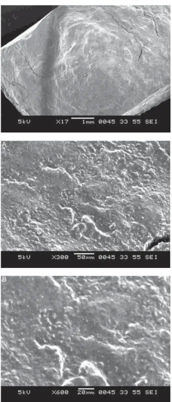
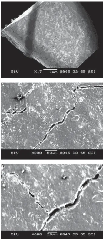
The images acquired from the SEM were used for descriptive analysis. The entire tooth sample as observed in all 3 groups showed cracks on the surface caused by dehydration procedures (Figures 3-5).
The SEM observation revealed that all the instruments managed to remove the calculus
#$$55,##05!#$6###7in vitro,#8+0
u Baseline Ra Value-PostInstrumentation Ra Value
deposits quite effectively. Large remaining deposits were rarely seen. Remnants of calculus were seen at !""=( !~@""= (Figures 3b, 4b, 5b). Presence of smear layer was
DISCUSSION
Studies investigating the differences between manual, piezoelectric and magnetostrictive
ultrasonic systems are inconclusive8,9. The same
has been corroborated by Lea and Walmsley’s = respect to powered instruments. It is worthwhile recapitulating that a large number of variables - # cross section and generator power, contact load, angle and duration, generator power, tip shape, instrumentation end point, vibration generation # 1 section and tip motion - associated with attempts to investigate such differences, make it practically # the method of instrumentation that causes the least
amount of root surface alteration13. Using both,
stereomicroscope and SEM evaluation, Breininger,
et al.2 (1987) compared curettes and ultrasonic
methods in removing plaque. They concluded that neither instrument removed all stained accretions. % ## tips were found on all surfaces. Evidently, some form of standardization is required to allow meaningful
comparisons to be made between studies.
Further, a number of studies have shown that one session of closed root instrumentation does not achieve the goal of total elimination of all calculus # ## scaling and root planing have failed to secure calculus free root surfaces. It is worth mentioning
Figure 2- Percentage reduction of surface roughness (Ra) values in group A, B and C
Figure 3- Scanning electron microscopy (SEM) view of group A sample. A: SEM view of group A sample at 300x
A
B
Figure 1- Mean Ra values (in μm) before and after instrumentation in all three groups
at this juncture that even under optimal conditions
in vitro it is not always possible to remove entire
calculus from all root surfaces. There are generally
two methods of evaluating tooth damage in vitro.
9 surface is clean and clear of calculus as deemed by the operator. The second is to instrument for a controlled length of time or number of strokes.
While the latter is often more controlled insofar as operating parameters such as load and contact angle
are concerned, clinically it is less appropriate13.
Employing the former method in the present study, teeth samples were instrumented until they were clean - clinically a more pertinent aim.
One of the highlights of this study is the fact that periodontally diseased teeth were selected.
A
B
Figure 4- Scanning electron microscopy (SEM) view of group B sample. A: SEM view of group B sample at 300x
Figure 5- Scanning electron microscopy (SEM) view of group C sample. A: SEM view of group C sample at 300x !
A
B
Results from such a work are more meaningful because that the sample mimics actual conditions in patients unlike works that are carried out on healthy
teeth extracted originally for orthodontic reasons6.
Moreover, through random allocation of teeth to their respective groups they were well matched as #6# initial calculus in each group. This was an important aspect given that these teeth were diseased and affected by calculus.
As aforementioned, mean Ra (μm) values were calculated after instrumentation. The teeth were then examined under a stereomicroscope >"=*
seen5. Subsequent SEM analysis showed that
surfaces with smaller mean Ra values exhibited less gouges and scratches than those with higher mean Ra values.
SEM observations in our study indicate that the use of hand instruments resulted in a smooth surface than obtained by ultrasonic instruments.
Ribeiro, et al.17 (2006) had also concluded that
diamond coated sonic tips and ultrasonic universal tips produced similar roughness of surface which was higher than that produced by hand curettes. # regarding instrumentation with curettes and
ultrasonic instruments12,14,15,21. However, Ewen and
% 10>??
Jones, et al.11 (1972) found slight difference. Such
variations in results can be attributed to methods and techniques of instrumentation. We discovered # the two ultrasonic instruments where we felt that the magnetostrictive instrument produced a # 8 However, caution should be exercised in interpreting the results too strictly given that a limited number of surfaces were examined and the interpretations were purely subjective.
The difference in Ra values between the two ultrasonic instruments could also be due to a difference in the power output. Thus, while medium power setting was used for both and the same operator conducted the procedure, there was no way of deducing if the two ultrasonic devices delivered similar power at the same settings. The power of the piezoelectric device could have been higher than that of the magnetostrictive device causing more root damage which was interpreted
as a higher Ra value4. In a study published in 20066
the authors assessed the manual and ultrasonic root surface scaling at low, medium and high power settings; roughness of the instrumented teeth was evaluated. The authors concluded that ultrasonic instrumentation at high power settings produces rougher root surfaces than
ultrasonic instrumentation at lower power; and that manual instrumentation with curettes produces lower roughness than ultrasonic instrumentation independent of power setting.
Notwithstanding these limitations, our results clearly indicate that all three instruments
considerably reduced calculus on the root surfaces8.
Both ultrasonic and manual instrumentation removed calculus quite effectively as seen by naked 9 ultrasonic methods produced greater disturbance on surface topography than hand instrumentation, given that the differences in surface roughness produced by the three different instruments were are operationally more viable since they ensure more patient comfort and cause less operator fatigue.
CONCLUSION
Within the limits of the present study, given that the manual, magnetostrictive and piezoelectric ultrasonic instruments produce the same surface # for creating a biologically compatible surface of periodontally diseased teeth is similar.
ACKNOWLEDGEMENTS
The authors thank the technical staff who ceded the surface roughness measuring instrument 7 = the Mechanical Engineering Department, Manipal Institute of Technology, Manipal University, Manipal; and, Department of Metallurgy, National Institute of Technology, Suratkhal, Karnataka for the SEM examination.
REFERENCES
1- Axelsson P, Lindhe J, Nyström B. On prevention of caries and periodontal disease. Results of a 15-year longitudinal study in adults. J Clin Periodontol. 1991;18:182-9.
2- Breininger DR, O’Leary TJ, Blumenshine RVH. Comparative effectiveness of ultrasonic and hand scaling for the removal of subgingival plaque and calculus. J Periodontol. 1987;58:9-18. 3- Buchanan SA, Robertson PB. Calculus removal by scaling/ root planing with and without surgical access. J Periodontol. 1987;58:159-63.
4- Busslinger A, Lampe K, Beuchat M, Lehmann B. A comparative
in vitro study of a magnetostrictive and a piezoelectric ultrasonic
scaling instrument. J Clin Periodontol. 2001;28:642-9.
~1+ B% B2/& + 7 1986;13:205-10.
7- Dragoo MR. A clinical evaluation of hand and ultrasonic # # > $7 B 4 1992;12:311-23.
8- Drisko CH. Root instrumentation. Power-driven versus manual scalers, which one? Dent Clin North Am. 1998;42:229-44. 9- Drisko CL, Cochran DL, Blieden T, Bouwsma OJ, Cohen RE, Damoulis P, et al. Position paper: sonic and ultrasonic scalers in periodontics. Research, Science and Therapy Committee of the American Academy of Periodontology. J Periodontol. 2000:71:1792-801.
>"1 % && of teeth following periodontal instrumentation. J Periodontol. 1977;48:92-7.
11- Jones SJ, Lozdan J, Boyde A. Tooth surfaces treated in situ
with periodontal instruments. Br Dent J. 1972;132:57-64. 12- Jotikasthira NE, Lie T, Leknes KN. Comparative in vitro studies
of sonic, ultrasonic and reciprocating scaling instruments. J Clin Periodontol. 1992;19:560-9.
13- Lea SC, Walmsley AD. Mechano-physical and biophysical properties of power-driven scalers: driving the future of powered instrument design and evaluation. Periodontol 2000. 2009;51:63-78.
14- Lee A, Heasman PA, Kelly PJ. An in vitro comparative study
of a reciprocating scaler for root surface debridement. J Dent. 1996;24:81-6.
15- Meyer K, Lie T. Root surface roughness in response to periodontal instrumentation studied by combined use of microroughness measurements and scanning electron microscopy. J Clin Periodontol. 1977;4:77-84.
>@1B 61787% B4 M, Rateitschak KH. Non-surgical periodontal treatment: where are the limits? An SEM study. J Clin Periodontol. 1992;19:240-4. >?1B# ()+B+)]('& &+& in vitro study of root roughness
after instrumentation with ultrasonic and diamond tip sonic scaler. J Appl Oral Sci. 2006;14:124-9.
18- Ritz L, Hefti AF, Rateitschak KH. An in vitro investigation on
the loss of root substance in scaling with various instruments. J Clin Periodontol. 1991;18:643-7.
19- Kepic TJ, O’Leary TJU, Kafrawy AH. Total calculus removal: an attainable objective? J Periodontol. 1990;61:16-20.
20- Tunkel J, Heinecke A, Flemmig TF. A systematic review of 1 # # in the treatment of chronic periodontitis. J Clin Periodontol. 2002;29(sp. Issue 3):72-81.
21- Wilkinson RF, Maybury JE. Scanning electron microscopy of the root surface following instrumentation. J Clin Periodontol. 1973;44:559-64.