ABSTRACT
Inluence of the apical enlargement size on the
endotoxin level reduction of dental root canals
Ariane Cassia Salustiano MARINHO1, Frederico Canato MARTINHO2, Alexandre Augusto ZAIA3, Caio Cezar Randi
FERRAZ3, Brenda Paula Figueiredo de Almeida GOMES4
1- BDS, Graduate student, Department of Restorative Dentistry, Endodontics Division, Piracicaba Dental School, State University of Campinas - UNICAMP, Piracicaba, SP, Brazil.
2- BDS, MSc, PhD, Research Professor, Department of Restorative Dentistry, Endodontics Division, Piracicaba Dental School, State University of Campinas - UNICAMP, Piracicaba, SP, Brazil.
3- BDS, MSc, PhD, Associate Professor, Department of Restorative Dentistry, Endodontics Division, Piracicaba Dental School, State University of Campinas - UNICAMP, Piracicaba, SP, Brazil.
4- DDS, MSc, PhD, Full Professor, Department of Restorative Dentistry, Endodontics Division, Piracicaba Dental School, State University of Campinas - UNICAMP, Piracicaba, SP, Brazil.
Corresponding address: Dr. Brenda PFA Gomes - Faculdade de Odontologia de Piracicaba - UNICAMP - Departamento de Endodontia - Av. Limeira, 901 - Areião - Piracicaba - São Paulo - Brasil - 13414-903 - E-mail: [email protected]
Phone: (55) 19 2106 5343 - Fax: (55) 19 2106 5218
Received: March 1, 2012 - Modiication: August 16, 2012 - Accepted: September 17, 2012
G
ram-negative bacteria play an essential role in endodontic infections because theyhave virulence factors such as endotoxin. Due to its potential cytotoxic activity, special attention has been given to the removal/neutralization of this endotoxin in the root canal system. Objective: The aim of this study was to evaluate the inluence of the apical enlargement size (AeS) by using rotary instruments on the endotoxin level reduction of dental root canals. Material and Methods: Forty root canals of the mandibular premolar
teeth were used. Escherichia coli endotoxin (055: B55) was inoculated into thirty root
canals. Ten teeth served as the negative control group. After the incubation period, the irst endotoxin samples were collected from the root canals with a sterile/apyrogenic paper point for the analysis of the endotoxin units (eU/mL) present before instrumentation (S1).
Specimen instrumentation was performed with the Mtwo® rotary system in the sequence
10/.04, 15/.05, 20/.06, 25/.06, 30/.05, 35/.04 and 40/.04. To monitor the effectiveness of increasing apical enlargement on endotoxin removal, the second endotoxin samples were collected from all the root canals after instrumentation with the following instruments:
#25/.06- (S2); #30/.05- (S3); # 35/.04- (S4); and #40/.04- (S5). Limulus amebocyte
lysate (LAL) was used to quantify the levels of endotoxin. The results were statistically
compared by using repeated measures of ANOVA with post hoc Tukey testing. Results:
Increasing levels of endotoxin removal was achieved by large sized apical enlargement: S2 (AeS #25/.06)- 89.2%, S3 (AeS #30/.05)- 95.9%, S4 (AeS #35/.04)- 97.8% and S5 (AeS #40/.04)- 98.2%. Substantial reduction of endotoxin content was obtained in S4 and S5 compared to S2 (p<0.05), however, the root canal preparation was not able to eliminate the endotoxin. Conclusions: Under the conditions of this study, it was concluded that the reduction of endotoxin levels of the dental root canals could be predicted by increasing the apical enlargement size.
Key words: Root canal. Bacterial infections. Instrumentation.
INTRODUCTION
Clinical investigations of endodontic infection have elucidated the strong correlation between oral bacterial lipopolysaccharide (LPS, known as endotoxin) present in the outer cell membranes of
Gram-negative bacteria12, and the presence of clinical
symptomatologyand apical periodontitis4,8,9,16,24.
Particularly in the periapical tissues, endotoxin
can stimulate cells to release pro-inlammatory
including bone resorption, even at very low concentrations8,10,14,16 . Due to its potential cytotoxic
activity in the pulp and periapical tissues, special attention has been given to the complete removal/ neutralization of endotoxin from infected root canals9,15.
The clinical eficacy of endodontic procedures
on bacterial LPS has been investigated in the endodontic literature8,10,13,15-16. Gomes, et al.7 (2009)
had evaluated the action of chemo-mechanical
preparation with hand iles and 2.5% NaOCl or
2% chlorhexidine (CHX) gel as auxiliary chemical substances for the removal of endotoxin from the infected root canals. Despite the high antimicrobial activity of these auxiliary chemical substances, neither 2.5% NaOCl nor 2% CHX gel were effective in eliminating endotoxin. The authors suggested that the reduction of only 47% of the endotoxin content was related to the mechanical action of the instruments in the dentinal walls, accomplished by
the low and backlow of the irrigants. Martinho,
et al.15 (2010) indicated a reduction of 96.27% of the endotoxin contents by using rotary iles
accomplished by sterile/apyrogenic saline solution as an irrigant.
With regard to the optimal shape and apical size of the root canal preparation, there is some controversy on whether apical enlargement is required1-2,8,14,18,23,28. As a rule, the most important
criteria for root canal enlargement should rely on
the clinical determination of the initial apical ile
size8. One recommended approach is to apically
enlarge the root canal up to three sizes larger than
the irst ile to bind.
The latest generation of root canal instruments includes nickel-titanium (Ni-Ti) rotary systems. The super-elastic property of Ni-Ti has allowed for a better centering ability of the instrument within the root canal lumen15. Besides, these instruments
have reduced operator fatigue, procedural errors, and the duration of the canal preparation25. Mtwo® rotary iles (VDW, Munich, Germany) stand out
as a new generation of rotary instruments20.The iles have two cutting edges with minimal radial
contact providing maximum space for dentinal debris removal. The system is comprised of 7 tip sizes (10-40) with constant tapers ranging between
0.04 - 0.07. Thereby, inal apical enlargement
can be perfomed up to #25/.0622, #30/.055,22-24,
#35/.0421,25 or #40/.0418,26,28.
In view of the widespread use of the rotary
iles, clinical investigations have evaluated the
performance of Mtwo® rotary iles for root canal
instrumentation regarding cleaning and shaping ability,transportation of debris into the apex, preservation of the original geometry of the root canal, fracture risk and bacterial reduction21.
However, no study evaluated the effectiveness of
the increased apical enlargement by using rotary
iles for endotoxin reduction.
Therefore, the aim of this study was to evaluate
the inluence of apical enlargement size (AES) in
reducing/eliminating endotoxin from root canals prepared with Mtwo® rotary iles.
MATERIAL AND METhODS
Specimen selection and preparation
This study was submitted to and approved by the ethics Committee of Research of the Piracicaba Dental School, State University of Campinas, UNICAMP (Protocol #186/09). Forty canals of freshly extracted human mandibular pre-molars were used in the study. These teeth were collected from a general dental practice and had been extracted for pulpal or periodontal reasons. The teeth were stored in a 0.9% saline solution until use.
In order to be included in this study, all teeth had to present a mature root apex with no evidence of external resorption and a single narrow straight root canal. Preoperative radiographs were taken to ensure that the teeth did not have root caries, fractures, multiples canals, lateral radicular canals,
calciications or curvatures. Specimen selection
was made on the basis of relative dimensions and similarity in root morphology. None of these teeth had received prior root canal treatment. Debris, calculus, and soft tissue remnants on the root surfaces were removed by using periodontal curettes (Golgran- Millennium, São Paulo, SP, Brazil). The crowns were transversely sectioned at the level of the cementum-enamel junction by using a water-cooled diamond disk (KG-Sorensen, Barueri, SP, Brazil). The root length was standardized to 15 mm. Apical patency was
determined by inserting a size 08 K-ile
(Dentsply-Maillefer, Ballaigues, Switzerland) into the root canal until the tip was visible at the apical foramen. The
root canals were instrumented up to a #20 K-ile
at the apical foramen in alternated rotation, under irrigation with 5 mL of distilled water after each
ile change. The smear layer was removed with
5.25% NaOCl (Drogal, Piracicaba, SP, Brazil) for 10 minutes under constant agitation (Agitador-Aquecedor Fanem, São Paulo, SP, Brazil), followed by 17% eDTA (Drogal, Piracicaba, SP, Brazil). The teeth were then washed thoroughly with distilled water for 1 hour6, and then the canals were dried
All specimens were sterilized with autoclave (Fanem, São Paulo, SP, Brazil) at 121°C for 20 minutes in distilled water and then randomly divided into 24-well cell culture plates (Corning Costar, Cambridge, MA, USA). To make both handling and instrumentation easier, the teeth were vertically
ixed at the cervical region with chemically activated
acrylic resin (Artigos Odontoĺgicos Clássico, São Paulo, SP, Brazil) inside the wells. For the degradation of preexisting endotoxins, all materials used in the experiment were sterilized by gamut radiation with cobalt 60 (20 KGy for 6 hours)3.
Specimen contaminated with endotoxin
Before starting the experiment, a pilot study was made in order to ensure the endotoxin contamination protocol by the Limulus amebocyte lysate assay (LAL). After this, 30 teeth were inoculated with 30 µL of a solution containing
Escherichia coli 055: B55 endotoxin (1.000 eU/mL)
and incubated for 24 hours at 37°C in a humidiied
atmosphere, according to Oliveira, et al.20 (2005).
As a negative control group, ten teeth were inoculated with 30 µL of LAL water (endotoxin-free)
and incubated for 24 hours at 37°C in a humidiied
atmosphere.
Sampling and instrumentation procedure
After the incubation period, all the root canals were sampled to determine the initial levels of endotoxin present before chemo-mechanical preparation (S1). A sterile/apyrogenic absorbent paper point was introduced into the full extension of the root canal and remained in position for 60 seconds. It was then placed in an endotoxin-free
glass and frozen at -20°C for further quantiication
of the endotoxin. Mtwo® was carried out according to
the manufacturer’s instructions (sequence 10/.04, 15/.05, 20/.06, 25/.06, 30/.05, 40/.04) into the full extension of the root canal with a gentle in-and-out motion. Once the instrument had achieved the end of the canal and rotated freely, it was removed25. The
working time of each instrument was monitored. In order to avoid possible cutting blade interference, the Mtwo® iles were used once. The canals were
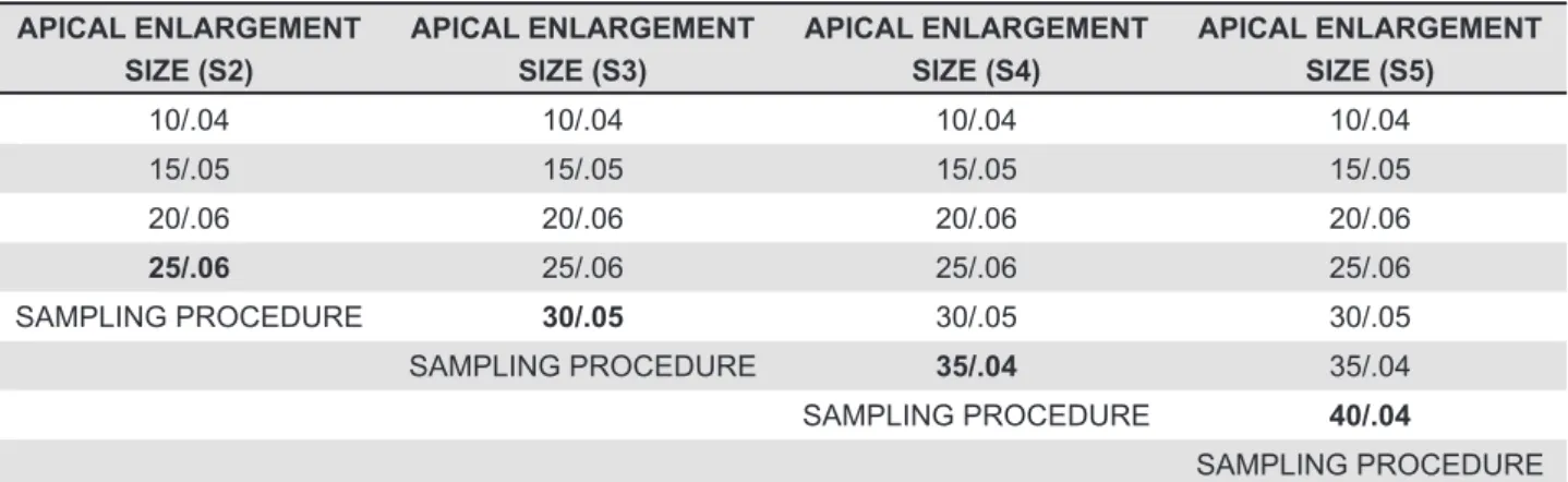
shaped by using 1 mL of non-pyrogenic water that was renewed with every change of instrument. All the experimental procedures were conducted by the same operator. To evaluate the effectiveness of increased apical enlargement on the endotoxin levels, the root canals were sampled with a sterile/ apyrogenic absorbent paper point, after using the following instruments: #25/.06- as S2 (basic series); #30/.05- as S3; #35/.04- as S4; and #40/.04- as S5 (shown in Table 1).
Determination of endotoxin concentration
The turbidimetric test (Pyrogent 5000®,
BioWhitaker, Cambrex Co, Walkersville, MD, USA) was used to measure the endotoxin levels in the root canals via the LAL technique, previously published by the authors13,15-16. It is a kinetic,
quantitative assay for the detection of endotoxin, in which the activated LAL converts the coagulogen into coagulin to produce turbidity in the sample. All the samples were mixed with the reconstituted LAL reagent, placed in a photometer, and automatically monitored over time until the appearance of turbidity. The time required before the appearance of turbidity (reaction time) was inversely proportional to the amount of endotoxin. In the presence of a large amount of endotoxin, the reaction occurred rapidly, in the presence of a small amount of endotoxin, the reaction time was increased. The concentration of endotoxin within the unknown samples was calculated from a standard curve. Its wide-range sensitivity [0.01-100 endotoxin units (eU/mL)] requires small amounts of dilutions of the clinical samples, thus optimizing the laboratory time. As a parameter for the calculation of the amount of endotoxins existing in the root canal samples, a standard curve was plotted by using the endotoxin of a known concentration supplied by the kit (100 eU/mL), with its dilutions reaching
APICAL ENLARGEMENT SIZE (S2)
APICAL ENLARGEMENT SIZE (S3)
APICAL ENLARGEMENT SIZE (S4)
APICAL ENLARGEMENT SIZE (S5)
10/.04 10/.04 10/.04 10/.04
15/.05 15/.05 15/.05 15/.05
20/.06 20/.06 20/.06 20/.06
25/.06 25/.06 25/.06 25/.06
SAMPLING PROCEDURE 30/.05 30/.05 30/.05
SAMPLING PROCEDURE 35/.04 35/.04
SAMPLING PROCEDURE 40/.04
SAMPLING PROCEDURE
Bold font highlights the inal apical instruments in each group
the inal concentrations (0.01, 0.1, 1, 10 EU/mL)
according to the manufacturer’s instructions. A 96-well microplate (Corning Costar, Cambridge, MA, USA) was used in a heating block at 37°C and maintained at this temperature throughout the assay. First, the endotoxin samples were suspended in 1 mL of LAL water supplied by the kit and agitated in vortex for 60 seconds. Immediately after, 100 mL of the blank, followed by the same volume of the standard endotoxin solutions (0.01, 0.1, 1, 10 eU/mL) and 100 mL of the samples, were added in duplicate to the 96-well microplate with their respectively positive controls. To avoid inhibition or enhancement of the LAL, a known concentration of
Escherichia coli endotoxin was added to the clinical samples, as recommended by the manufacturer’s instructions (spike procedure). Both the unspiked and spiked samples (control) had their endotoxin concentrations automatically calculated. The endotoxins recovered were equal to the known spike concentration ranging from 50% to 200%. All reactions were performed in duplicate to validate the test. The test procedure was performed according to the manufacturer’s instructions. The absorbance of endotoxins was individually measured by using an enzyme-linked immunosorbent assay plate reader (Ultramark, Bio-Rad Laboratories Inc, Hercules, CA, USA). During the calculation of the endotoxin concentration, the microplate reader, by using the WinKQCL Software (BioWittaker, Cambrex Co, Walkersville, MD, USA), monitors the absorbance of each well of the microplate continuously at 340 nm throughout the assay. By using the initial absorbance reading of each well as its own blank, the reader determines the time required for absorbance at 0.03 absorbance units. This time is termed as the reaction time. The WinKQCL Software automatically performs a log/log linear correlation of the reaction time of each standard curve with its corresponding endotoxin concentration, including printing the standard curve parameters. If the
absolute value of the correlation coeficient (r) is
0.980, a polynomial model can be used to construct
a standard curve and in turn predict the endotoxin concentrations of the test samples.
Statistical analysis
The data obtained with the LAL assay was statistically analyzed by using the SAS Software, version 9.1 (SAS Institute, Cary, NC, USA). The Shapiro-Wilk test was used to verify the normal data distribution. Both the mean and standard deviation regarding the endotoxin levels after each sampling procedure were determined. A one way repeated measures ANOVA was applied. When
signiicant differences were found between the
groups, the Tukey’s test was subsequently used.
The signiicance levels were always set at 5%
(p<0.05).
RESULTS
The Shapiro-Wilk test showed data normality (p>0.266). For the validation of the LAL assay, the
standard curve fulilled the criteria of linearity for all
running assays, as reported by the guidelines. The LAL assay indicated that endotoxins were present in 100% of the root canals investigated (30/30) with a median value of 630 eU/mL. Sterility of the
specimens was conirmed with the negative control
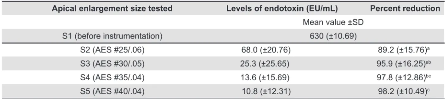
group showing an absence of endotoxin in all the root canals (10/10). As a result of the chemo-mechanical preparation, the LPS content was reduced to a mean value of 10.8 eU/mL. The mean values and the standard deviation of the different levels of endotoxin reduction are shown in Table 2.
DISCUSSION
The present study evaluated the inluence of
the apical enlargement size (AeS) in reducing/ eliminating endotoxin in the root canals prepared with Mtwo® rotary iles. Analyses of the results
indicated that aroot canal preparation larger
than a #30/.05 ile showed the best performance
regarding endotoxin removal. Additionally, the size
Apical enlargement size tested Levels of endotoxin (EU/mL) Percent reduction
Mean value ±SD
S1 (before instrumentation) 630 (±10.69)
S2 (AES #25/.06) 68.0 (±20.76) 89.2 (±15.76)a
S3 (AES #30/.05) 25.3 (±25.65) 95.9 (±16.25)ab
S4 (AES #35/.04) 13.6 (±15.69) 97.8 (±12.86)bc
S5 (AES #40/.04) 10.8 (±12.31) 98.2 (±10.49)c
Mean values followed by different letters indicate statistical signiicant difference among the sequences tested (p<0.05)
of the apical enlargement implicated signiicantly
in the removal of endotoxins from the root canals. At the base line samples, after 24 hours of incubation time, endotoxins were recovered from 100% of the root canals sampled, validating the contamination protocol in the present study. Nissan, et al.19 (1995) evaluated the diffusion capacity of
bacterial endotoxin through human dentin in the apical direction and concluded that endotoxin was capable of passing through 0.5 mm of dentin in a period that ranged from 15 min to 4½ h, suggesting that the dentin may allow the passage of bacterial products to the pulpal space. Oliveira, et al.20
(2005) showed that endotoxin can diffuse through the dentinal tubules towards the cement in 24 h.
In clinical practice, the most frequently Mtwo®
sequence used, the basic series, ending in an apical enlargement size of #25/.06, was able to reduce endotoxin contents by 89.2% (S2). In fact, considering the apical diameter of the lower premolars, which corresponds to that of a #25 or
a #30 ile27, it was expected that the enlargement to a #25 ile-size would not signiicantly improve
the disinfection of the root canal system, as demonstrated by the present study.
Clinically, as a rule, the root canal enlargement
relies on the determination of the initial apical ile
size8. Then the canal should be apically enlarged up to three sizes larger than the irst ile to bind
the working length. In order to achieve a more accurate measurement of the initial apical diameter,
the removal of interferences by cervical pre-laring
should be considered previously8,18.
Our data indicated that the root canal
enlargement achieved by a inal apical ile larger
than #30/.05, showed the best performance in removing endotoxin from the dentinal walls. In agreement, Martinho, et al.15 (2010) evaluating the
ability of the root canal procedures on eliminating endotoxins, demonstrated a 96.27% reduction of endotoxin, achieved by a root canal enlargement
up to a #30 ile size. In fact, an apical enlargement
wider than ISO #30 is recommended to remove not only endotoxin (as shown in the present work), but also the more infected dentin. Also, this approach allows deeper irrigation and contributes to enclosing the canal cross-sections, accessory canals and apical deltas5,8,11,17,23.
Regardless of the apical enlargement size, endotoxins were still detected in 100% of the root canals after instrumentation. Whether the residual amount of endotoxins is able to induce a periapical
inlammatory response is not known. Theoretically,
in the absence of living Gram-negative cells to maintain the LPS levels in the periapical tissues, the effect of these residual levels of endotoxins left behind after the root canal instrumentation, would only temporarily modulate the host defenses,
without impairing the endodontic treatment outcome.
Overall, our study demonstrated that a root
canal preparation, with a ile larger than #30/.05,
achieves a better removal of endotoxins from the root canals. Moreover, the determination of the
initial apical ile size is one of the most important
steps to determine a correct apical enlargement diameter to be achieved during root canal instrumentation, particularly, when approaching a root canal enlargement based on the ‘‘three sizes
up from the irst ile to bind’’ rule.
CONCLUSION
Based on the results of this research, endotoxin levels of dental root canals could be predicted by increasing the apical enlargement size. Root canal preparation was effective in reducing the amount of endotoxin within the infected root canals. Further studies should be performed with different rotary systems associated with irrigation devices to establish protocols for a more effective root canal cleaning.
ACKNOwLEDgMENTS
We would like to thank Ana Regina de Oliveira Polay for technical support. We are also thankful to Lonza for the Kinetic-QCL equipment. This work was supported by the Brazilian agencies FAPeSP (10/51113-1; 10/17877-4; 10/19136-1; 11/50510-0) and CNPq (302575/2009-0; 150557/2011-6).
The authors deny any conlicts of interest.
REFERENCES
1- Baugh D, Wallace J. The role of apical instrumentation in root canal treatment: a review of the literature. J endod. 2005;31:333-40.
2- Coldero LG, McHugh S, MacKenzie D, Saunders WP. Reduction in intra-canal bacteria during root canal preparation with and without apical enlargement. Int endod J. 2002;35:437-46.
3- Csako G, elin RJ, Hochstein HD, Tsai CM. Physical and biological properties of U.S. standard endotoxin eC after exposure to ionizing radiation. Infect Immun. 1983;41:190-6.
4- Dahlén G, Bergenholtz G. endotoxic activity in teeth with necrotic pulps. J Dent Res. 1980;59:1033-40.
5- Falk KW, Sedgley CM. The inluence of preparation size on the mechanical eficacy of root canal irrigation in vitro. J endod. 2005;31:742-5.
6- Ferraz CC, Gomes BP, Zaia AA, Teixeira FB, Souza-Filho FJ.
In vitro assessment of the antimicrobial action and mechanical ability of chlorhexidine gel as an endodontic irrigant. J endod. 2001;27:452-5.
7- Gomes BP, Martinho FC, Vianna Me. Comparison of 2.5% sodium hypochlorite and 2% chlorhexidine gel on oral bacterial lipopolysaccharide reduction from primarily infected root canals. J endod. 2009;35:1350-3.
9- Horiba N, Maekawa Y, Abe Y, Ito M, Matsumoto T, Nakamura H. Correlations between endotoxin and clinical symptoms or radiolucent areas in infected root canals. Oral Surg Oral Med Oral Pathol. 1991;71:492-5.
10- Horiba N, Maekawa Y, Matsumoto T, Nakamura H. A study of the distribution of endotoxin in the dentinal walls of infected root canals. J endod. 1990;16:331-4.
11- Khademi A, Yazdizadeh M, Feizianfard M. Determination of the minimum instrumentation size for penetration of irrigants to the apical third of root canal systems. J endod. 2006;32:417-20. 12- Lüderitz O, Tanamoto K, Galanos C, McKenzie GR, Brade H, Zähringer U, et al. Lipopolysaccharides: structural principles and biologic activities. Rev Infect Dis 1984;4:428-3.
13- Martinho FC, Gomes BP. Quantiication of endotoxins and
cultivable bacteria in root canal infection before and after chemo-mechanical preparation with 2.5% sodium hypochlorite. J endod. 2008;34:268-72.
14- Martinho FC, Chiesa WM, Leite FR, Cirelli JA, Gomes BP. Antigenic activity of bacterial endodontic contents from primary root canal infection with periapical lesions against macrophage in the release of interleukin-1beta and tumor necrosis factor alpha. J endod. 2010;36:1467-74.
15- Martinho FC, Chiesa WM, Marinho AC, Zaia AA, Ferraz CC,
Almeida JF, et al. Clinical investigation of the eficacy of
chemo-mechanical preparation with rotary nickel-titanium files for removal of endotoxin from primarily infected root canals. J endod. 2010;36:1766-9.
16- Martinho FC, Chiesa WM, Zaia AA, Ferraz CC, Almeida JF, Souza-Filho FJ, et al. Comparison of endotoxin levels in previous studies on primary endodontic infections. J endod. 2011;37:163-7. 17- McGurkin-Smith R, Trope M, Caplan D, Sigurdsson A. Reduction of intra-canal bacteria using GT rotary instrumentation, 5.25% NaOCl, eDTA, and Ca(OH)2. J endod. 2005;31:359-63.
18- Mickel AK, Chogle S, Liddle J, Huffaker K, Jones JJ. The role of apical size determination and enlargement in the reduction of intra-canal bacteria. J endod. 2007;33:21-3.
19- Nissan R, Segal H, Pashley D, Stevens R, Trowbridge H. Ability of bacterial endotoxin to diffuse through human dentin. J endod. 1995;21:62-4.
20- Oliveira LD, Carvalho CA, Valera MC, Koga-Ito CY, Jorge AO. Diffusion ability of endotoxin through dentinal tubules. Braz Oral Res. 2005;19:5-10.
21- Paqué F, Ganahl D, Peters OA. effects of root canal preparation on apical geometry assessed by micro-computed tomography. J endod. 2009;35:1056-9.
22- Parris J, Wilcox L, Walton R. effectiveness of apical clearing: histological and radiographical evaluation. J endod. 1994;20:219-24.
23- Pitts DL, Williams BL, Morton TH Jr. Investigation of the role
of endotoxin in periapical inlammation. J Endod. 1982;8:10-8.
24- Schein B, Schilder H. endotoxin content in endodontically involved teeth. 1975. J endod. 2006;32:293-5.
25- Sonntag D, Ott M, Kook K, Stachniss V. Root canal preparation with the NiTi systems K3, Mtwo and ProTaper. Aust endod J. 2007;33:73-81.
26- Usman N, Baumgartner JC, Marshall JG. Inluence of instrument
size on root canal debridement. J endod. 2004;30:110-2.
27- Wu MK, Barkis D, Roris A, Wesselink PR. Does the irst ile to
bind correspond to the diameter of the canal in the apical region? Int endod J. 2002;35:264-7.
28- Yared GM, Bou Dagher FE. Apical enlargement: inluence