359
Horseshoe lung: a case report
Radiol Bras 2007;40(5):359–361 Case Report
HORSESHOE LUNG: A CASE REPORT*
João Paulo Kawaoka Matushita1
, Guilherme Carvalho Missiaggia2
, Roberto Márcio A. Peixoto2
, Hilton Muniz Leão Filho2
, Ricardo Saraiva Dias2
, Wilson Campos Tavares Junior2 , Cristina S. Matushita3
, José Nelson Mendes Vieira4
, João Paulo K. Matushita Junior5
The authors report a case of a newborn term infant with horseshoe lung associated with supracardiac total anomalous pulmonary venous drainage; such association remains unreported in the Western literature. Keywords: Horseshoe lung; Chest radiography; Computed tomography; Angiography.
Pulmão em ferradura: relato de caso.
Os autores relatam um caso de um recém-nascido a termo, com pulmão em ferradura associado a drenagem anômala total de veias pulmonares supracardíacas, cuja associação não encontramos em nenhum relato na literatura ocidental.
Unitermos: Pulmão em ferradura; Radiografia do tórax; Tomografia computadorizada; Angiografia. Abstract
Resumo
* Study developed in the Service of Radiology and Diagnostic Imaging – Hospital das Clínicas da Faculdade de Medicina da Universidade Federal de Minas Gerais (UFMG), Belo Horizonte, MG, Brazil.
1. Associate Professor, PhD, Faculdade de Medicina da Uni-versidade Federal de Minas Gerais (UFMG), Full Professor, Dis-cipline of Radiology and Imaging Diagnosis in the Course of Physiotherapy – Faculdades Biológicas da Saúde/Centro Univer-sitário Newton Paiva, Belo Horizonte, MG, Brazil.
2. MDs, formely Residents at the Service of Radiology and Diagnostic Imaging – Hospital das Clínicas da Faculdade de Medicina da Universidade Federal de Minas Gerais (UFMG) Belo Horizonte, MG, Brazil.
3. Former Graduate Student of Medicine at Universidade do Vale do Sapucaí (Univás), Pouso Alegre, MG, Brazil.
4. MD, Assistant Professor at Faculdade de Medicina da Uni-versidade Federal de Minas Gerais (UFMG), Belo Horizonte, MG, Brazil.
5. Graduate Student of Medicine at Universidade do Vale do Sapucaí (Univás), Pouso Alegre, MG, Brazil.
Mailing address: Prof. Dr. João Paulo K. Matushita. Rua Caetés, 530/216, Centro. Belo Horizonte, MG, Brazil, 30120-080. E-mail: [email protected]
Received April 23, 2005. Accepted after revision January 8, 2007.
INTRODUCTION
Horseshoe lung was first described by Spencer in 1962(1), as a malformation in
which an isthmus of pulmonary paren-chyma extends from the right to the left lung bases across the midline posterior to the heart. The term “horseshoe lung” was coined in analogy to “horseshoe kidney”(2).
Horseshoe lung can be defined as hypo-plastic right lung and fusion of the pos-teroinferior segments of both lungs poste-riorly to the heart and anteposte-riorly to the esophagus and spinal column. The result-ing isthmus of pulmonary parenchyma re-ceives the blood supply from the right pul-monary artery, and the respective bronchi, from the right bronchial tree. So, the ves-sels and bronchus of the isthmus cross the
midline, and this particular feature is found both in the angiography and bronchogra-phy. In cases associated with scimitar syn-drome, the right hypoplastic lung presents with bronchial and vascular anomalies de-scribed in this syndrome.
The pulmonary parenchyma is histo-logically normal(3). The isthmus presents its
own pleural layer that is frequently incom-plete, allowing communication between the right and left pleurae(3).
CASE REPORT
A male newborn term infant who pre-sented at birth with respiratory distress and persistent cyanosis. A chest radiographic image showed hypotransparency of the whole right hemithorax and hyperinflation of the left lung.
A chest computed tomography was per-formed demonstrating a hypoplastic right lung and hyperinflation of the left lung with signs of air trapping in the lower lobe, with an image of continuity between the lower lobes, suggesting pulmonary herniation or horseshoe lung.
The echodopplercardiogram showed dextrocardia and signs of pulmonary hyper-tension with severe dilatation of right chambers and a suspicion of anomalous pulmonary venous drainage.
The hemodynamic study demonstrated pulmonary artery hypertension with total anomalous pulmonary venous drainage into the superior vena cava, a branch of the right pulmonary artery supplying the left
lower pulmonary lobe, and na anomalous branch of descending aorta supplying the right lower pulmonary lobe.
Because of the pulmonary artery hyper-tension, the patient was submitted to sur-gical intervention for correction of the to-tal anomalous pulmonary venous drainage. The child died, and the initial diagnosis was confirmed by means of necropsy.
DISCUSSION
360
Matushita JPK et al.
Radiol Bras 2007;40(5):359–361
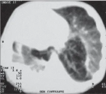
Figure 2. Computed tomog-raphy of pulmonary window demonstrating right nary hypoplasia, left pulmo-nary hyperinflation and an isthmus bridging the lower lobes, posteriorly to the heart.
Figure 3.A: Posteroanterior injection with anomalous drainage of the pulmonary veinsinto the superior vena cava. B: Posteroanterior injection demonstrating right branch of pulmonary artery with a branch across the hemithorax. C: Posteroanterior injection demonstrating left and right branches of pulmonary artery, with a branch across the left hemithorax. D: Ventriculography demonstrating anomalous blood supply to the base of the right lung by an artery originating from the descending aorta.
C D
A B
deformity cannot be observed(4,5). In
pa-tients with right pulmonary hypoplasia, associated or not with scimitar syndrome, the frontal view on a chest radiograph dem-onstrates dextrocardia, a variable degree of right hemithorax hypoplasia and a hyperin-flated left lung. These findings are typical of horseshoe lung in association or not with scimitar syndrome, and with posterior fu-sion of the basal segments of the right and left lungs(6–8).
In the setting of this anomaly, the right lung is abnormal in terms of size, lobe and bronchial tree(9,10). The right pulmonary
361
Horseshoe lung: a case report
Radiol Bras 2007;40(5):359–361
Scimitar syndrome is characterized by a partially anomalous right pulmonary venous drainage into the systemic vein, usually bellow the diaphragm. This vein presents an oblique caudal course, resem-bling the short, curved sword called scimi-tar, and in 15% of cases is associated with horseshoe lung(12,13).
In the present study, horseshoe lung was not observed in association with scimitar syndrome, but with total anomalous pul-monary venous drainage into the superior vena cava, with severe pulmonary hyper-tension. Association with other cardiovas-cular abnormalities, such as atrial septal defect, interventricular septal defect, per-sistent arteriosus ductus, single left ven-tricle, etc., may occur.
In the suspicion of horseshoe lung, pul-monary arteriography should be performed. This method can detect variable degrees of right pulmonary artery hypoplasia and ab-normal distribution of its segmental branches. Chest computed tomography is the best non-invasive method to confirm this diagnosis, demonstrating the pulmo-nary isthmus between the posterior region of the pericardium and the thoracic column. The angiographic study of the pulmonary artery is useful for diagnostic confirmation since this method allows the visualization of the right pulmonary artery with variable
degrees of hypoplasia as well as a decrease in this artery segmentation. Also the level of the pulmonary artery pressure can be detected.
The prognosis in cases of horseshoe lung is dependent on the pulmonary artery pressure. Usually, the types of horseshoe lung associated with normal pulmonary artery pressure are asymptomatic(2).
Angio-cardiography and echodopplercardiogram are utilized for evaluating associated car-diovascular abnormalities.
Pathognomonic features related to horseshoe lung cannot be found on conven-tional chest radiographic images. The diag-nosis may be reached by means of pulmo-nary arteriography and/or computed to-mography. Currently, depending on the lo-cal diagnostic resources, an excellent as-sessment of the pulmonary vascularization can be performed by computed tomogra-phy and magnetic resonance imaging (angiotomography and angioresonance) or even helical and multiplanar computed to-mography reconstruction techniques.
REFERENCES
1. Spencer H. Pathology of the lung. 3rd ed. Oxford: Pergamon Press, 1977;75–76.
2. Dupuis C, Remy J, Remy-Jardin M, Coulomb SM, Breviere GM, Ben Laden S. The “horseshoe” lung: six new cases. Pediatr Pulmonol 1994;17: 124–130.
3. Haworth SG, Sauer U, Buhlmeyer K. Pulmonary hypertension in scimitar syndrome in infancy. Br Heart J 1983;50:182–189.
4. Dische MR, Teixeira ML, Wincherster PH, Engle MA. Horseshoe lung associated with a variant of the “scimitar” syndrome. Br Heart J 1974;36: 617–620.
5. Thilenius OG, Ruschhaupt DG, Replogle RL, Bharati S, Herman T, Arcilla RA. Spectrum of pulmonary sequestration: association with anoma-lous pulmonary venous drainage in infants. Pe-diatr Cardiol 1983;4:97–103.
6. Felson B. Chest roentgenology. Philadelphia, PA: Saunders, 1973;87–92.
7. Felson B. Pulmonary agenesis and related anoma-lies. Semin Roentgenol 1972;7:17–30. 8. Richter K, Boch G. Dieanomalie der
epibronchie-len rechten. Pulmonaiarterieais. Litsymptom eines pulmo-kardiovakuioren syndromes. Rofo 1967;107:31–38.
9. Kittle CF, Crockett JE. Vena cava bronchovascular syndrome: a triad of anomalies involving the right lung: anomalous pulmonary vein, abnormal bron-chi and systemic pulmonary arteries. Ann Surg 1962;156:222–233.
10. Neill CA, Ferencz C, Sabiston DC, Sheldon H. The familial occurrence of hypoplastic right lung with systemic arterial supply and venous drain-age “scimitar syndrome”. Bull Johns Hopkins Hosp 1960;107:1–21.
11. Farnsworth AE, Ankeney JL. The spectrum of the scimitar syndrome. J Thorac Cardiovasc Surg 1974;68:37–42.
12. Frank JL, Poole CA, Rosas G. Horseshoe lung: clinical, pathologic, and radiologic features and a new plain film finding. AJR Am J Roentgenol 1986;146:217–226.