252
Yamada AM et al. Breast swelling secondary to central vessels stenosis
Radiol Bras. 2013 Jul/Ago;46(4):252–254
Bilateral breast swelling secondary to superior vena cava
obstruction and subclavian vein thrombosis
*
Edema bilateral das mamas secundário a obstrução da veia cava superior e trombose de veia subclávia
Ariadne Mayumi Yamada1, Ana Lucia Kefalas Oliveira Melo2, Gesner Pereira Lopes3, Genesio Borges
de Andrade Neto1, Valesca Bizinoto Monteiro1, Renato Santos Soares1
Superior vena cava syndrome is defined by a set of signs and symptoms secondary to superior vena cava obstruction caused principally by malignant diseases. The present report describes the case of an unusual clinical manifestation of this syndrome with bilateral breast swelling, and emphasizes the relevance of knowledge on mammographic signs of systemic diseases.
Keywords: Mammography; Superior vena cava syndrome; Subclavian vein thrombosis.
A síndrome da veia cava superior é definida por um conjunto de sinais e sintomas secundários a uma obstrução da veia cava superior, causada principalmente por neoplasias malignas. Este relato de caso demonstra uma manifestação clí-nica incomum dessa síndrome, o edema bilateral das mamas, e destaca a importância do conhecimento dos sinais mamográficos de doenças sistêmicas.
Unitermos: Mamografia; Síndrome da veia cava superior; Trombose de veia subclávia.
Abstract
Resumo
* Study developed at Universidade Federal do Triângulo Mineiro (UFTM), Uberaba, MG, Brazil.
1. MDs, Residents in Radiology and Imaging Diagnosis, Universidade Federal do Triângulo Mineiro (UFTM), Uberaba, MG, Brazil.
2. PhD, Titular Member of Colégio Brasileiro de Radiologia e Diagnóstico por Imagem CBR), Associate Professor, Universi-dade Federal do Triângulo Mineiro (UFTM), Uberaba, MG, Brazil.
3. Master, Titular Member of Colégio Brasileiro de Radiolo-gia e Diagnóstico por Imagem (CBR), Head of Division of Radiol-ogy and Imaging Diagnosis, Universidade Federal do Triângulo Mineiro (UFTM), Uberaba, MG, Brazil.
Mailing Address: Dra. Ariadne Mayumi Yamada. Avenida Leopoldino de Oliveira, 579, Bloco 23, ap. 103, Parque do Mi-rante. Uberaba, MG, Brazil, 38081-000. E-mail: kenkoyamada@ hotmail.com.
Received November 1st, 2012. Accepted after revision March 7, 2013.
Yamada AM, Melo ALKO, Lopes GP, Andrade Neto GB, Monteiro VB, Soares RS. Bilateral breast swelling secondary to superior vena cava obstruction and subclavian vein thrombosis. Radiol Bras. 2013 Jul/Ago;46(4):252–254.
0100-3984 © Colégio Brasileiro de Radiologia e Diagnóstico por Imagem CASE REPORT
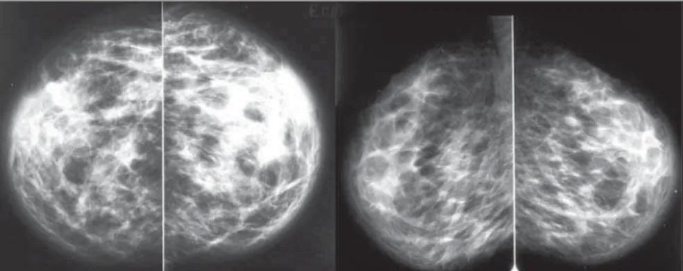
no evidence of nodules or microcalcifi-cations (Figures 1 and 2). Contrast-en-hanced chest computed tomography dem-onstrated the presence of an ill-defined mediastinal mass causing deviation and invasion, particularly of the superior vena cava (Figure 3). Doppler ultrasonography of upper limbs demonstrated total occlu-sion of the subclavian, axillary and brachial veins at left, caused by the presence of a thrombus attached to the luminal surface. The patient progressed with facial swelling, anasarca, worsening of her clinical condi-tion and death due to respiratory failure. Anatomopathological analysis revealed the presence of a carcinoma of the upper lobe facial plethora, visual symptoms, dyspnea
and cough(2). The breast swelling described in the present case report is an uncommon manifestation.
CASE REPORT
A 38-year-old female patient, smoker, presented with dyspnea and chest pain for nine days, left upper limb edema and bilat-eral breast swelling for three days. The ini-tial diagnosis was pneumonia. Antibiotic therapy was subsequently initiated. The patient was submitted to bilateral mam-mography which demonstrated skin and subcutaneous thickening bilaterally, with
INTRODUCTION
Superior vena cava syndrome is defined by a set of signs and symptoms secondary to superior vena cava obstruction. Such a syndrome was first described in 1757 by William Hunter, in a patient with syphilitic aortic aneurysm(1). Currently, malignant diseases represent the main etiologic fac-tors associated with this syndrome. Throm-botic conditions also constitute frequent causes, reflecting an increase in the utiliza-tion of intravascular catheters. Most com-mon clinical manifestations include facial and upper limbs swelling, presence of col-lateral circulation in the neck and chest,
253
Yamada AM et al. Breast swelling secondary to central vessels stenosis
Radiol Bras. 2013 Jul/Ago;46(4):252–254
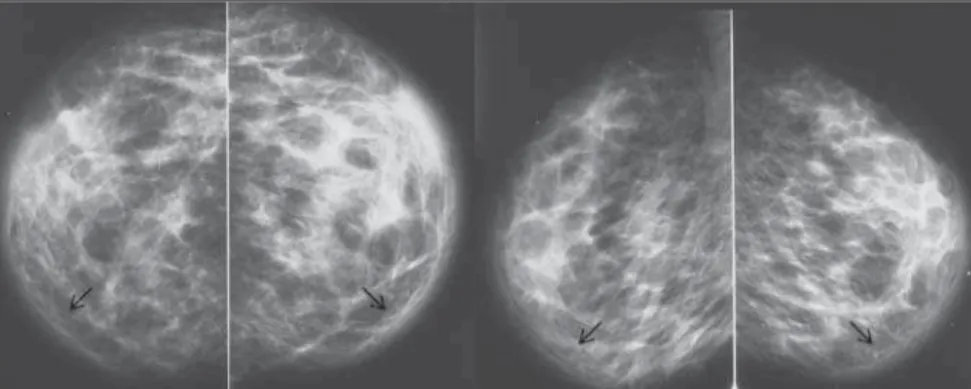
ness of the cranial and lateral skin is < 2.5 mm; on the other hand, the thickness of the medial and caudal skin may be > 3 mm(14). In the present case, bilateral breast swell-ing represented by skin thickenswell-ing was ob-served. The deep venous drainage of the breast occurs through the internal mam-mary and axillary veins, which drain into the pulmonary capillary vasculature, and intercostals veins, which drain into the su-perior vena cava and lungs (Figure 4)(15,16). The anatomical site of the subclavian oc-clusion will determine if the clinical con-dition will be marked either by upper limb
Figure 3. Contrast-enhanced chest computed tomography, coronal and sagittal sections. Ill-defined mediastinal mass with invasion of the superior vena cava (arrows).
of the left lung, left subclavian vein throm-bosis, and metastases to the liver, kidneys and small bowel.
DISCUSSION
The Brazilian radiological literature has recently reflected a great preoccupation with the role played by imaging methods in the improvement of breast diseases diagno-sis(3–12). Although mammography is utilized mainly for detecting breast cancer, it can also reveal vascular, lymphatic, skin or parenchymal abnormalities related to extramammary diseases(13). Skin thickening generally occurs due to increased dermal thickness resulting from edema, collagen accumulation or tumor infiltration. On craniocaudal and mediolateral oblique
mammographic views, the normal thick- Figure 4. Breast anatomy. The venous drainage is highlighted. Figure 2. Mammography, bilateral craniocaudal and mediolateral oblique views with skin evaluation
254
Yamada AM et al. Breast swelling secondary to central vessels stenosis
Radiol Bras. 2013 Jul/Ago;46(4):252–254 edema (the most common manifestation),
or by breast swelling characterized by the presence stenosis proximal to the junction between the mammary and subclavian veins(17). In the present case report the as-sociation of the extrinsic compression of the superior vena cava by a lung carcinoma with thrombosis of other central vessels such as the subclavian vein contributed to an increase in the venous pressure and fluid extravasation into the interstitial space, re-sulting in a bilateral presentation of breast congestion. Breast swelling related to cen-tral vessels stenosis has been scarcely re-ported in the literature, most of times in patients with arteriovenous fistula(18). In the present case report, the authors evaluated the relevance of breast skin edema for de-termining the causes of venous obstruction.
REFERENCES
1. Parish JM, Marschke RF Jr, Dines DE, et al. Etio-logic considerations in superior vena cava syn-drome. Mayo Clin Proc. 1981;56:407–13. 2. Lepper PM, Ott SR, Hoppe H, et al. Superior vena
cava syndrome in thoracic malignancies. Respir Care. 2011;56:653–66.
3. Miranda CMNR, Santos CJJ, Maranhão CPM, et
al. A tomografia computadorizada multislice é ferramenta importante para o estadiamento e se-guimento do câncer de mama? Radiol Bras. 2012;45:105–12.
4. Calas MJG, Gutfilen B, Pereira WCA. CAD e ma-mografia: por que usar esta ferramenta? Radiol Bras. 2012;45:46–52.
5. Azevedo AC, Canella EO, Djahjah MCR, et al. Conduta das funcionárias de um hospital na ade-são ao programa de prevenção do câncer de mama. Radiol Bras. 2012;45:215–8.
6. Alvares BR, Freitas CHA, Jales RM, et al. Mam-mographic density in asymptomatic menopausal women: correlation with clinical and sonographic findings. Radiol Bras. 2012;45:149–54. 7. Barra FR, Barra RR, Barra Sobrinho A. Novos
métodos funcionais na avaliação de lesões mamá-rias. Radiol Bras. 2012;45:340–4.
8. Criado DAB, Braojos FDC, Torres US, et al. Aes-thetic breast augmentation with hyaluronic acid: imaging findings and implications for radiologi-cal assessment. Radiol Bras. 2012;45:181–3. 9. Urban LABD, Schaefer MB, Duarte DL, et al.
Re-comendações do Colégio Brasileiro de Radiolo-gia e Diagnóstico por Imagem, da Sociedade Bra-sileira de Mastologia e da Federação BraBra-sileira das Associações de Ginecologia e Obstetrícia para rastreamento do câncer de mama por métodos de imagem. Radiol Bras. 2012;45:334–9. 10. Calas MJG, Alvarenga AV, Gutfilen B, et al.
Ava-liação de parâmetros morfométricos calculados a partir do contorno de lesões de mama em ultrasso-nografias na distinção das categorias do sistema BI-RADS. Radiol Bras. 2011;44:289–96.
11. Marques EF, Medeiros MLL, Souza JA, et al. In-dicações de ressonância magnética das mamas em um centro de referência em oncologia. Radiol Bras. 2011;44:363–6.
12. Lykawka R, Biasi P, Guerini CR, et al. Avaliação dos diferentes métodos de medida de força de compressão em três equipamentos mamográficos diferentes. Radiol Bras. 2011;44:172–6. 13. Cao MM, Hoyt AC, Bassett LW. Mammographic
signs of systemic disease. Radiographics. 2011; 31:1085–111.
14. Silva AO, Oliveira ALK, Macedo ACS, et al. Es-tudo por imagem das patologias mamárias com espessamento de pele: ensaio pictórico. Radiol Bras. 2008;41 Supl 1:11.
15. Bauab S. Anatomia, histologia e fisiologia da mama feminina relacionadas com os aspectos de imagem. In: Aguillar V, Bauab S, Maranhão N. Mama – diagnóstico por imagem. 1ª ed. Rio de Janeiro, RJ: Revinter; 2009. p. 8–9.
16. Moore KL. Mama. In: Moore KL, Dalley AF, Agur AMR. Anatomia orientada para a clínica. 6ª ed. Rio de Janeiro, RJ: Guanabara Koogan; 2011. p. 100.
17. Gadallah MF, el-Shahawy MA, Campese VM. Unilateral breast enlargement secondary to hemo-dialysis arteriovenous fistula and subclavian vein occlusion. Nephron. 1993;63:351–3.