Effects of omega-3 fatty acids on regulatory T cells in hematologic neoplasms
Dayanne da Silva Borges Betiati Paula Fernanda de Oliveira Carolina de Quadros Camargo Everson Araújo Nunes
Erasmo Benício Santos de Moraes Trindade
Universidade Federal de Santa Catarina - UFSC, Florianópolis, SC, Brazil
Conlict-of-interest disclosure:
The authors declare no competing inancial interest
Submitted: 9/11/2012 Accepted: 12/23/2012
Corresponding author: Dayanne da Silva Borges Betiati Universidade Federal de Santa Catarina - Programa de Pós-Graduação em Nutrição Campus Universitário Reitor João David Ferreira Lima
Centro de Ciências da Saúde - Departamento de Nutrição - Bairro Trindade
88040-900 Florianópolis, SC, Brazil [email protected]
www.rbhh.org or www.scielo.br/rbhh
DOI: 10.5581/1516-8484.20130033
Abstract
The development of leukemia and lymphomas is related to the increase in inlammatory process modulators. These, in turn, have divergent actions on the neoplastic process. Populations of T cells have different roles in the neoplastic environment; while interferon-gamma positive T cells have antitumor activity, the FoxP3+interleukin-10 positive population present a pro-tumor activity. Simultaneously, the inlammatory process promotes the mobilization of fatty acids from the cell membrane to produce lipid mediators, which also participate of the inlammatory response. Eicosapentaenoic (EPA) and docosahexaenoic (DHA) omega-3 fatty acids, when incorporated in the plasmatic membrane, decrease the arachidonic acid (AA) metabolism and the production of eicosanoids derived from it. Thus, an alternative family of lipid mediators are produced that are often less inlammatory than those produced from arachidonic acid. Fatty acids can also inluence the production of peptide mediators such as cytokines, and the expression of transcription factors, which can determine the production patterns of eicosanoids and cytokines as well as cell differentiation. Due to these properties, the objective of this literature review was to investigate studies published over the last 15 years on the effects of using omega-3 fatty acids on inlammatory markers in leukemia and lymphomas.
Keywords: Fatty acids, omega-3; Inlammation; Hematologic neoplasms; Leukemia; Lymphoma; Eicosapentaenoic acid; Docosahexaenoic acids; Tumor markers, biological
Introduction
The development of leukemia and lymphomas causes changes in the concentrations of inlammatory modulators and cell proportions, which in turn, have divergent actions on the neoplastic process. Although some populations of immune cells produce antineoplastic cytokines, neoplastic cells can produce cytokines that will act on immune cells, causing suppression of anticancer immunity(1).
Regulatory T cells (Tregs) can suppress the activity of other immune cells, such as T CD8+ cells, dendritic cells, monocytes/macrophages, B cells, natural killer (NK) cells and T
natural killer (TNK) cells. In some oncologic situations, Tregs have been related to the escape of tumor cells from anticancer immunity(2).
A higher number of Tregs was found in an analysis of the tissue of colorectal cancer compared to the number found in the intestinal mucous membranes of healthy individuals(3).
In gastric cancer, the progression of the disease is directly related to the increase in tumor iniltrating Tregs(4).
Immune suppression by Tregs occurs via the expression of interleukin (IL)-10 and transforming growth factor-beta (TGF-β) as well as through stimulation of the receptor cytotoxic T-lymphocyte-associated protein-4 (CTLA-4) which inhibits the response of T lymphocytes to antigens. The activity of Tregs is important to maintain the immune homeostasis, preventing the development of autoimmune diseases, but can be subverted by neoplastic cells in the evasion of antitumor immune responses(5).
In the inlammatory process, omega-3 (ω-3) fatty acids are usually considered important because, when incorporated into membrane phospholipids, they can be used as a substrate in the production of inlammatory mediators (e.g. eicosanoids and resolvins) during cellular stress. This metabolic process occurs in detriment of the arachidonic acid (AA) metabolism, triggering the production of an alternative family of lipid mediators, which, are often less inlammatory than those produced from AA. Additionally, ω-3 fatty acids can inluence the production of peptide mediators (cytokines) and the expression of transcription factors, which can determine the production pattern of cytokines and eicosanoids(6,7). This body of information suggests that the use of omega-3 fatty acids could modulate the inlammatory response, and thus, assist in the combat against diseases with inlammatory backgrounds, including neoplasms(7).
Methods
On September 3rd, 2012 a search of the Scopus and PubMed
databases was carried out of publications since 1998.
The search terms used were: (Leukemia OR lymphoma OR “hematological neoplasia” OR “hematological cancer”) AND (“ish oil” OR DHA OR EPA OR “Omega 3”). Review articles and publications that did not evaluate the effects of ω-3 fatty acids or that were applied in healthy populations or in other disease situations were excluded. Additionally a general overview about the effects of ω-3 fatty acids on inlammation was conducted; the inlammatory process in neoplasias as well as the supplementation of omega-3 fatty acids in these situations.
Omega-3 fatty acids and inlammation
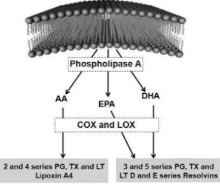
The utilization of membrane phospholipid components to produce mediators occurs during cellular stress, whether physical, biochemical or functional. Inlammation causes constant cellular stress in both immune cells and host tissue cells with reductions in the production of adenosine triphosphate (ATP) and a resulting increase in the calcium channel permeability leading to a great inlux of this ion into the cell. The inlow of calcium ions into cells activates the cytosolic enzyme phospholipase A2 or C metabolic pathways, which, in turn, act on the phospholipids of cell membranes, making molecules of these fatty acids available to the cytosol. In inlammatory cells, these free fatty acid molecules in the cytosol can be submitted to the action of the cyclooxygenase (COX) or lipoxygenase enzymes (LOX) which in most cells will lead to the formation of prostaglandins (PGs). Thromboxanes (TXs) and prostacyclins are synthesized when this metabolism occurs inside platelets and endothelial cells, respectively(8).
The eicosanoids [including PGs, TXs and leukotrienes (LTs)] derived from AA (an ω-6 fatty acid family component) are from the even series (e.g. 2 and 4 series) and mostly have a pro-inlammatory effect. The intake of ω-3 polyunsaturated fatty acid (PUFA) increases the incorporation of ω-3 fatty acids in the cell membrane and increases the production of PGs, TXs and LTs derived from the fatty acids (the odd series e.g. 3 and 5 series)(6). In one study,
healthy volunteers who took a supplement containing ish oil had increased incorporation of eicosapentaenoic fatty acid (EPA) in the phospholipids of white blood cells after one week and, at the same time, decreased the concentrations of ω-6 fatty acids (e.g. AA)(9).
The LTs have important biological functions including a monocyte chemoattractant action, polymorphonuclear leukocyte aggregation and degranulation, leukocyte stimulation and adhesion to the endothelial wall during the formation of inlammatory iniltrations. They are synthesized mainly by leukocytes and all cells have COX, but only inlammatory cells have LOX(8).
AA, used as the main substrate to the synthesis of eicosanoids, is one of the most abundant fatty acids in inlammatory cells. The ω-3 fatty acids, EPA and docosahexaenoic (DHA), when incorporated into the plasma membrane, reduce the metabolism of AA and thus reduce the eicosanoids derived from it, because they have higher afinity as substrates for COX-2 and LOX-5. Thus, an alternative family of eicosanoids starts to be formed which are often less inlammatory than those produced from AA. D and
E series resolvins are also produced from ω-3 fatty acids; these are lipid mediators that have a powerful anti-inlammatory action (Figure 1). These fatty acids can inluence the production of peptide mediators (cytokines) and the expression of transcription factors (such as nuclear factor kappa B - NF-kB) which can determine the production pattern of cytokines and eicosanoids(6,7). Faber et al.(9) reported that healthy individuals who take oral supplements
containing 2.4 g EPA and 1.2 g of DHA for one week, have an increased ex vivo production of IL-1β, tumor necrosis factor alpha
(TNF-α), IL-6, IL-8 and interferon-gamma (IFN-g).
Figure 1 - Synthesis of lipid mediators from arachidonic acid (AA), eicosapentaenoic (EPA) and docosahexaenoic (DHA) omega-3 fatty acids
(Adapted from Calder (6))
The COX-1 and COX-2 enzymes are important in the regulation of the immune response and have a key role in the inhibition of apoptosis, angiogenesis and cell proliferation, as well as in cell mobilization. COX-2 (an induced isoform of the enzyme) is expressed constitutively by all malignant and pre-malignant cells and has been correlated with local chronic inlammation and tumor neovascularization(10).
A reduction in the expression of COX-2 and, consequently, a decrease in the plasma concentrations of prostaglandin E2 (PGE2) was observed in an experiment in which rats inoculated with tumor cells (Walker 256) were supplemented with ish oil (1 g/kg/day). This was associated with an increase in the number of apoptotic cells inside the tumor(11). COX-2 is particularly
associated with the production of (PGE2). This prostaglandin can bind to its receptor on T cells inducing the production of adenosine 3’,5’-cyclic monophosphate (cAMP), leading to the inhibition of the proliferation of T helper (Th)-1 lymphocytes and stimulation of the production of Th2 cells. Evidence shows that deiciency in Th1 response can be linked to predisposition to cancer(10).
Neoplasias: inlammatory process
The participation of TGF-β in the differentiation of Th17 puts this cell lineage in a close relation with Tregs and so TGF-β also induces the differentiation of naive T cells to Tregs in peripheral immune compartments(16). Moreover, a plasticity phenomenon
has been observed between the T cell phenotypes; for example the T CD8+ cells can express IL-17 (CD8+IL-17+ T) or Tregs can
take the phenotype of Th17 and vice versa, by reprogramming and changes in gene expression in T cells(1). According to Yang et
al.(3), hypoxia, a common feature in the tumor microenvironment,
induces IL-17 expression in FOXp3+ Tregs. CD4+FOXp3+IL17+
T cells are cells that are in an intermediate point of differentiation between Tregs and Th17(3).
The relation between neoplasms and the inlammatory process is complex and is frequently discussed from two perspectives, intrinsic and extrinsic. The intrinsic perspective states that genetic changes, established from cell genesis or triggered by carcinogenic agents, provoke neoplastic transformation. These events include the activation of several types of oncogenes by mutation, rearrangement or chromosomal ampliication and inactivation of tumor suppressor genes. The affected cells produce inlammatory mediators thus creating an inlammatory microenvironment in tumors for which there was no previous underlying inlammatory condition (e.g. breast tumors). On the other hand, from the extrinsic perspective, local and systemic inlammatory response (especially when chronic) and infections promote an environment that increases the risk to develop cancer in a determined anatomical locus (e.g. colon, prostate and pancreas)(17,18).
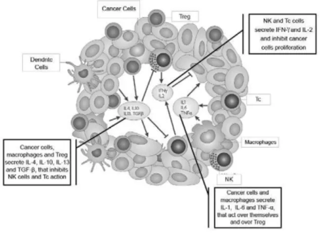
Additionally, there is coexistence between the malignant cells and the cells of the immune system. While NK cells and cytotoxic T cells produce IFN-γ and IL-2 in order to inhibit growth of cancer cells (inhibiting the production of IL-1, IL-6 and TNF-α), the malignant cells themselves secrete cytokines (IL-1, IL-6 and TNF-α) that promote their own proliferation. Furthermore, these cytokines stimulate macrophages and Tregs to secrete IL-4, IL-10, IL-13 and TGF-β that will inhibit the production of cytokines by NK and T cells, causing a suppressant effect in anticancer immunity(Figure 2)(19).
cells that acts as a growth factor for myeloma cells(12). Ovarian
clear cell adenocarcinomas present increases in the expression of IL-6, which may be related to the stimulation of the production of inlammatory cytokines, the promotion of tumor angiogenesis and to the migration of macrophages to the iniltrated tumor(13).
Cytokines are released by different cell types however the immune cells have an important role in the release of these agents in the neoplastic microenvironment. While the T cell population, such as IFN-γ+ T cells, can have antitumor activity,
another population, for example FoxP3+IL-10+ cells present
pro-tumor activity. Another mediator, IL-17, secreted by T helper lymphocytes, stimulates the inlammatory process and hence can promote both growth or regression of the tumor(1).
A study of patients with classic Hodgkin’s lymphoma (HL) showed that plasma concentrations of IL-2 were inversely correlated to levels of albumin (higher concentrations of IL-2 were correlated with albumin at less than 4 g/dL) and disease staging (higher concentrations of IL-2 in Stages I and II of the disease) This suggests that IL-2 decreases gradually with the progression of the disease. In these patients, the 4 and IL-10 cytokines (representatives of Th2-mediated response) were positively correlated with prognostic variables, such as lymphopenia (lymphocyte count less than or equal to 600 cells/mm3), hemoglobin
lower than 10.5 g/dL, higher advanced stages of the disease (Stages III and IV), and high-risk patients according to International Prognostic Index (IPI) classiication(14).
Malignant cells can use a variety of methods to evade immunological response, including overregulation of the expression of major histocompatibility complex (MHC) molecules and overregulation of mechanisms involving T cell death. The production of immunosuppressive cytokines (TGF-β and IL-10) by malignant cells can be responsible for most of the inhibition of cell-mediated immunity and has been related to several types of cancer, for example lymphomas(10). T cell anergy is common in malignant diseases, with these cells frequently presenting abnormalities in their signal transduction pathways. Among the alterations observed in T lymphocytes in malignant diseases is a reduced ability to produce IL-2 and IFN-g by Th1. In addition,
some types of malignant cells produce a potent tumor growth stimulant and immunosuppressant factor, known as TGF-β(10).
More recently, the crucial role that Th17 lymphocytes play in the development of inlammatory autoimmune diseases, bone marrow transplant rejection and pro-tumor function with the production of IL-1 family members was described. Th17 cells participate in the inlammatory process of numerous immunological reactions through the production of IL-17 and IL-22. The differentiation of cells from T0 (naïve T cells) to Th17 has been related to the presence of the TGF-β, IL-1 and IL-6 cytokines whereas stabilization of the differentiation is related to IL-23, mainly produced by dendritic cells and macrophages(1,15).
There is evidence that the number of Th17 cells is higher in the tumor microenvironment with these cells being found in several tumors, including in hematological neoplasias. Initially, Th17 cells seem to have antitumor activity, but with changes in the microenvironment, they promote neoplastic growth through the synthesis of inlammatory mediators (TGF-β, IL-6, IL-23, IL-21, PGE2, IL-1β, TNF-α), many of which are induced by IL-17(1).
Figure 2 - Role of the main cytokines in the tumor microenvironment
An accumulation of Th17 and Tregs was found in the tumor lymphocytic iniltration of patients with early stages of gastric cancer. However, with the progression of the disease there was a change in this balance characterized by a decrease in the Th17 cells and an increase in Tregs. This alteration may be a consequence of the production of cytokines, such as, IL-21, IL-23, IL1β and TGF-β, in the local microenvironment of gastric cancer. However, a direct correlation between the production of IL-21 and IL-23 and the prevalence of Th17 cells was not observed in these patients. A predominance of the Th2 cell-mediated inlammatory response was observed in this neoplasia, with no changes between the different disease stages. However, the balance between Th17 and Tregs was clearly modiied(4).
The proportion of Th17 cells is higher in non-treated patients with acute myeloid leukemia (AML) when compared with healthy individuals. However, in the same patients, this proportion decreased after treatment and complete remission of the disease. The production of IL-17, IL-6 and TGF-β1 cytokines is also greater in non-treated patients with AML compared to healthy individuals(15).
Higher percentages of CD3+CD4+ and CD3+CD8+ T
lymphocytes were observed in peripheral blood samples from patients with classical HL compared to healthy individuals, as well as an increase in the concentrations of TNF, IFN-g, IL-4, IL-5
and IL-10 while no difference was observed in the concentrations of IL-2. The increase of serum levels of IL-10, was correlated with a worse prognosis according to an association with clinical variables(14).
In a study of AML, patients with chemotherapy-induced cytopenia presented higher levels of Tregs and lower levels of T cytotoxic-1 lymphocytes (Tc1) and Th1 compared to healthy individuals. The levels of Th17 were similar in patients with untreated AML and those with induced cytopenia. The levels of CD4+CD25+FOXp3+ T cells were elevated in all patients with
AML. Additionally, a correlation between Th17 and Th1 was observed in both untreated and treated AML(20).
In malignant diseases, the Th2-mediated immune response seems to promote neoplastic development, while simultaneously inhibits Th1 response, the type of response with a predominant antineoplastic activity(6). Additionally, neoplastic cells produce
TGF-β which stimulates neoplastic growth and differentiation of Th17 and Tregs. These cells apparently promote neoplastic development and the suppression of immune antitumor activity(1,14,21).
Neoplasias: supplementation with polyunsaturated fatty acids (ω-3)
Fritschi et al.(22) investigated the ingestion of ish in
individuals who had developed leukemia, multiple myeloma and non-Hodgkin lymphoma (NHL). They observed an association between higher consumption of fresh ish and a protective effect against all neoplasias of the study. Moreover, a higher consumption of energy and fat is associated with increased risk for the development of these neoplasias. However, when the largest proportions of energy and fat come from fresh ish, the risk to develop these diseases is lower. In a similar study, Cvetkovié et al.(23) investigated the proile of fatty acids in serum phospholipids
of patients with NHL and observed higher concentrations of
palmitic acid (16:0), oleic acid (18:1 n-9), dihomo-γ-linolenic acid (DGLA, 20:3 n-6), AA (20:4 n-6) and docosatetraenoic acid (22:4 n-6) and lower serum concentrations of EPA (20:5 n-3), DHA (22:6 n-3) and docosapentaenoic acid (DPA, 22:5 n-3) when compared to the control group.
Some studies have reported the clinical effects of the use of omega-3 in neoplasias. In one study with several types of cancer patients, there was no decline in the number or function of polymorphonuclear cells when the volunteers were supplemented with 2 g of ish oil for eight weeks during chemotherapy(24).
Murphy et al.(25) and Van Der Give Meij et al.(26) offered ish oil as a
dietary supplement in 16 and 20 lung cancer patients, respectively during chemotherapy and observed that their body weight and fat free mass were maintained. Similar results were observed in 10 colorectal cancer patients under chemotherapy supplemented with 2 g of ish oil daily over nine weeks(27), as well as in 19 patients after tumor resection from multiple locations, receiving the same dosage as the former study during eight weeks of chemotherapy(24).
Recently, the National Consensus on Oncology Nutrition(28)
reported that immunomodulatory diets containing ω-3 fatty acids showed beneits in cancer patients including reduction in the incidence of post-operative infectious complications, reductions in the intensity of the inlammatory response and severity of infectious complications, and a reduction in hospital length of stay with a consequent decrease in the cost of treatment.
Thus, ω-3 fatty acids could help in the treatment of neoplasms by acting on the production of lipid mediators with attenuated inlammatory activity or acting at the resolution phase of the inlammatory process(4), on the expression of
COX-2 enzyme with consequent inhibition of PGECOX-2(11), and in the
production of cytokines as well as transcription factors(19,20).
Hematological Neoplasias: supplementation with polyunsaturated fatty acids (ω-3)
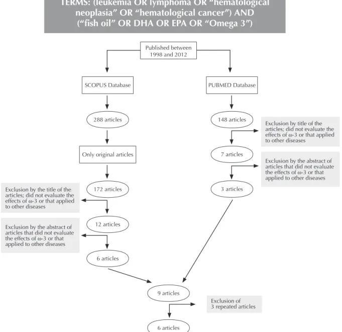
Nine articles on ish oil and hematological neoplasias with in vivo
and in vitro methodological designs were found during the literature
review however three were repeated in both databases. Studies regarding this speciic subject in humans were not found (Figure 3).
This systematic review of the literature shows that there is a lack of studies that evaluate the immunomodulatory effects of ω-3 fatty acids on hematological neoplasias. Among the six studies identiied, four were in vitro experiments and two used animal models (in vivo). The in vitro studies found that neoplastic cells incubated with certain
ω-3 fatty acids, in isolation or in combinations, showed higher apoptosis induction rates. In the study of Gillis et al.(29), leukemia
cells (HL60) were incubated with EPA, GLA (g-linolenic fatty acid
In an animal model, Ogilvie et al.(33) provided supplements of
ω-3 fatty acids to dogs submitted to chemotherapy for lymphoblastic lymphoma and observed that the association of ω-3 fatty acids increased overall survival and disease free survival. In another study, Johansson et al.(34) gave diets enriched with ish oil (source of ω-3)
or corn (source of ω-6) to mice with lymphoma over 12 months and found that ish oil was signiicantly more effective than corn oil to delay the progression of lymphoma during the irst 8 months.
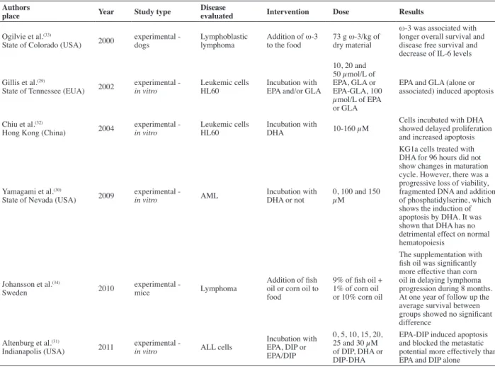
The experimental studies on ish oil and hematological neoplasias published in the Scopus and PubMed databases between 1998 and August 2012 are listed in Table 1. The authors of this paper are developing research on humans that evaluates inlammatory markers of individuals with hematological neoplasias (acute Leukemia and non-Hodgkin Lymphomas) supplemented with ish oil.
TERMS: (leukemia OR lymphoma OR “hematological
neoplasia” OR “hematological cancer”) AND
(“ish oil” OR DHA OR EPA OR “Omega 3”)
Figure 3 - Flowchart of the inclusion of articles from the Scopus and PubMed databases
Published between
1998 and 2012
SCOPUS Database PUBMED Database
288 articles 148 articles
Only original articles
172 articles
12 articles
6 articles
7 articles
3 articles
9 articles
6 articles Exclusion by the title of the
articles; did not evaluate the
effects of ω-3 or that applied
to other diseases
Exclusion by the abstract of
articles that did not evaluate
the effects of ω-3 or that
applied to other diseases
Exclusion by title of the
articles; did not evaluate the
effects of ω-3 or that applied
to other diseases
Exclusion by the abstract of
articles that did not evaluate
the effects of ω-3 or that
applied to other diseases
apoptosis by DHA. In addition, the DHA had no detrimental effect on normal hematopoiesis.
In the in vitro study of Altenburg et al.(31), acute
lymphocytic leukemia (ALL) cells were incubated with EPA, 2,6-diisopropylphenol-docosahexaenoate (DIP) or EPA-DIP. After 48 hours the EPA-DIP association induced apoptosis by activating the caspases cascade with consequent degradation of the poly (ADP ribose) polymerase (DNA repairing enzyme). Additionally, the EPA-DIP conjugation decreased the metastatic potential more eficiently than EPA and DIP alone. In a similar study, Chiu et al.(32) incubated
leukemic cells (HL-60) with DHA synthesized by a microalgae (Crypthecodinium cohnii) and observed a delay in cell proliferation,
as well as induction of phosphorylation and inactivation of Rb protein thereby promoting apoptosis of leukemic cells.
Conclusions
The current literature regarding ω-3 fatty acids presents a solid body of putative positive effects regarding the application of this nutritional strategy in patients with hematological neoplasias. The data suggest that, in these diseases, ω-3 fatty acids can potentially have indirect effects by promoting immunomodulation or direct effects on neoplastic cells. Nevertheless, with this review we conclude the importance of performing further clinical research to assess the possible immunomodulatory effects of ω-3 fatty acid supplementation in individuals with the diagnosis of hematological neoplasias.
References
1. Murugaiyan G, Saha B. Protumor vs. Antitumor Functions of IL-17. J Immunol 2009 183:4169-75.
2. Issazadeh-Navikas S, Teimer R, Bockermann R. Inluence of dietary components on regulatory T cells. Mol Med. 2012;18:95-110.
3. Yang S, Wang B, Guan C, Wu B, Cai C, Wang M, et al. Foxp3+IL-17+ T
cells promote development of cancer-initiating cells in colorectal cancer. J Leukoc Biol. 2011;89(1):85-91.
4. Maruyama T, Kono K, Mizukami Y, Kawaguchi Y, Mimura K, Watanabe M, et al. Distribution of Th17 cells and FoxP3(+) regulatory T cells in tumor-iniltrating lymphocytes, tumor-draining lymph nodes and peripheral blood lymphocytes in patients with gastric cancer. Cancer Sci. 2010;101(9):1947-54. 5. Allen CT, Judd NP, Bui JD, Uppaluri R. The Clinical implications
of antitumor immunity in head and neck cancer. Laryngoscope. 2012;122(1):144-57.
6. Calder PC. Polyunsaturated fatty acids and inlammatory processes: new twists in an old tale. Biochimie 2009;91(6):791-5.
7. Perini JA, Stevanato FB, Sargi SC, Visentainer JE, Dalalio MM, Matshushita M, et al. Ácidos graxos poli-insaturados n-3 e n-6: metabolismo em mamíferos e resposta imune. Rev Nutr. 2010;23(6):1075-86.
8. Consolaro A. Inlamação e reparo: um sílabo para a compreensão clínica e implicações terapêuticas. Maringá: Dental Press; 2009.
9. Faber J, Berkhout M, Vos AP, Sijben JW, Calder PC, Garssen J, et al. Supplementation with a ish oil-enriched high-protein medical food leads to rapid incorporation of EPA into white blood cells and modulates immune responses within one week in healthy man and women. J Nutr. 2011;141(5):964-70.
10. Dalgleish AG,O’byrne K. Inlammation and cancer: the role of the immune response and angiogenesis. In: Dalgleish AG, Haefner BS. The link between inlammation and cancer: wounds that do not heal. Boston, MA: Springer Science; 2006. p.1-48.
Table 1 - Studies published in the Scopus and PubMed databases between 1998 and August 2012 on ish oil (omega-3) and hematological neoplasias
Authors
place Year Study type
Disease
evaluated Intervention Dose Results
Ogilvie et al.(33)
State of Colorado (USA) 2000 experimental - dogs Lymphoblastic lymphoma Addition of ω-3 to the food 73 g ω-3/kg of dry material
ω-3 was associated with longer overall survival and disease free survival and decrease of IL-6 levels
Gillis et al.(29)
State of Tennessee (EUA) 2002 experimental - in vitro Leukemic cells HL60 Incubation with EPA and/or GLA
10, 20 and 50 µmol/L of EPA, GLA or EPA-GLA, 100 µmol/L of EPA or GLA
EPA and GLA (alone or associated) induced apoptosis
Chiu et al.(32)
Hong Kong (China) 2004 experimental - in vitro Leukemic cells HL60 Incubation with DHA 10-160 µM
Cells incubated with DHA showed delayed proliferation and increased apoptosis
Yamagami et al.(30)
State of Nevada (USA) 2009 experimental - in vitro AML Incubation with DHA or not 0, 100 and 150 µM
KG1a cells treated with DHA for 96 hours did not show changes in maturation cycle. However, there was a progressive loss of viability, fragmented DNA and addition of phosphatidylserine, which shows the induction of apoptosis by DHA. It was shown that DHA has no detrimental effect on normal hematopoiesis
Johansson et al.(34)
Sweden 2010 experimental - mice Lymphoma
Addition of ish oil or corn oil to food
9% of ish oil + 1% of corn oil or 10% corn oil
The supplementation with ish oil was signiicantly more effective than corn oil in delaying lymphoma progression during 8 months. At one year of follow up the average survival between groups showed no signiicant difference
Altenburg et al.(31)
Indianapolis (USA) 2011 experimental - in vitro ALL cells
Incubation with EPA, DIP or EPA/DIP
0, 5, 10, 15, 20, 25 and 30 µM of DIP, DHA or DIP-DHA
EPA-DIP induced apoptosis and blocked the metastatic potential more effectively than EPA and DIP alone
11. Mund RC, Pizato N, Bonatto S, Nunes EA, Vicenzi T, Tanhoffer R, et al. Decreased tumor growth in Walker 256 tumor-bearing rats chronically supplemented with ish oil involves COX-2 and PGE2 reduction associated with apoptosis and increased peroxidation. Prostaglandins Leukot Essent Fatty Acids. 2007;76(2):113-20.
12. Balkwill FR, Mantovani A. Cancer-related inlammation: common themes and therapeutic opportunities. Semin Cancer Biol. 2012;22(1):33-40. 13. Anglesio MS, George J, Kulbe H, Friedlander M, Rischin D, Lemech C,
Power J, Coeard J, Cowin PA, House CM, Chakravaert P, Gorringe KL, Campbell IG; Australian Ovarian Cancer Study Group, et al. IL6-STAT3-HIF signaling and therapeutic response to the angiogenesis inhibitor sunitinib in ovarian clear cell cancer. Clin Cancer Res 2011;17(8):2538-48. 14. Mitelman AK, Buccheri V, Pracchia LF, Rubens CV, Poppe S, Cavalcante
AM, et al. Quantiicação das citocinas séricas Th1/Th2 por citometria de luxo no linfoma de Hodgkin clássico. Rev Bras Hematol Hemoter. 2009;31(4):260-6.
15. Wu C, Wang S, Chen Q, Peng S, Zhang Y, Qian J, et al. Increased frequencies of T helper type 17 cells in the peripheral blood of patients with acute myeloid leukaemia. Clin Exp Immunol. 2009;158(2):199-204. 16. Korn T, Bettelli E, Oukka M, Kuchroo VK. IL-17 and Th17 Cells. Annu
Rev Immunol. 2009;27:485-517.
17. Colotta F, Allavena P, Sica A, Garlanda C, Mantovani A. Cancer-related inlammation, the seventh hallmark of cancer: links to genetic instability. Carcinogenesis. 2009;30(7):1073-81.
18. Mantovane A, Allavena P, Sica A, Balkwill F. Cancer-related Inlammation. Nature. 2008; 454(7203):436-44.
19. Seruga B, Zhang H, Bernstein LJ, Tannock IF. Cytokines and their relationship to the symptoms and outcome of cancer. Nature 2008; 8(11):887-99. Comment in: Nat Rev Cancer. 2009;9(3):224.
20. Ersvaer E, Liseth K, Skavland J, Gjertsen BT, Bruserud O. Intensive
chemotherapy for acute myeloid leukemia differentially affects
circulating Tc1, Th1, Th17 and Tregs. BMC Imunnol. 2010;11:38. 21. Rubin E, Rubin R, Aaronson S. Neoplasia. In: Rubin E. Patologia:
bases clínicopatológicas da medicina. 4th ed. Rio de Janeiro: Guanabara Koogan; 2006. p.169-95.
22. Fritschi L, Ambrosini GL, Kliewer EV, Johnson KC, Canadian Cancer Registries Epidemiologic Research Group. Dietary ish intake and risk of leukaemia, multiple myeloma, and non-Hodgkin lymphoma. Cancer Epidemiol Biomarkers Prev 2004;13(4):532-37.
23. Cvetkovié K, Vucic V, Cvetkovic B, Petrovic M, Ristic-Medic D, Tepsic J, et al. Abnormal fatty acid distribution of the serum phospholipids of patients with non-Hodgkin lymphoma. Ann Hematol. 2010;89(8):775-82.
24. Bonatto SJR, Oliveira HHP, Nunes EA, Pequito D, Iagher F, Coelho I, et al. Fish oil supplementation improves neutrophil function during cancer chemotherapy. Lipids. 2012;47(4):383-9.
25 .Murphy RA, Mourtzakis M, Chu QS, Baracos VE, Reiman T, Mazurak VC. Nutritional intervention with ish oil provides a beneic over standard of car for weight and skeletal muscle mass in patients with non-small cell lung cancer receiving chemotherapy. Cancer. 2011;117(8):1775-82. 26. van der Meij BS, Langius JA, Smit EF, Spreeuwenberg MD, Blomberg
BM, Heijboer AC, et al. Oral nutrition supplements containing (n-3) polyunsaturated fatty acids affect the nutritional status of patients with stage III non-small cell lung cancer during multimodality treatment. J Nutr. 2010;140(10):1774-80.
27. Silva JA, Trindade EB, Fabre ME, Menegotto VM, Gevaerd S, Buss ZS, et al. Fish oil supplementation alters markers of inlammation and nutritional status in colorectal cancer patients. Nutr Cancer. 2012;64(2):267-73.
28. Brasil. Ministério da Saúde. Instituto Nacional do Câncer. Consenso Nacional de Nutrição Oncológica. Rio de Janeiro: INCA; 2011. v.2. Available from: http://bvsms.saude.gov.br/bvs/publicacoes/inca/ Consenso_Nutricao_vol_2.pdf
29. Gillis RC, Daley BJ, Enderson BL, Karlstad MD. Eicosapentaenoic acid and γ-linoleic acid induce apoptosis in HL-60 cells. J Surg Res. 2002;107(1):145-53.
30. Yamagami T, Porada CD, Pardini RS, Zanjani ED, Almeida-Porada G. Docosahexaenoic acid induces dose dependent cell death in an early undifferentiated subtype of acute myeloid leukemia cell line. Cancer Biol Ther. 2009;8(4):331-7. Comment in: Cancer Biol Ther. 2009;8(4):338-9. 31. Altenburg JD, Harvey KA, McCray S, Xu Z, Siddiqui RA. A novel
2,6-diisopropylphenyl-docosahexaenoate conjugate induces apoptosis in T cell acute lymphoblastic leukemia cell lines. Biochem Biophys Res Commun. 2011;411(2):427-32.
32. Chiu LC, Wong EY, Ooi VE. Docosahexaenoic acid modulates different genes in cell cycle and apoptosis to control growth of human leukemia HL-60 cells. Int J Oncol. 2004;25(3):737-44.
33. Ogilvie GK, Fettman MJ, Mallinckrodt CH, Walton JA, Hansen RA, Devenport DJ, et al. Effect of ish oil, arginine, and doxorubicin chemotherapy on remission and survival time for dogs with lymphoma: a double-blind, randomized placebo- controlled study. Cancer. 2000;88(8):1916-28.
34. Johansson AS, Norén-Nystrom U, Larefalk A, Holmberg D, Lindskog M. Fish oil delays lymphoma progression in the TLL mouse. Leuk Lymphoma. 2010; 51(11):2092-7.