Depth-scanning confocal Raman for rapid in vivo
determination of testosterone concentration profiles
in human skin
Marco Antonio Botelho,I,IIStela Julia Guerreiro,I,IVDinalva Brito Queiroz,I,IVGisele Barros,I,IV
Monaliza Cavalcante,IJuliana M. O. Souza,IIIAndre´ M. Silva,IIITelma L. G. Lemos,III Lucindo Quintans JrV
IPost-graduation Program in Biotechnology, Laboratory of Nanotechnology, University Potiguar, Natal, RN 59010-060, BrazilIILaboratory of
Biotechnology, Federal Institute of Science and technology of Ceara, Fortaleza, CE BrazilIIIDepartment of Organic and Inorganic Chemistry, Federal
University of Ceara, 60470 Fortaleza, BrazilIVNanotechnology Department, Applied Biotechnology Institute INBIOS, 60125-100 Fortaleza, Brazil VDepartment of Physiology, Federal University of Sergipe, Sa˜o Cristo´va˜o, Brazil
OBJECTIVE: The present study aimed to measurein-vivothe depth and the percentage of testosterone in human skin.
METHOD: Two healthy young Brazilian volunteers were evaluated through a Confocal Raman Spectroscopy probe on the right volar forearm. Testosterone spectroscopy of was performed on the Stratum Corneum, Viable Epidermis and Dermis; the percentage concentration of testosterone was compared between the baseline and one hour after local application of a transdermal nanostructured testosterone (5%) formulation.
RESULTS: To the best of our knowledge, this is the first time that the depth and percentage of testosterone has been evaluated non-invasively in-vivo. No adverse events were attributed to this protocol. The method is effective in differentially measuring testosterone in the skin layers.
CONCLUSION: This protocol may serve as a good choice for rapid hormone quantification for forensic or medical purposes.
KEYWORDS: Nanotechnology; Confocal Raman Spectroscopy; Transdermal Delivery; Testosterone;
Nanoparticles.
Botelho MA, Guerreiro SJ, Queiroz DB, Barros G, Cavalcante M, Souza JMO, Silva AM, Lemos TLG, Quintans Jr L. Depth-scanning confocal Raman for rapid in vivo determination of testosterone concentration profiles in human skin. MEDICALEXPRESS. 2014;1(1):31-35.
Received for publication onJanuary 19 2014;First review completed onJanuary 21 2014;Accepted for publication onJanuary 23 2014 E-mail: [email protected]
’ INTRODUCTION
Over the years, there is an increasing interest and a permanent debate about which is the best and most accurate method for anabolic steroids quantification.1,2
Different strategies have shown that doping methods are becoming more sophisticated and difficult to detect by the conventional analytical methods used in sports medicine. Recently, special emphasis has been given to androgenic steroids and its derivatives to detect strategies on misuses of sports-forbidden substances.2
Recently many protocols have been evaluated by different laboratories to detect different compounds in saliva or blood samples.3Determining hormone amounts is not an easy task with the currently used methods and because of difficulties in sample preparation.4
Confocal Raman Spectroscopy is a safe and efficient technique that permits an accurate depth-scanning of the skin; thus it can be used to detect different compounds through the skin layers.5-8It is a non-invasive method that
enables precise and accurate technological detection for individual measurements.9,10
The present study aimed to evaluate an alternative approach in order to detect minimal amounts of androgen hormones; this may enhance doping detection and thus contribute to prevent this negative strategy frequently used by athletes around the world.
’ MATERIALS AND METHODS
Ethics
This protocol was approved by the Ethical Committee of Universidade Paulista UNIP, Sao Paulo/Brazil (UNIP #437/09), according to the Helsinki Declaration guidelines. A written informed consent was obtained from the individuals willing to participate.
Study Protocol
Experiments were performed twice on the volar forearms of 2 male healthy volunteers aged 29 and 34 years. In the present study, the subjects received a single transdermal dose in the right forearm of a nanoemulsion Biolipid/B2TM DOI:
31 Copyrightß2014 MEDICALEXPRESS. This is an open access article distributed under the terms of the creative commons attribution
testosterone (Evidence Pharmaceuticals, Sao Paulo/SP, Brazil).
An evaluation was performed before the local application of the transdermal nanoemulsion in order to determine the baseline concentration of testosterone in the stratum corneum (SC), the Viable Epidermis (VE) and the Dermis (D) during the trial. A second evaluation was performed 1 hour after emulsion administration.
Nanostructured Emulsion Preparations
The nanoemulsion was developed at the Department of Nanotechnology at Institute of Applied Biotechnology (inBIOS) in association with the Laboratory of Advanced Materials at Federal University of Ceara´ and the Laboratory of Biotechnology at Potiguar University. Testosterone was purchased from Sigma Aldrich. The hormone + BIOLIPID/ B2TM formulation was prepared and the following mass
ratio was obtained: Testosterone (5%) + Biolipid/B2TM. The main composition of this emulsion is based on the nanoparticulated testosterone hormone and a transdermal penetration enhancer vehicle BIOLIPID B2TM (Evidence Pharmaceuticals, Sao Paulo/SP, Brazil). The testosterone nanoparticles were prepared using a water-oil emulsion method.5
HPLC analysis of testosterone
The analyses were carried out on a HPLC (Shimadzu), model LC/20AT, equipped with a detector UV-Vis SPD-M20A, oven 301C and a 20 mL loop injector (Shimadzu). Experiments were conducted using a CLC-ODS, C-18 column (5mm particle size, 150 mm x 4.6 mm I. D.) with a flow rate of 1 mL/min for 100 ppm of testosterone.
The mobile phase used was a mixture of CH3CN/H2O in
different concentrations, with isocratic elution and UV detection (SPD-20A/UV-VIS). The acetonitrile used was of HPLC grade from Tedia. The water was distilled using a Milli-Q system (Millipore) and mobile phase was filtered through a 0.49mm nylon filter. The retention times, mobile phase, and UV-Vis for the testosterone was (65:35, v/v) Rt-5.4 min, and UV-Vis 244 nm.
Clinical procedures
The clinical consultation consisted on a brief lecture about risks and benefits of transdermal testosterone application. During the transdermal procedure both volunteers were asked to respond questions about any adverse effects in order to monitor any adverse event.
Raman spectrometer assay
Raman Confocal spectroscopy (CRS) measurements were performed according to Botelho et al5 through Confocal Raman Spectroscopy Analysis (CRSA) (River Diagnostics, Skin Composition Analyzer/3510, Rotterdam, The Netherlands).
Statistical Analysis
All statistical analyses were performed using GraphPad Prism version 3.0 (GraphPad Software, Inc. San Diego, CA, USA). The data are presented as means at baseline and 1 hour after transdermal testosterone formulation. Statistical differences between baseline and after treatment were evaluated by F test to compare variances. Values of less than or equal to 0.05 were considered statistically significant.
’ RESULTS
Confocal Raman Spectroscopy testosterone depth and concentration on skin layers
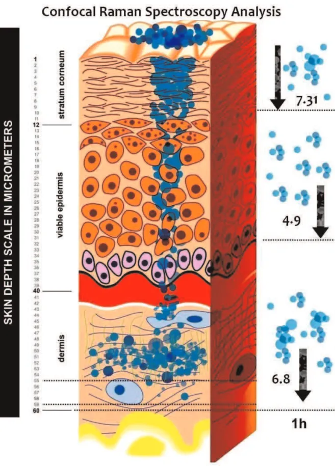
Confocal Raman Spectroscopy showed relevant and significant data analysis about the depth and concentration of testosterone particles in skin layers one hour after transdermal application (Figure 1). Depth measurements included Stratum Corneum (SC), Epidermis (E) and Dermis (D) assessed before and 1h after testosterone nanoemulsion application.
Figure 1 shows a diagram describing the flow of testosterone particles through the three layers over the first hour after testosterone transdermal application.
Both volunteers were submitted to Confocal Raman Spectroscopy for testosterone at baseline and 1 hour after hormone administration. Figure 2 shows the mean of testosterone percentage in the Stratum Corneum of both volunteers. Before the transdermal testosterone application, hormone was undetectable.
Figure 3 shows testosterone levels in the Viable epider-mis. In both volunteers, no testosterone was detected before administration and the levels 1 hour later were statistically significant.
Figure 4 shows testosterone nanoparticle concentrations in the Dermis. We show that the Depth-Scanning Confocal Raman was able to detect nanoparticles of testosterone after the one hour after transdermal application.
Adverse events
No adverse effects were reported by the volunteers at any time during the study.
’ DISCUSSION
In this study, the Confocal Raman Spectroscopy (CRS) analysis was shown to be capable of detecting minimal amounts of testosterone in the three skin layers one hour after transdermal application.
Testosterone can be administered by different routes, including oral or intramuscular injection, generally leading to many side effects.5 A number of studies have been conducted in order to detect small amounts of hormones and other compounds for different purposes.4,7However,
only recently have short-term studies provided strong evidence about the possibility of detecting nanoparticles of steroid hormonesin vivo.4,6
The use of steroids in sport configures a worldwide phenomenon with side effects and adverse events for users.2,4The most recent studies on doping detection have
been trying to evaluate differences in the regular physiologic pattern in order to detect minimal variations thereof. So far, there are few clinical studies focused on the detection of anabolic hormones for doping or forensic purposes.
In this study a transdermal hormone formulation was locally applied to the volar forearm of volunteers. The Raman equipment was able to evaluate the depth profiling of testosterone through the Stratum Corneum into the Viable Epidermis and Dermis.
To the best of our knowledge, this is the first time that Confocal Raman Spectroscopy was used as a method to describe testosterone profiling in human skin in vivo. This
and for surveillance at sports competitions. Through this technique, it was possible to measurein vivothe depth and
percentage of testosterone after 1 hour of follow-up. Previous studies have reported on different methods to detect small amounts of different substances.4,6,9When CRS is used, a consistent and accurate analysis is provided, probably due to the unique fingerprint-like spectra that can be obtained for different compounds in living organisms.
This study indicates the efficacy of this methodology, since it is a rapid information system that ensures testosterone detection in the Dermis through 1 up to 59mm (Figure 1).
The protocol used in this study can be indicated as an effective strategy to detected small amounts of other steroids,
especially those used to enhance sports performance. The findings demonstrate that after 1 hour of follow-up, the enhancer used to deliver testosterone was able to detect nanoparticles of testosterone reaching significant differences when compared to the baseline (Figure 2). In the Rio de Janeiro 2016 Olympics, this technique could be used to enhance the conventional methods by the Olympic Games accreditation laboratories.3
Within the limitations of this trial, we have shown that Confocal Raman Spectroscopy is effective in detecting nanoparticles of testosterone with statistically significant differences. These findings support and justify future studies with larger samples in order to validate this strategy as an alternative method for doping and forensic purposes.
’ CONCLUSIONS
In this study we have shown that Confocal Raman Spectroscopy can be used for monitoring in vivo testosterone through skin layers. To the best of our knowledge, this is the first study to show the depth profile of testosterone by CRS. Confocal Raman Spectroscopy was able to detect small amounts of testosterone in the skin sites, thus providing an alternative strategy to evaluate other compounds in the skin. The technique can be used as an efficient strategy for detecting drugs in sports competitions and in other forensic or clinical scenarios.
’ ACKNOWLEDGEMENTS
We gratefully acknowledge the financial support of The Federal Institute of
Science and Technology of Ceara´ (EDITAL No%13/2013-PRPI/PRO´ -INFRA/
IFCE#136) Pro-Rector Research Auzuir Ripardo and Jose´ Wally Menezes. We would like to thank Jorge Bachur for graphical Design and the Conselho Nacional de Desenvolvimento Cientı´fico e Tecnolo´gico - CNPq Brazilian Agency for Scientific and Technological Development (Post-Doctoral Scholar-ship Proc #202316/2011-4 and Research ScholarScholar-ship #310483/2012-3).
We also thank Rector Virgı´lio Augusto Sales Araripe (IFCE).
’ AUTHOR CONTRIBUTIONS
Botelho MA performed the statistical analysis. Queiroz DC was responsible
Figure 2 - Confocal Raman Spectroscopy Analysis shows show
individual results of the percentage of testosterone nanoparti-cles in the Stratum Corneum. BIOLIPID/B2TM-Testosterone (5%)
formulation was administered topically to two young men on the right forearm. *Po0.05 was considered a significantly
different value compared to baseline(F test).
Figure 4 -Confocal Raman Spectroscopy Analysis shows
indivi-dual results of the percentage of testosterone nanoparticles in the Dermis. BIOLIPID/B2TM-Testosterone (5%) formulation was
administered topically to two young men on the right forearm. *Po0.05 was considered a significantly different value compared
to baseline(F test).
Figure 3 - Confocal Raman Spectroscopy Analysis shows show
individual results of the percentage of testosterone nanoparti-cles in the Viable Epidermis. BIOLIPID/B2TM-Testosterone (5%)
the project and wrote the manuscript. Guerreiro SJ, Barros G and Cavalcante M were responsible for the bibliographic review. Souza J.M.O, Silva AM and Lemos TLG were responsible for acquiring the HPLC analysis.
’ RESUMO
OBJETIVO: O presente estudo teve como objetivo avaliar in-vivo a
profundidade e a porcentagem de testosterona na pele humana.
ME´ TODO:Dois jovens volunta´rios brasileiros sauda´veis foram avaliadas atrave´s de uma sonda confocal de Espectroscopia Raman na face volar do
antebrac¸o direito. A espectroscopia de testosterona foi realizada no estrato
co´rneo, na epiderme via´vel e na derme; a concentrac¸a˜o percentual de testosterona foi comparada entre a linha de base e uma hora apo´s a aplicac¸a˜o
transde´rmica de uma formulac¸a˜o de testosterona nanoestruturada (5%).
RESULTADOS: Tanto quanto sabemos, esta e´ a primeira vez que a profundidade e a porcentagem de testosterona foi avaliada de forma na˜o
invasivain-vivo. Nenhum evento adverso foi associado a esse protocolo. O
me´todo e´ eficaz na medic¸a˜o diferencial da testosterona nas camadas da pele.
CONCLUSA˜ O:Este protocolo pode servir como uma boa alternativa para a ra´pida quantificac¸a˜o hormonal com finalidade forense ou me´dica.
’ REFERENCES
1. Badoud F, Boccard J, Schweizer C, Pralong F, Saugy M, Baume N. Profiling of steroid metabolites after transdermal and oral administration of testosterone by ultra-high pressure liquid chromatography coupled to quadrupole time-of-flight mass spectrometry. J Steroid Biochem Mol Biol. 2013;138(11):222-35.
2. Cirimele V, Kintz P, Ludes B. Testing of the anabolic stanozolol in human hair by gas chromatography-negative ion chemical ionization mass spectrometry. J Chromatogr B Biomed Sci Appl. 2000;740(2):265-71. 3. Botre` F, Wu M, Boghosian T. Preparation and accreditation of anti-doping
laboratories for the Olympic Games. Bioanalysis. 2012;4(13):1623-31. 4. Gonzaga LW, Botelho MA, Queiroz DB, Fechine P, Freire R, Azevedo E,
et al. Nanotechnology in Hormone Replacement Therapy: Safe and Efficacy of Transdermal Estriol and Estradiol Nanoparticles after 5 Years Follow-Up Study. Lat Am J Pharm. 2012;31(3):442-50.
5. Botelho MA, Queiroz DB, Freitas A, Guerreiro S, Umbelino S, Barros G, et al. Effects of a new testosterone transdermal delivery system, Biolipid B2-testosterone in healthy middle aged men: a Confocal Raman Spec-troscopy Study. J Pharm Sci Innov. 2013;2(2):1-7.
6. Botelho MA, Queiroz DB, Barros G, Guerreiro S, Fechine P, Umbelino S, et al. Nanostructured transdermal hormone replacement therapy for relieving menopausal symptoms: a confocal Raman spectroscopy study. Clinics. 2014;69(2):1-8.
7. Me´lot M, Pudney PD, Williamson AM, Caspers PJ, Van Der Pol A, Puppels GJ. Studying the effectiveness of penetration enhancers to deliver retinol through the stratum cornum by in vivo confocal Raman spectroscopy. J Control Release. 2009;138(1):32-9.
8. Badoud F, Boccard J, Schweizer C, Pralong F, Saugy M, Baume N. Profiling of steroid metabolites after transdermal and oral administration of testosterone by ultra-high pressure liquid chromatography coupled to quadrupole time-of-flight mass spectrometry. J Steroid Biochem Mol Biol. 2013;138(6):222-35.
9. Caspers PJ, Lucassen GW, Bruining HA, Puppels GJ. Automated depth-scanning confocal Raman microspectrometer for rapid in vivo determi-nation of water concentration profiles in human skin. J Raman Spect. 2000;31(8):813-8.