463 463 463 463 463 Mem Inst Oswaldo Cruz, Rio de Janeiro, Vol. 101(5): 463-491, August 2006
Evolution and pathology in Chagas disease - A Review
Antonio RL Teixeira/
+, Rubens J Nascimento, Nancy R Sturm*
Laboratório de Pesquisa Multidisciplinar em Doença de Chagas, Faculdade de Medicina, Universidade de Brasília, Caixa Postal 04536, 70919-970 Brasília, DF, Brasil *Department of Immunology, Microbiology and Molecular Biology,
David Geffen School of Medicine, University of California at Los Angeles, LA, US
Trypanosoma cruzi acute infections often go unperceived, but one third of chronically infected individuals die of Chagas disease, showing diverse manifestations affecting the heart, intestines, and nervous systems. A common denominator of pathology in Chagas disease is the minimal rejection unit, whereby parasite-free target host cells are destroyed by immune system mononuclear effectors cells infiltrates. Another key feature stemming from T. cruzi
infection is the integration of kDNA minicircles into the vertebrate host genome; horizontal transfer of the parasite DNA can undergo vertical transmission to the progeny of mammals and birds. kDNA integration-induced mutations can enter multiple loci in diverse chromosomes, generating new genes, pseudo genes and knock-outs, and resulting in genomic shuffling and remodeling over time. As a result of the juxtaposition of kDNA insertions with host open reading frames, novel chimeric products may be generated. Germ line transmission of kDNA-mutations determined the appearance of lesions in birds that are indistinguishable from those seen in Chagas disease patients. The production of tissue lesions showing typical minimal rejection units in birds’ refractory to T. cruzi infection is consistent with the hypothesis that autoimmunity, likely triggered by integration-induced phenotypic alterations, plays a major role in the pathogenesis of Chagas disease.
Key words: Trypanosoma cruzi - kinetoplast DNA - horizontal transfer - genome growth - kDNA-mutation - pathology
Eons and interplay
The enzootic known as Chagas disease or American trypanosomiasis (Chagas 1909, 1911) is presented here as a hallmark of the interplay among extant organisms from different taxon of increasing complexity united by circum-stance. In the geological time scale of evolutionary his-tory, play initiated with the undulipodia acquiring a nucleus (Lake & Rivera 1994), resulting from a radical revo-lution and a major discontinuity between the prokaryotic and eukaryotic organisms during the proterozoic, 1500 million years ago (mya) (Margulis & Sagan 2002). In the absence of fossil records, the history of protozoa has been written mainly by morphological and life cycle data. How-ever, by the end of the twentieth century the availability of automated DNA sequencing enabled deduction of the evolutionary relationships of extinct species from the ge-nomes of their extant relatives (Lake et al. 1988, Stevens et al. 2001). DNA sequence analysis necessitated the engi-neering of a molecular clock that permitted evolutionary dating (Douzery et al. 2003, Delsuc et al. 2004). The clock advances such that mutations underlying evolution be-have as radioactive atoms, for similar reasons: the tauto-meric shifts of purine and pyrimidine nucleotides, although an unpredictable stochastic process can be prognosti-cated with reasonable accuracy at regular intervals (Klein & Takahata 2002). In practice, however, some species ac-cumulate mutations faster than others, and, like a grand-father clock, the molecular clock requires calibration based
Financial support: CNPq, Finep, FAP-DF +Corresponding author: [email protected]
Received 16 March 2006 Accepted 7 June 2006
on the empirical sequence data from extant species. In addition, the Earth’s development exerts an influence on biological evolution through subtle changes in climate and environment, or cataclysms such as tsunamis, neces-sitating adjustment of the clock to explain discontinuities beyond the Gondwanaland partition (Krause & Bonaparte 1993, Salgado-Laborieau 2001). However, chasms will re-main wherever horizontal DNA transfer (HDT) among dis-parate species occurs (Nitz et al. 20041, Simonson et al. 2005, Babushok 2006), complicating reconstruction of the universal tree of life. The expected shifts (e.g., mutations) are essentially normalized, and DNA accumulates substi-tutions at a stable pace. Since mutations appear at more-or-less regular intervals, time can be estimated propor-tionally (Klein & Takahata 2002, Podlipaev et al. 2004). The constant accumulation of mutations affecting amino-acid change in proteins during evolution is thus account-able (Douzery et al. 2004). Molecular clock data used to reconstruct evolutionary history can be calibrated with congruent morphological and lifecycle data of existing organisms (Table I).
The protoctist (Eukaryota, Excavata, Euglenozoa) an-cestors of the protozoa are dated to the pre-phanerozoic
1 The manuscript Nitz et al. appeared in a July 2004 issue of
464 464 464 464
464 Chagas pathology and genetics • Antonio RL Teixeira et al.
(Margulis et al. 2000). Protozoa belonging to the Class Zoomastigophorea include the most interesting Order: Kinetoplastida. Included in the Family Trypanosomatidae are parasites of major medical and veterinary importance:
Trypanosoma cruzi, which produces Chagas disease in the Americas, Trypanosoma brucei, the agent of sleeping sickness in Africa, and Leishmania species, responsible for leishmaniases worldwide. No clear division separates the Stercorarian trypanosomes and representatives have been found in lower vertebrates, such as the crocodile parasite Trypanosoma gray (Hoare 1972). Their closest relatives are the bodonids and cryptobiids that parasitize fish and amphibians (Donelson et al. 1999). Phylogenetic analyses based on small subunit ribosomal RNA (SSU rRNA) and heat-shock protein (Hsp90)first- and second-position codon nucleotides support placement of the root for the kinetoplastids next to the bodonids (Simpson et al. 2002), suggesting that trypanosomatids descended from the bodonids and that Boldo saltans is the closest extant relative. The interlocking network organization of kineto-plast DNA seen in trypanosomatids therefore is a derived condition from open-conformation minicircles found early in kinetoplastid evolution (Lukes et al. 2002).
The molecular analysis of SSU rRNA to determine the phylogenetic relationship between Trypanosoma chelodina from tortoise (Emidura signata, Elseya latis-ternum and Chelodina longicollis) and Trypanosoma binneyi from platypus (Ornithorhyncus anatinus) ex-cluded co-evolution of these trypanosomes with the ver-tebrate mammal host (Jakes et al. 2001). However, early evolutionary acquisition was achieved by exposing clean bullfrog tadpoles (Rana catesbiana) to leeches (Desserdobella picta) that had fed on frogs infected with
Trypanosoma pipientis (Siddall & Desser 1992). The pres-ence of trypanosomatids in the blood of aquatic inverte-brates (leech) and verteinverte-brates (fish) suggested that evo-lution of the former required secondary acquisition of host and habitat (Davies & Johnston 2000, Hamilton et al. 2005) during the phanerozoic, 570 mya.
The evolution of the Stercorarian T. cruzi likely re-quired stepwise adaptation to invertebrate and vertebrate hosts. A direct line of evidence showing a phylogenetic relationship between trypanosomatids of leeches, fish, and amphibians, and those of mammals cannot be drawn, but the close relationships between lizards and triatomines in an ecosystem located in Baja California, Mexico is sug-gestive. In this environment various triatomine bugs (Dipetalogaster maximus) and lizards (Sauromalis aus-tralis) dwell in burrows of exposed rocks in the absence of mammals. The complete T. cruzi lifecycle was observed in lizards that were infected by ingestion of the proto-zoan-infected D. maximus, and thereafter clean D. maxi-mus acquired T. cruzi upon feeding from that lizard (re-viewed in Teixeira 1987). The ultimate reservoirs of these trypanosomes may not be mammalian.
The origin of multicellular animals as assessed through topology of 18S rRNA indicates that the Annelida-Mol-lusca are the closest relatives of arthropods. There are a million species of arthropod in the Class Insecta: Hemi-ptera.Using amino acids, nucleotides, and the mitochon-drial molecular clock calibrated by Blattaria (cockroaches),
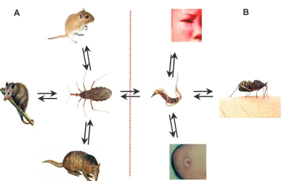
Orthoptera (crickets and locusts), Hemiptera (true bugs), Diptera, and Lepidoptera (butterflies and moths), dates in accord with available insect fossils and biogeographical history were obtained (Gaunt & Miles 2002). The terres-trial transition of arthropod ancestors to insect ancestors coincided with the earliest vascular plant megafossil 434 to 421 mya, and the emergence of triatomine bugs oc-curred at 99.8 to 93.5 mya (Table I). By this date mammals harboring hemoparasitic trypanosomes presented the best intracellular niche for further differentiation and mul-tiplication, thus fulfilling the current T. cruzi lifecycle growth requirements (Fig. 1).
Hematophagy is the lifestyle of 14,000 insects depen-dent on the ionized iron [Fe++]-bound heme protein core in the hemoglobin molecule. The obligate hematophagy of triatomine bugs represents a primary factor in their bi-ology, distribution, and evolution (Lent & Wygodzinsky 1979). The success of T. cruzi and of Triatomine spp. depends on the availability of [Fe++] in its environment, as limiting heme availability inhibits reproduction (Paiva-Silva et al. 2002, Maya-Monteiro et al. 2004), and, there-fore successful adaptation resulted from this biochemical requirement of both partners. Among the hematophagous bugs belonging to the Family Reduviidae are the strictly hematophagous insects of the Subfamily Triatominae that became adapted to terrestrial ecoregions limited by paral-lels 42º in the Northern United States and 42º in Southern Argentina. The enormous diversification represented in the five families of triatomines (Carcavallo et al. 1997) has occurred within the major ecosystem habitats of the Ameri-cas (Dinerstein et al. 1995), fulfilling the specific bug’s lifecycle requirements. South America’s broad-leaf, hu-mid tropical forests are a major dwelling of the branching tribe Rhodniini, mainly adapted to palm trees,whereas the tribe Triatomini adapted to rocky habitats and tree cavities(Lyman et al. 1999, Gaunt & Miles 2000), dwelling in the major dry ecosystems cerrado and savanna.
Triatomines further adapted to specialist niches have the opportunity to select trypanosomes and mammalian hosts over the length of evolutionary history: In the palm niches dwell marsupials harboring trypanosomes defined as zymodeme 1 (Z1/DTU I), whereas in the tree cavities, ground burrows, and rocky outcrops are rodents and eden-tates (armadillos and anteaters) harboring trypanosomes of zymodemes Z2 and Z3 (DTU II subgroups a-to-e) (Yeo et al. 2005, Elias et al. 2005). Molecular clock phylogeny has suggested that genera Rhodnius and Triatoma
branched off 40 mya (Gaunt & Miles 2000). At that time oral contamination was probably the most common route of infection of insectivorous mammals, including early primates.
465 465 465 465 465 Mem Inst Oswaldo Cruz, Rio de Janeiro, Vol. 101(5), August 2006
TABLE I
Early evolution of the lifeand Chagas disease
Time before
Eon Era/Epoch present (mya) Appearance of biological forms
Archean Pre-Cambrian 4600 Life appears
Proterozoic 2500 Eucharia and Archaea Bacterium (prokaryote)
1500 Euchariarchaea; undulipodia (eukaryote)
Cambrian 570 Earliest fishes, leeches and trypanosomatids
Ordovician 480 Amphibians
Silurian 434 Earliest vascular plants
Devonian 360 Winged insects
Carboniferous 320 Earliest reptiles
Permian 245 Marsupials
Triassic 208 Earliest birds and small mammals
Jurassic 144 Earliest flowering plants
Cretaceous 100 Trypanosoma cruzi; Triatomines
Paleocene 60 Earliest large mammals
Oligocene 23.7 Platyrrhini: earliest hominoids
Pliocene 5.3 Catarrhini: earliest hominids
Holocene 0.13 Homo sapiens
Last minute 0.05 Melanesians arrive in South America 0.009 Atacama mummies showing Chagas disease 0.0005 Europeans and Africans arrive in Americas
The data are in accord with chronostratographic scale for Earth’s history. The scale in years deciphered from fossil dates is adapted from Calendar of Earth’s History (Klein & Takahata 2002). Mya: millions of years ago.
Phanerozoic
Paleozoic
Mesozoic
Cenozoic
Fig. 1: sylvatic and peri-domicile lifecycles of Trypanosoma cruzi in early mammalian hosts and in man. A: hematophagous bugs
466 466 466 466
466 Chagas pathology and genetics • Antonio RL Teixeira et al.
maritmus) died of acute Chagas disease acquired at the Guadalajara Zoo in Jalisco, Mexico (Jaime-Andrade et al. 1997). By contrast, unaccountably broad mammalian in-tra-specific diversity probably adds some glitter to the interplay and permissiveness to T. cruzi infection. These enzootic infections were probably common long before human speciation.
Last minute
The Tanzania Mumba Rock Shelter fossil of Homo sapiens sapiens has been dated to 0.13 mya (Brauer & Mehlman 1988). Radiation theory states that Homo erectus
originated in Africa and migrated to other continents. Humans had developed primitive culture and consider-able mental capacity before they reached the American continents. An alternative hypothesis holds that pre-his-toric Asians reached North America through the Bering Isthmus during glaciations 0.04 to 0.03 mya (reviewed in Salgado-Laborieau 2001). However, a human fossil located at Toca do Boqueirão, Serra da Capivara, in the northeast state of Piauí, Brazil, dates the arrival of Homo sapiens sapiens to the American continent 0.048 mya (Guidon & Delibras 1986, Bahn 1993). A third hypothesis supports the idea that Melanesians arrived in South America by boat (Salgado-Laborieau 2001).
The Atacama Desert situated in Northern Chile and Southern Peru, possibly the driest place on Earth, was occupied by hunter-gatherer Amerindians 0.011 mya, at which time it was a route from the coast to the mountains (Neves et al. 1998). The environmental conditions in the desert (Berenguer et al. 1985) favored the conservation of mummified organic remains. Some specimens from desic-cated human mummies showed the gross lesions typical of Chagas disease, and T. cruzi was identified by histo-logical analysis (Rothhammer et al. 1985, Fornaciari et al. 1992). Tissue extracts yielded a PCR product that hybrid-ized with a specific probe for the kinetoplast DNA of T. cruzi. The prevalence of Chagas disease in the Atacama Amerindian population reached 41% during a Holocene interval, ranging from 9,000 years to approximately the time of the Spanish conquest 500 years ago (Aufderheide et al. 2004). In this Andean region camelids and rodents were domesticated, and T. infestans had adapted to primi-tive human dwellings (Guhl et al. 2000). Thus, long before Colombus conquered the Americas T. cruzi infectionwas prevalent in the wild, and close proximity of triatomines increased the risk of secondary acquisition of the infec-tion by humans.
The five tribes of triatomines, including 130 species, are widely distributed from the Northern US to Patagonia in Southern Argentina (Carcavallo et al. 1999). At least 40 triatomine species can harbor T. cruzi, thus they are all potential transmitters of infection. Sympatry and syntopy, common features associated with transmission of flagel-lates to invertebrate and vertebrate hosts, are readily ob-served in the evolutionary history of T. cruzi infecting mammalian hosts in the Americas.
The clock goes round
Chagas disease was acquired easily by settlers of the Americas in the post-Colombus days. A disease that
at-tacked newly-arrived individuals of European or African ancestry leading to sudden death or to a consumptive heart disease was known: A medical dictionary published in the 19th century registered “mal de engasgo”, dysph-agia related to Chagas mega-esophagus (reviewed in Teixeira 1987, Miles 2004). Nowadays, Chagas disease is hyperendemic in the regions of Latin America where the human population lives in close proximity to triatomines carrying T. cruzi. Countrywide surveys estimate that 25% of Latin Americans (100 million people) are at risk of con-tracting the disease, which today affects 18 million people (WHO 2002). On the basis of field studies conducted over the course of several decades, it has been estimated that 30% of the infected human population (5.4 million cases) will develop clinically overt disease. Mortality has been estimated at 0.56%, thus 100.8 thousand Chagas patients are expected to succumb to the disease per year (Prata 1975).
The infectious agent and the host organism
The trypomastigote forms of T. cruzi invade phago-cyte cells at the site of entry in the body. Some of the invading flagellates may be destroyed, but many of the internalized parasites replicate and complete the cycle, differentiating into new forms that invade other cells and tissues. The intracellular amastigotes can persist dormant in the host body for decades, hidden in muscle cells with-out causing significant damage to the tissues.
Target cell invasion involves microtubule-dependent lysosome recruitment and fusion at the site of attachment to the target cell plasma membrane (Tardieux et al. 1992, Andrade 2004, Chakrabarti et al. 2005). Upon internaliza-tion, the resulting acidic environment at the fusion site activates a secreted porin-like protein promoting trypomastigote escape from the phagocytic vacuole and differentiation into the dividing amastigote persisting in the cytoplasm (Andrews 2002) to initiate another cycle of infection. The fate of the infection depends on the ability of the virulent trypomastigotes to escape from the diges-tive enzymes in the phagolysosomes (Rao et al. 2004). Including a replicative lag period of at least 18 h and a mean population doubling time of 15 h, the complete in-tracellular cycle lasts four days (Engel et al. 1985). Cells harboring a high-density parasite load burst open and release differentiated forms that initiate new cycles of in-fection. Host defense mechanisms can control the rein-fection process in moderate or low-density phagocyte invasion (reviewed in Teixeira et al. 1996).
Although vertebrate monocytes and macrophages are remarkably effective in overriding microbial infections, some kinetoplastids have evolved to survive in phago-cyte cells possessing active NADPH oxidases through contributions of respiratory burst-derived reactive oxy-gen (O2-, H
467 467 467 467 467 Mem Inst Oswaldo Cruz, Rio de Janeiro, Vol. 101(5), August 2006
Conserved signal transduction pathways related to activation of glucose metabolism, energy consumption, protein phosphorylation, and oxidative burst activity play a major role in parasite-host relationships. The stimulus-response coupling through protein-kinase C (PKC), which is defective in mononuclear phagocytes, reflects attenua-tion of PMA-induced translocaattenua-tion of enzyme to the par-ticulate fraction of infected cells (Olivier et al. 1992). The ability of flagellates to avoid protein kinases (MAPK)-, NF-κB, and extra-cellular signal-regulated kinase 1/2 acti-vation of macrophages through a specific lipophospho-glycan-unit virulence factor is part of the strategy evolved to elude the host’s innate defense. Significant modifica-tion in the phosphorylamodifica-tion status of tyrosine-containing protein in response to heat stress suggests that phos-phorylation/dephosphorylation plays a role in signal trans-duction pathways for parasite entry and differentiation in the host cell (da Cunha et al. 2005).
Various signal transduction events are triggered in the host cell during invasion by T. cruzi, which appear to regulate phosphatydilinositol 3-kinase (PI3K) and protein kinase B (PKB/Akt). Strong activity of PI3K and PKB/Akt was detected when macrophages are incubated with trypomastigotes or their isolated membranes, and this early invasion signal has been associated with successful para-site internalization (Todorov et al. 2000, Wilkowsky et al. 2001). The intracellular protozoan lifecycle appears to be regulated by phosphatidylinositol-specific phospholipase C and by protein tyrosine phosphates. Altogether, mul-tiple defense mechanisms associated with protein phos-phorylation networks play a major role in the control of T. cruzi growth and differentiation (Chakrabarti et al. 2005). A balance between these signal transduction events im-plies that parasite-cellular ‘cross-talk’ exists regarding defense mechanisms and regulation of growth.
Virulent T. cruzi isolates express an unusual family of glycoinositolphospholipids on their surface, closely re-lated to glycosylphosphatydil anchors (Previato et al. 1990, 1995, Lederkremer et al. 1991, Almeida et al. 2000). These molecules fall into two categories, depending on the sub-stitution of the third mannosyl residue in the conserved glycan sequence Man4-(AEP)-GleN-InsP04 by ethanola-mine phosphate or b-galactofuranose (reviewed in Previato et al. 2005). The role of these molecules in promoting patho-genicity and parasite survival in the intracellular environ-ment remains unknown.
Interlocking networks
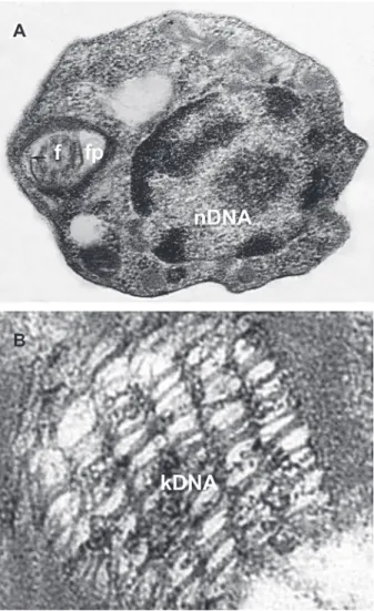
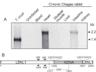
The kinetoplast DNA (kDNA) contains a few dozen maxicircles (23 kb) and thousands of minicircles (1.4 kb) catenated into a complex network (Fig. 2A, B), comprising 10-15% of the total cell DNA (Lukes et al. 2002, Junqueira et al. 2005). Maxicircles encode the structural genes nec-essary for mitochondrial function (Westenberger et al.
2006), many of which are modified post-transcriptionally by a unique uridine insertion/deletion process called RNA editing (Simpson et al. 2004). In T. cruzi, each minicircle contains four equally-spaced conserved regions thought to contain origins of replication (Avliyakulov et al. 2003); the four intervening variable regions each have the po-tential to encode an individual guide RNA (Avila & Simpson
1995), the small transcripts that specify the location and number of uridines to be added to or deleted from the mRNA. The structure of the kDNA complex is not under-stood, nor have the intricacies of kDNA replication been fully elucidated. Replication involves doubling of the number of minicircles and maxicircles and distributing the progeny into two daughter networks that are each identi-cal to the parent network (reviewed in Liuet al. 2005). Prior to their replication, minicircles are individually re-leased from the network into the kinetoflagellar zone situ-ated between the kDNA disk and the flagellar basal body (Ferguson et al. 1992). The proteins that initiate minicircle replication, including the universal minicircle sequence binding protein, primase, and replicative polymerase are localized within the kinetoplast flagellar zone (Li & Englund 1997, Abu-Elneel et al. 2001, Das et al. 2004).
The revelation that minicircles are integrating into the host genome (Teixeira et al. 1991, 1994a,b, Simões-Barbosa et al. 1999) and correlated to the pathology of Chagas disease (Nitz et al. 2004) has renewed interest in
under-Fig. 2: ultrastructure of Trypanosoma cruzi and its
468 468 468 468
468 Chagas pathology and genetics • Antonio RL Teixeira et al.
standing the composition of minicircles within the known sub-groups of T. cruzi. Minicircle kDNA integrations be-have as mutagens in the host. kDNA has been used for the identification of subgroups of T. cruzi populations (Avila et al. 1990). The relationships among distinct para-site populations have been characterized using largely nuclear markers into six discrete typing units (DTUs) (Brisse et al. 2000a,b, 2001). The definition of these DTUs allows a systematic approach to the analysis of kDNA in order to assess any direct links with pathogenicity, and three clades have been defined based on maxicircles se-quences (Machado & Ayala 2001), however the minicircle population dynamics that could vary from isolate to iso-late.
Genetic exchange and diversity
So it is necessary, at least intermittently…, this thing called sex. As of course you and I knew it must be. Otherwise surely, by now, we mammals and dragonflies would have come up with something more dignified
(Quammen 1985)
The sexual lifecycle may reflect the history of life’s programming and adaptation to an oxygenated biosphere. Sex is an ancient cellular capacity present in primitive eu-karyotes. So vital, it may have originated with an advan-tageous symbiotic union (Margulis & Sagan 1995) of two bacteria with complementary metabolic strategies. Many organisms thought to be exclusively asexual also repro-duce sexually (Maynard-Smith 1998). Continuous refine-ment of this magnificent engineering results in an unre-solved problem, a missing link to sexual fertilization (Redfield 1999, Jan et al. 2000). The contribution of a-karyotic genomes and membranes in the evolution from two pro-karyotes to one eu-karyote (Margulis et al. 2000) is evident, with each gamete sharing common descent with extant microbial species in two moneric domains: oocyte to eu-bacteria (since eukaryotes have eubacterial membranes), and sperm to archea (Walther et al. 1999). It seems likely that the sexual reproduction process used by early prokaryotes was a prerequisite to large evolu-tionary leaps through cycles of cell fusion and chromo-some segregation, possibly providing selective recombi-nation advantages to molecular parasites.
Character compatibility of molecular markers used to distinguished asexual and sexual reproduction represents a potent tool (Mes 1998) complementary to the use of high variability dominant markers such as random ampli-fied polymorphic DNA fragments (RAPDs) and ampliampli-fied fragment length polymorphisms in genetic analyses of trypanosomatids. Genetic exchange is now known to oc-cur during the life cycle of many parasitic protozoa, in-cluding trypanosomes. Among the African trypanosomes, crosses between T. brucei individuals resulted in hybrids formed during tsetse fly transmission. The hybrids ap-pear mainly diploid, but various traits in some chromo-somes were inherited in a non-Mendelian fashion (Walliker 1989).
Isoenzyme and RAPD analyses of T. cruzi isolates from Central and South America showed two homozy-gous and the corresponding heterozyhomozy-gous phenotypes
consistent with genetic exchange (Carrasco et al. 1996). Similarly, two T. cruzi stocks that carried different drug-resistance markers were co-passaged through an entire life cycle. Six double-drug (hygromicin and neomycin) resistant trypanosomes were recovered from the mamma-lian stage of the lifecycle, showing fusion of parental geno-types, loss of alleles, homologous recombination, and uniparental inheritance of kDNA. These results are con-sistent with hybrid genotypes among natural isolates of
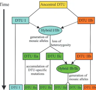
T. cruzi that show aneuploidy and recombination across vast genetic distances, consistent with non-Mendelian genome duplication (Gaunt et al. 2003). An analysis of multiple nuclear markers has led to the proposal (Fig. 3) that the extant lines of T. cruzi can be described by a grand total of two such genetic exchange events (Sturm et al. 2003, Westenberger et al. 2005), which are consis-tent with the distribution of the three clades defined for the maxicircle lineages (Machado & Ayala 2001).
The demonstration of genetic recombination result-ing in marked polymorphisms requires the use of appro-priate nomenclature. Here the term stock rather than strain will be used to designate the wildtype T. cruzi isolates. This term is in keeping with the fact that T. cruzi stocks kept in the laboratory will not maintain stable physiologi-cal and biochemiphysiologi-cal markers after serial passage in vitro
and in vivo. Since T. cruzi populations show broad plas-ticity due to the capacity to transpose lineage, the word ‘clone’ will not be used to designate the progeny of a single T. cruzi individual either, because over time the identity of the ancestor fades.
Polymorphisms of T. cruzi populations isolated from Chagas heart patient hSLU239 and cloned subpopulations
h1 and h2 and from Chagas megacolon patient mSLU142 and cloned subpopulations m1, m2, m3 and m4 were stud-ied. Subpopulations h1 and h2 differed from those de-rived from the subpopulations m1-m4 in some 13 enzymes analyzed. RFLP analysis showed polymorphisms in four glycolytic enzymes and separate the T. cruzi stocks and their subpopulation isolates into three groups: I, formed by hSLU239 and isolate m4, which was classified as ho-mozygous CC and BB. II, composed by mSLU142 and isolates h1 and h2, which was classified heterozygous for aldolase; III, including isolates m1, m2, and m3 classified as homozygous for each of five enzymes. Thus T. cruzi
infection in each Chagas patient is produced by geneti-cally diverse, polymorphic parasite populations (Lauria-Pires et al. 1996). Furthermore, the particular growth kinet-ics, doubling time and differentiation in axenic liquid me-dium of each of those parental stocks and derived sub-population isolates revealed broad behavioral diversity (Lauria-Pires et al. 1997). These observations are consis-tent with several studies (Dvorak 1984, Engel et al. 1985) showing significant inter- and intra-group differences between growth rates, varying amount of total DNA/para-site subpopulation and marked heterogeneity of paraDNA/para-site populations to the intracellular cycle of T. cruzi.
469 469 469 469 469 Mem Inst Oswaldo Cruz, Rio de Janeiro, Vol. 101(5), August 2006
and pathogenicity in mice and, therefore, clinical and pathologic manifestations of disease could not be asso-ciated with intrinsic characteristics of T. cruzi populations (Lauria-Pires & Teixeira 1996). Moreover, to determine the role of T. cruzi superinfection on the outcome of disease, groups of BALB/c mice were prime-infected with low viru-lence isolates h1 and h2 and challenged with high viru-lence isolates m3 and m4. All mice injected with the m3
and m4 isolates succumbed before or at 16 days post-infection. In contrast, all mice injected with the h1 and h2
isolates survived the prime infection and were super-in-fected with the m3 and m4 parasites. Low-level parasitemias were observed after challenge with virulent parasites, and the histopathological lesions and mortality ratios were not different from those seen in mice that re-ceived a single T. cruzi injection. Thus morbidity and mortality in BALB/c mice infected with T. cruzi subpopu-lations are not associated with the frequency or parasite burden (Lauria-Pires & Teixeira 1997).
The protective effect of primary infection with non-virulent T. cruzi subpopulations against subsequent chal-lenge with virulent parasites was determined in groups of BALB/c mice. Low degrees of parasitemia were observed in mice challenged with the highly virulent clones and their survival ratios were not different from those seen in mice that received a single injection of non-virulent T. cruzi (Lauria-Pires & Teixeira 1997). Regardless of the
in-tra-group genetic diversity, infection with non-virulent T. cruzi prevented severe infection in mice subsequently challenged with highly virulent parasites.
Acquired immunity to T. cruzi infection
The basis of acquired immunity to T. cruzi has been reviewed extensively (Teixeira 1987, Brener & Gazinelli 1997). Thus, we revisit the protective immunity concomi-tant to T. cruzi infection in mammal hosts, which bears features resembling those described for other intracellu-lar chronic infectious diseases such as leishmaniasis, tu-berculosis, and leprosy. Initial infection may initiate un-perceived by the hosts in a large majority of cases and full-blown acute infections are occasionally seen in chil-dren below 15 years of age. Regardless of the mode of initiating invasion of the host organism, the infection per-sists in the body life-long, albeit with a continuously de-creasing parasite load. As infections progress to the chronic stage they tend to become cryptic and demon-stration of the infectious agent becomes progressively more difficult, although its presence can usually be con-firmed by immunologic and molecular assays. The ongo-ing biological process follows a course that favors spe-cies survival in a great majority of cases: The host-para-site relationship persists with the absence of disease mani-festation, and the host usually dies of causes unrelated to Chagas disease. The roles played by host humoral and cell-mediated immune reactions are crucial in disease out-come. We discuss aspects of host resistance not reviewed previously.
CD4+ Th 1 lymphocytes are the main conducters for induction of partially protective immunity against T. cruzi
(Hoft et al. 2000). CD8+ T lymphocytes, interferon-(INF)γ, and macrophages are important elements controlling para-site replication during the acute phase of infection. In the chronic stage of the disease, parasite-specific antibodies that fix complement and lyse the blood trypomastigotes are thought to be the main effector molecules to secure latency of infection. LTh type 1 cells that secrete inter-leukin(IL)-2 and γ-IL seem to be involved in Tc-, mac-rophage-, and IgG2-producing cells. LTh type 2 secreted IL-4, IL-5, and IL-10 are implicated in the humoral immune response and inhibition of LTh 1, Tc and macrophages (O’Garra & Murphy 1993). The depletion of CD8+ cells before inoculation resulted in increasing parasitemia and mortality in mice. Knock-out of CD4 and CD8 genes aug-mented the inflammatory response and parasite release in affected tissues. In inflammatory infiltrates CD4– and CD8– lymphocytes were seen (Sun & Tarleton, 1993). T. cruzi
inoculation in resistant and susceptible mice showed that LTh cells are critical in determining important features of the protective immune response. The dominant TLh1 re-sponse was associated with resistance mediated by LTc and γ-INT that induce the macrophage L-arginine meta-bolic pathway and free radical production. LTc lympho-cytes were associated with elimination of T. cruzi-infected host cells and with inflammatory heart lesions (Laucella et al. 1996). Nevertheless, Martin & Tarleton (2005) have observed vigorous anti-parasite response from both CD4+ and CD8+ T cells. It was shown that mice with variant natural killer T (NKT) cells bear striking inflammatory
in-Fig. 3:a schematic of the evolution of Trypanosoma cruzi groups.
470 470 470 470
470 Chagas pathology and genetics • Antonio RL Teixeira et al.
filtrates of dendritic, natural killer, B, and T cells. These inflammatory cells produce increasing concentrations of gamma interferon, tumor necrosis factor alpha and NO, show a diminished antibody response, and usually die. A subpopulation of invariant NKT cells appeared to dampen the inflammatory response and mice survival. The pivotal role of IL-12, γ-INT, and iNOS in the control of parasitemia, inflammation, and host resistance is antagonized by the IL-4 (Michailowsky et al. 2001). NKT cells play a role in the regulation of the immune responses during infection and autoimmune disease (Duthie & Kahn 2005).
In human Chagas heart disease there is a predomi-nance of CD8+ lymphocytes, which are many-fold more frequent than CD4+ T cells (Reis et al. 1993). In the gas-trointestinal form, however, a significant decrease in the absolute number of CD3+ lymphocytes and in CD19+ B lymphocytes as well as an inversion of the CD4/CD8 ratio was seen, whereas this ratio remained unchanged in the heart patients as compared to that of control (Lemos et al. 1998). These T cells showed down-regulation of CD62L, which has been associated with effector memory pheno-type. Further characterization of gene expression in Chagas heart disease showed immune response, lipid metabolism, and mitochondrial oxidative phosphorylation genes selectively up-regulated. The γ-INT signaling path-way up-regulating chemokines could be involved in heart hypertrophy (Cunha Neto et al. 2005).
Regulation of the partially protective immune response in naturally infected humans and in experimental T. cruzi
infection in mice is not understood precisely (Marino et al. 2005). Partially protective immune responses associ-ated with host resistance to T. cruzi infection may be in-volved with severe inflammatory infiltrates and pathol-ogy in the heart, digestive tract, and other tissues (Correa-Oliveira et al. 1999, Gomes et al. 2003). Thus, these studies contraindicate the prospect for a successful vaccine, de-spite enthusiasm for the effort (Levin 1996, Kierszenbaum 1999).
Clinical presentations of Chagas disease
Key practical points in human and veterinary medi-cine are those associated with emerging symptoms and signals that can be recognized as clinical features of a disease. Any persistently infectious biological process can be divided into as many segments as required for facilitating measures that leads to palliation of symptoms and treatment of signals. T. cruzi infections in mammalian hosts have been divided into successive acute and chronic stages.
Acute Chagas disease
Most cases of acute T. cruzi infection are ascribed to triatomine bugs, the insect vector that transmits the pro-tozoan. A delayed-type indurate skin lesion may appear at the portal of parasite entry in immune-competent hosts, but not in immune-compromised patients lacking cell-me-diated immune response (Teixeira et al. 1978, Teixeira & Teixeira 1995). Acute infection in the latter case goes un-perceived by the patient and/or by the physician in the absence of diagnostic signs and symptoms. A field study showed that approximately 75% of the acute cases were
seen in children less than 10 years of age (reviewed in Teixeira 1987).
Death in acute Chagas disease patients (possibly one case in 1000 acutely infected) is usually related to heart failure and/or meningitis and encephalitis. Sinus tachy-cardia, first-degree AV block, low QRS voltage and pri-mary T wave changes can be recorded by electrocardio-graph (ECG). X-rays in such cases show an increased cardiac silhouette of varying degrees. Interestingly, all symptoms and signals that correlate with irrevocable in-volvement of different organs cede spontaneously with-out apparent sequel (WHO 2002).
Indeterminate phase
The chronically infected individual remains a life-long source of the parasite as an indeterminate phase reser-voir. Approximately one third of all individuals with inde-terminate infections will develop chronic Chagas disease. The indeterminate phase of infection has been defined based on criteria of (i) positive specific IgG antibody test and/or parasitological demonstration; (ii) absence of symptoms and signs of Chagas disease; (iii) absence of ECG abnormalities; (iv) regular size of heart, esophagus and colon by X-ray. Using these criteria, regardless of the geographic area where field studies have been conducted, it has been estimated that two thirds of T. cruzi-infected individuals will remain in this condition during their eco-nomically-productive lives. Thus most patients with in-determinate infections are 20 to 50 years of age, compris-ing approximately 12 million people with positive immu-nologic tests for the parasite. Their life span is similar to that observed for the general population (Macedo 1999). Usually indeterminate phase individuals are identified as such during job application or blood bank screening. Denial of work is unjustified, but the candidate is dis-qualified from blood donation.
Chronic cardiac disease
471 471 471 471 471 Mem Inst Oswaldo Cruz, Rio de Janeiro, Vol. 101(5), August 2006
were many-fold more frequent in Chagas patients than in controls (p < 0.001). The progressively shifting right bundle branch blocks in particular were recorded more frequently than the remaining ECG alterations in the Chagas patient population. ECG revealed ventricle wall hypokinesis and intracavitary thrombus. The heart en-largement in Chagas patients is an ominous sign, leading to a poor prognosis (Lauria Pires et al. 2000). A word of caution is needed because the severely compromised Chagas patient may die during a Holter-type ECG moni-toring of arrhythmias and other heart disturbances. More-over, patients having apparently stable ECG changes may show sudden flare-ups of an underlying defect leading to heart failure. The variability of the ECG and clinical mani-festations is remarkable: Some patients showing cumula-tive changes in successive ECG recordings apparently halt the evolving life-threatening arrhythmias and lead a normal life.
In one population of chronic patients, 57% mortality was related to Chagas disease, of which 58% had heart insufficiency, and 37.5% died suddenly (Prata 1999). The remaining deaths were related to the digestive forms as-sociated with chronic disease. The average time between acute infection and development of severe chronic Chagas lesions has been estimated at 28 ± 7 years (Prata 2001). However, Chagas cardiomyopathy rapidly progresses to-ward death no longer than 5 years after signs of heart failure. A common finding in death resulting from heart failure is cerebral infarction as a consequence of a throm-bus detached from the left ventricle.
Digestive form of Chagas disease
Gastrointestinal disorders are among the most com-mon clinical manifestations of chronic Chagas disease. A clinical study aimed at the evaluation of autonomic func-tion in chagasic patients showed esophageal alterafunc-tions usually occurred earlier in the course of the chronic infec-tions when compared to similar abnormalities in the heart (reviewed in Macedo 1999). A digestive disease patient may complain of difficulty swallowing and regurgitation of food, clinical symptoms related to megaesophagous. Alternatively, some complain of constipation due to a fe-cal bolus in the rectal ampoule as a result of megacolon. Megaesophagous and megacolon are seen frequently in endemic areas; patients showing these conditions bear specific antibodies to T. cruzi and consistently positive nucleic acid tests (NAT) assays. These ‘mega’ conditions can affect individual patients independently, in associa-tion with each other or in associaassocia-tion with heart disease. Megaesophagus can manifest clinically in chagasics as early as 2 years of age or any time throughout life, although the majority of cases are seen in men between 20 and 40 years old. Megaesophagus anticipates heart trouble in many patients. The disease manifests by dys-phagia, heartburn, hiccups, regurgitation of food, and in-creasing salivation. This clinical picture associates chagasics megaesophagus and caquexis resulting from difficulty to eat. The disease appears to evolve during periods of dysphagia followed by long periods during which symptoms are absent.
Megacolon is seen considerably later in the course of
Chagas disease in comparison with megaesophagus. The main symptom of chagasic megacolon is constipation. The progressive retention of hardened bolus leads to dilation and thickening of the walls of the colon, usually compro-mising the sigmoid colon and rectum. Difficulty with pass-ing of bolus leads to dilation of the remainpass-ing intestines, increasing bowel movements, pain, and constant physi-cal distress. The long-term use of laxatives can cause ul-cerations of mucosal surfaces, septicemia, rupture of the wall of the intestine and peritonitis. Typical complications of megacolon are intestinal obstruction and rupture (re-viewed in Prata 1999).
Pathology in human Chagas disease
Data on gross and microscopic pathology of acute Chagas disease was derived from two cases (Teixeira et al. 1970). The access to slides with sections from the two patients was kindly provided by Dr Moysés Sadigursky, from the Hospital of the Federal University of Bahia, Bra-zil. Data on chronic Chagas disease stems from ARLT’s records of 20 post-mortem studies carried out personally at the Hospital of the Federal University of Bahia, Brazil, and at the Hospital of the University of Brasília, Brazil.
Acute Chagas disease
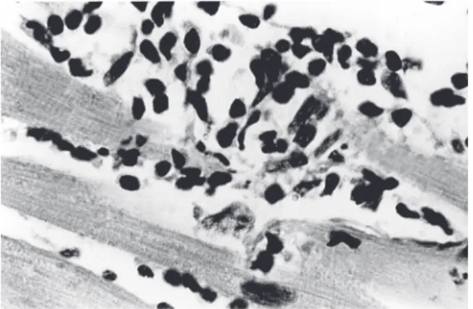
In the human body T. cruzi can parasitize any tissue derived from embryonic mesoderm, endoderm, and neu-roectoderm. However, the intensity ofinfection in the body varies from case to case, likely depending on the genetics of host and parasite (Campbell et al. 2004). Mesoderm-derived conjunctive tissues smooth and striated muscles, bone marrow and phagocytic mononuclear systems, and gonadal cells can be heavily parasitized. The histopathol-ogy of an 18 month-old boy and of a 4 month-old girl who died of acute Chagas disease (Teixeira et al. 1970) revealed niches ofamastigotes inside the theca cells of the ovary and inside the goniablasts of seminiferous tubes of the testes. Reproductive apparati were rarely examined previ-ously in the course of T. cruzi infections. Endodermal tis-sue structures are eventually parasitized byamastigotes; epithelial cells of liver, kidneys, thyroid, pancreas, and other glands are spared infection. Neuroectoderm cells are parasitized less frequently than other embryonic-de-rived tissues; if the infection reaches the nervous sys-tem, glial cells, usually astrocytes, are invaded.
472 472 472 472
472 Chagas pathology and genetics • Antonio RL Teixeira et al.
and glial cells, but not in neurons. Nevertheless, neuronal lysis associates adherence of mononuclear cells of the immune system and depopulation of target neurons. In-flammatory infiltrates associate with glial and neuronal cells, secondarily compromising parasite-free neurons.
Typically the heart of a deceased acute chagasic pa-tient is enlarged, flabby, dilated, and congested. Lymph nodes situated between the aorta and the pulmonary ar-tery, are engorged. The epicardial surface shows wide-open coronary vessels accompanied by whitish lymphatic vessels with tiny pearl-like nodules. These morphologi-cal changes presage the striking inflammatory infiltrates running through the walls of the heart. Microscopically, many muscle fibers and occasionally interstitial histio-cytes show nests of dividingamastigotes. Mononuclear cells, mainly small and large lymphocytes with expanded cytoplasmic processes, and macrophages infiltrate the myocardium and adhere to the membrane of target heart cells. Several aspects of characteristic target cell destruc-tion can be observed. Notably, some parasitized cells are isolated from the destructive inflammatory infiltrates. Para-site-free heart cells are mostly rejected or destroyed by immune system mononuclear cells. Confluence of mul-tiple rejection units comprises the overall microscopic pic-ture of lesions in acute Chagas disease. Inflammatory in-filtrates invade cardiac parasympathetic ganglia where glial or Schwann cells can be parasitized, but neurons are spared. Adherence of inflammatory mononuclear cells to neurons leads to lysis and loss of several of these neural units in the acute phase. Additionally, inflammation ex-tends into sympathetic nerves in the epicardial and intra-mural heart structures.
Involvement of central nervous system structures in the acute phase can be frequent, as T. cruzi can be recov-ered from the cerebral spinal fluid in 72.7% of cases (Hoff et al. 1985). However, in half of these cases there is a lack of alteration of fluid components and absence of neuro-logical damage. In acute cases showing clinical manifes-tation of neurological involvement, lesions are related to meningitis and encephalitis. Overall, the brain can show congestion of blood vessels and edema. Brain tissue is damaged sparsely by inflammation around small blood vessels, vascular hemorrhages, and microglial cell nodu-lar proliferation in the white and gray matter. Typically inflammatory cells invade the meningeal leaflets and en-circle blood vessels inserted deep in the brain. T. cruzi
nests can be seen in brain astrocytes.
Indeterminate phase
Microscopic substrates for changes in the indetermi-nate phase were drawn from 20 biopsies revealing mini-mal inflammatory heart lesions (Mady et al. 1982) that were generally focal and small. Skeletal muscle biopsies showed spotty inflammation, target cell lyses and degeneration (Sicca et al. 1995). Inflammatory lesions in the heart, di-gestive tract, and skeletal muscle are similar to those seen in clinically manifest chronic disease patients, but to a much lower degree. Inflammatory infiltrates surround muscle fibers, resembling the minimal rejection unit of the target heart cell. In some cases multiple rejection units compromise a bundle of fibers in a single muscle; any
nerve or sympathetic ganglion in the region can be af-fected by inflammatory infiltrates. In the digestive tract, lesions reaching the parasympathetic ganglia and neu-ronal cell depopulation have been observed (Lopes 1999). Progressive clinical-pathological lesions present in chroni-cally infected chagasics classify the disease according to the affected organ.
Chronic Chagas heart disease
Chronic Chagas heart disease affects individuals of both sexes equally, usually between 30 and 45 years of age. In patients showing progressive ECG alterations unexpected deaths occur at a rate of 37.5% (Prata et al. 1986, Lopes 1999), whereas 58% develop ominous signs of heart failure and usually die within 7 to 24 months (Dias 2000). Congestive failure involves the right and left cham-bers of the heart, thus affecting all blood circulation. The heart increases in size, occupying the base of the thoracic cavity and bulging against the chest wall. The average heart weight reaches 540 ± 90 g, in patients dying of con-gestive failure, whereas in those undergoing sudden death the weight reaches 390 ± 50 g. At the endocardial surface, the chambers of the heart become dilated and the walls can be thickened. A typical gross feature is the efface-ment of the apex of the left ventricle, showing aneurismal dilation. The presence of thrombus at different stages is seen frequently in the apex of the left ventricle and in the right atrium, and can be associated with thrombus embo-lism in the lung, brain, spleen, and kidney. Thrombus em-bolic phenomena in the brain and lungs are associated often with the ultimate cause of death in chronic heart disease. The epicardial surface of the heart shows dilated coronary vessels accompanied by lymph vessels with periodic, small, pearl-like nodules indicative of the drain-age system from the subjacent myocardium inflammatory process.
evanes-473 473 473 473 473 Mem Inst Oswaldo Cruz, Rio de Janeiro, Vol. 101(5), August 2006
cent inflammatory infiltrates. Inflammatory cells infiltrate specialized myofibers of the heart conductive system in the same manner that they infiltrate contractile myocar-dium. The intensity of this self-destructive inflammatory process varies from site to site in the myocardium; when some lesions are initiated, others are intermediate or phas-ing out. Some areas of the heart may be spared while oth-ers are severely damaged by inflammation; the intensity of these processes reaching the entire heart simulta-neously would be fatal. At the ultra-structural level, in addition to association of mononuclear infiltrates with target cells undergoing lysis, myofibers show features of hypertrophy, mitochondrial swelling, necrosis, hyaline degeneration, disruption, and loss of myofibrils (Tafuri et al. 1973). As lesions increase in age loose fibrous tissue and inflammatory cells are replaced by dense fibrous scars that can be seen scattered throughout the heart walls (Rossi 2001).
Chronic Chagas mega syndromes
Pathology of the esophagus and colon associated with chronic Chagas syndromes is dependent essentially on inflammatory lesions upon smooth muscle fibers of the walls of the hollow viscera in the digestive system, affect-ing particularly the intramural parasympathetic neurons. Cases of mega syndromes affecting the stomach, duode-num, gall bladder, vesicle bladder, and bronchus have been reported also (Adad et al. 2001). For each of these condi-tions the pathology shows a common denominator de-scribed for most prominent esophagus and colon condi-tions (Hagger et al. 2000): Inflammatory lesions in para-sympathetic ganglia lying between the smooth muscle layers (Auerbach’s plexus) and in the sub-mucosal (Meissner’s plexus) of the hollow viscera lead to gangli-onitis, and neuron drop-outs as described for intracar-diac ganglia.
Although the presence of parasite nests in peri-gan-glion fibroblasts or intra-ganperi-gan-glion glial cells has been re-ported, there are no accounts of parasympathetic neuron invasion (Tafuri 1971, Da Mata et al. 2000), nor is there evidence for a hypothesized neurotoxin secreted by the parasite (Andrade & Andrade 1966) and, therefore, neu-ronal depopulation does not correlate with parasite-in-duced host cell death. To the contrary, neuron death is associated clearly with immune system mononuclear cell adherence and lyses, as for that lesion type the minimal rejection unit correlates with destruction of heart target cells in Chagas disease. Active ganglionitis resolves with neuronal depopulation and fibrous tissue replacement. In summary, the most conspicuous lesions in Chagas mega syndromes parallel those in the heart and associate in-flammatory mononuclear cell infiltrates with the minimal rejection unit that appears to be a common pathological denominator in human Chagas disease.
The comparative pathology of Chagas disease
At some stage, blood-sucking invertebrates (leeches) feeding upon fish, amphibians, and reptiles, acquired ver-tebrate trypanosomes and subsequently passed them on to terrestrial birds and mammals. Trypanosome infections in aquatic vertebrates could improve our understanding
of the host-parasite relationships (Cox & Moore 2000, Hamilton et al. 2005). The absence of a fully-developed immune system in aquatic vertebrates could correlate to some extent with their lack of symptoms or minor patho-genic manifestations. This area demands further explora-tion, potentially leading to establishment of the ontog-eny of the innate mechanism of resistance and/or suscep-tibility of vertebrate hosts to trypanosomatids parasites.
Birds
Birds are refractory to T. cruzi infection.Upon intra-venous or intramuscular injections T. cruzi disappears immediately from the site of inoculation (Teixeira 1987) and cannot be recovered from bird blood by any means. However, inoculation of T. cruzi infective trypomastigotes in the air chamber of fertile chicken eggs results in intrac-ellular growth of parasite amastigotes in embryo cells until the 10th day post-fertilization. Thereafter, the infection is eliminated by the embryo’s innate immune mechanism (Nitz et al. 2004).
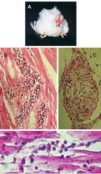
Host-specific trypanosomes of birds can produce se-vere infections (Chandenier et al. 1988, Sehgal et al. 2001). Avian-specific Trypanosoma bouffardi experimental in-fections produce severe pathology in canaries. Enlarge-ment of the spleen coincided with peak parasitemia in the absence of other gross lesions. Histopathology revealed lymphoid tissue hyperplasia and focal myocarditis. A ca-nary that became blind and died of a systemic disease was examined (ARLT received slides for review from Gene Hubbard, VMD, Southwest Foundation for Biomedical Research, San Antonio, Texas): Heart and skeletal muscles showed typical inflammatory infiltrates and target cell ly-sis. The eyeball showed severe inflammation, with nests of dividing kinetoplastids in ciliary muscles. Pathology of the disease, caused by a natural trypanosome infection in birds, was similar to that of mammals infected with T. cruzi.
Marsupialia
The Metatheria (Marsupialia: Didelphidae) and Eu-theria (Edentata: Dasypodidae; Rodentia: Muridae) are considered the earliest mammals to become involved in the enzootic cycle of T. cruzi infection. Didelphidae and Dasypodidae, opossums and armadillos spp., respec-tively, are major sylvatic reservoirs (Legey et al. 1999, Yeo et al. 2005) The eco-epidemiology of enzootic Chagas dis-ease in North America is dependent largely on the rela-tionships of triatomine vectors with opossums and arma-dillos (Yaeger 1988, Karsten et al. 1992, Pung et al. 1995).
474 474 474 474
474 Chagas pathology and genetics • Antonio RL Teixeira et al.
The pathology seen in heart sections from these natu-rally-infected marsupials showed myocarditis, character-ized by mononuclear cell infiltrates and target cell lysis. Furthermore, inflammatory infiltrates were seen in the heart, skeletal muscle and in smooth muscle of the esophagus, and small and large intestines. Histopathological study of representative tissue sections taken from three opos-sums free of parasite showed an absence of tissue le-sions (Teixeira et al. 2001). Araujo Carreira et al. (1996) found T. cruzi nests in the scent glands, hearts, and di-gestive tracts of 10 naturally-infected D. marsupialis. An inflammatory infiltrate of moderate to severe intensity was present in smooth and striated skeletal and heart muscles. Despite the presence of tissue lesions in wild T.
cruzi-infected marsupials, armadillos, and rodents, some re-searchers have proposed that these animals “learned to live in harmony” with T. cruzi and, therefore they do not display apparent disease (Legey et al. 1999). No long-term study has shown ratios of morbidity and mortality or the relative lifespans of wild mammalian reservoirs in the presence or absence of T. cruzi infections.
Rodentia
Naturally T. cruzi-infected rodents (Rodentia: Echi-myidae; Rodentia: Cricetidae; and Rodentia: Muridae) have been captured in various ecosystems on the Ameri-can continents. In one study, the prevalence of parasite infection reached 9.1% of captured wild rodents (Raccurt 1996). Calomys callosus (Rodentia: Cricetidae) are resis-tant to T. cruzi, surviving inocula that normally kill mice (Borges et al. 1992). The histopathological analysis of sections from T. cruzi-infected rodents showed parasit-ism of liver cells and of striated muscles. Inflammatory infiltrates in heart and skeletal muscles were moderate or absent (Borges et al. 1983). Resistance to chronic infec-tion appeared to correlate with interferon gamma serum levels and H2O2 release by peritoneal macrophages. C. callosus may have developed immune mechanisms for survival and thus acts as a reservoir (Borges et al. 1995). The interaction of T. cruzi with the caviomorph rodent
Trichomys apereoides (Rodentia: Echimyidae) revealed features suggestive of an ancient adaptation to T. cruzi
infection. Chronic infection in T. apereoides remained pathologically cryptic for 5 months, despite persistence of infection (Herrera et al. 2004).
Naturally occurring T. cruzi infection of sylvatic Rat-tus ratRat-tus and Rattus novergicus (Rodentia: Muridae) have
been described (Herrera & Urdaneta-Morales 1997). Some
T. cruzi-infected wild rats showed parasitemia and numer-ous nests of amastigotes in striated skeletal and cardiac muscle, and in smooth muscle of the digestive tract. In addition to showing myocarditis and myositis, inflamma-tory infiltrates could be seen occasionally in the proxim-ity of amastigote nests. Over 9% of offspring showed transplacental T. cruzi infection in infected rats (Moreno et al. 2003). Several laboratory rat strains have been ex-perimentally-infected with different T. cruzi stocks to as-sess their ability to produce myocarditis, histopathologic alterations in organs of the digestive system, and auto-nomic nervous systems denervation (Camargos et al. 2000). Extensive lesions in the heart of acutely-infected rats,accompanied by destruction of noradrenergic nerve terminals, were complement-independent (Machado et al. 1994). Chronicinfection of rat revealed intra-cardiac de-structive ganglionitis; the inventory of ED1 and ED2 mac-rophages and immune-competent cells infiltrating the gglion was consistent with the fact that abundance of an-tigen presenting cells correlates with permeability of the blood-brain barrier and tissue lesions. Within the brain, astrocytes (Fig. 4A, B) are target cells for parasite prolif-eration (Da Mata et al. 2000). Peri-ganglionitis and gangli-onitis were seen in 62.5% of the para-vertebral, sympa-thetic cervical ganglion frominfected rats (Camargos & Machado 1988). Qualitative and quantitative morphology analyses suggest that infection of Wistar rats causes myelin damage and axonal swelling of myelinated fibers of the vagus nerve (Fazan & Lachat 1997).
Among the Class Rodentia, small rodents in the Fam-ily Muridaeare the most utilized laboratory animals for studies aimed at unraveling features of immunology and associated pathology in Chagas disease. The albino Swiss mouse (Mus musculus) is by far the most common animal in the study of parasite-host relationships in the course of experimental T. cruzi infection. Genetic control of im-mune responses in mice has been studied extensively, and this knowledge is useful for understanding the mecha-nisms of resistance and of susceptibility to T. cruzi. Exist-ing knock-out mouse strains are important tools for deter-mining the role of a selected gene in regulation of ac-quired immune responses in the course of infection (Araujo-Jorge 2000).
Features of Chagas disease pathology have been un-raveled in the laboratory mouse model. Briefly, myocardi-tis, myosimyocardi-tis, sympathetic and parasympathetic
ganglioni-TABLE II
Histopathological findings in Trypanosoma cruzi infected Didelphis marsupialis
Cases a Parasite detection
(Xeno, Hemo, and NAT) Histopathology b
Heart Muscles Digestive tube
1 to 9 Positive + to ++ + to +++ + to ++
10 to12 Negative Negative Negative Negative
475 475 475 475 475 Mem Inst Oswaldo Cruz, Rio de Janeiro, Vol. 101(5), August 2006
tis, and central nervous system inflammatory infiltrates have been reported (Rossi Destetti 1995, Waghabi et al. 2002). In lesions at target tissues, the majority of mono-nuclear cells in inflammatory infiltrates bear surface mark-ers characterized as CD8+ cells (Leavey & Tarleton 2003). Both CD4+ and CD8+ T cells participate in this process (Fuenmayor et al. 2005). Aberrant T-cell responses in Chagas disease are required to initiate immune responses that damage the heart (DosReis et al. 2005).
Specifically, lesions of the autonomic sympathetic and parasympathetic nervous systems have been studied (Tafuri et al. 1971, 1979). At the level of optic and electron microscopy, Schwann and glial cells of nerve structures were found parasitized in the course of severe acute in-fections, while neurons were spared. The ultrastructural studies showed typical peri-ganglionitis, ganglionitis, and neuronolyses that were carried out by inflammatory mono-nuclear cell infiltrates (Tafuri 1971). Secretion of a
hypo-thetical neurotoxin by T. cruzi that would kill neurons (Koeberle 1963) was discarded in acutely super-infected animals; mice receiving high doses of the immunosup-pressor hydrocortisone showed increasing numbers of parasitic forms of T. cruzi in Schwann and glial cells, but neurons were spared. In the absence of immunosuppres-sion, acutely infected mice showed intense perigan-glionitis and ganperigan-glionitis, resulting in neuronolysis by inflammatory cells in infiltrates (Andrade & Andrade 1996). The advantage of using numerous isogeneic mouse lineages to reproduce features of human Chagas disease has been in jeopardy by a lack of definition of stable bio-chemical and physiological markers to characterize T. cruzi
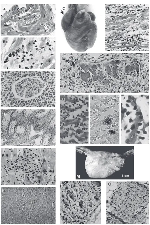
isolates and stocks kept in the laboratory (reviewed in Teixeira 1987). Furthermore, the events of genetic exchange in T. cruzi (Gaunt et al. 2003) underscore the instability of genetic features on the part of the parasite; reproducibil-ity of some specific features of the infection could not be achieved, despite the use of isogenic mice. These fea-tures should not be considered as a caveat for using mice in experimental studies of Chagas disease, because in the wild, highly polymorphic parasite populations infect out-bred mammalian hosts and, therefore diversity is expected to be the common denominator of disease presentation. Nevertheless, the main pattern of T. cruzi infection in the laboratory mouse, although showing some variability, is characterized by a fulminating acute phase in which most infected animals, if not all, die within a few weeks of para-site inoculation. The infected mice attain high parapara-sitemias and amastigotes can be easily found by histological exam of tissue sections. Although tissue parasitism can be in-tense, affected tissues may not show inflammatory infil-trates and target cell destruction. The highly virulent ar-chetype Berenice and Tulahuen T. cruzi stocks, produc-ing intense parasitism of the heart (Fig. 5A), skeletal and smooth muscles, and of the mononuclear phagocytic sys-tem, respectively, kill infected mice in two to three weeks. The cause of death in T. cruzi-infected mice is associated with high parasitemias and necrosis of the spleen. A vari-able percentage of acutely-infected animals may survive the acute phase, and then enter the chronic stage (Fig. 5B). Some consider mouse a suitable model for chronic Chagas disease (Marinho et al. 2004). The usefulness of this host cannot be underestimated; unquestionably, the mouse is suitable for the initial pre-clinical trials to deter-mine the anti-trypanosomal activity and toxicity of candi-date chemotherapeutic agents (Teixeira et al. 1994a,b).
Lagomorpha
The usefulness of the rabbit (Lagomorpha: Leporidae) has been recognized from the earliest experimental stud-ies on Chagas disease (Chagas 1909). The rabbit ( Ory-ctolagus cuniculus), inhabiting burrows in the ground or among rocks in the wild, can co-habit with triatomines, and is an important reservoir host in the cycle of T. cruzi
transmissionin some regions of the South American con-tinent where they are domesticated. Rabbit scarcely had been used as a laboratory animal in experimental studies of Chagas disease, probably because animals are expen-sive to maintain in individual cages, and the lifespan is triple that of mouse. However, rabbits are highly resistant
Fig. 4:parasitism in the brain of a rat infected with virulent
476 476 476 476
476 Chagas pathology and genetics • Antonio RL Teixeira et al.
to T. cruzi infection and usually do not die in the acute phase, but later (20 ± 8 months) of chronic Chagas dis-ease (Teixeira et al. 1975, 1983, Teixeira 1986); this advan-tageous feature of the rabbit has been recognized (Figueiredo et al. 1986, Silva et al. 1996).
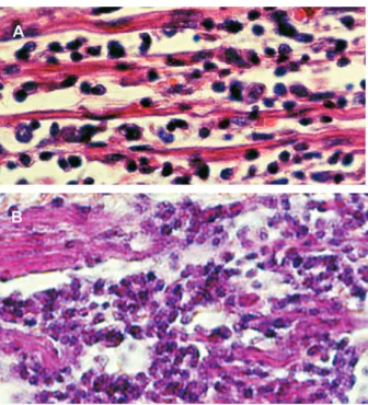
In one study, 34, one-month-old, out-bred New Zealand white rabbits received T. cruzi infections either intrader-mally, intravenously, or by drop instillation in the eye con-junctiva (Teixeira et al. 1983). Regardless of the route of infection the rabbits showed patent parasitemia by xeno-diagnoses up to the 4thmonth post-infection. Thereafter, the xenodiagnoses were negative. Typical chagoma signs developed in two rabbits one week after receiving para-site inoculation in the skin, however the acute phase of the infection ran asymptomatically. In absence of direct demonstration of the parasite, the persisting cryptic in-fections were detected by serologic tests and delayed-type skin reactions to T. cruzi antigens. However, ECG alterations consistent with enlargement and overload of cardiac chambers, alterations of ventricular repolarization, S-T changes and bundle branch blocks were often re-corded in the chronic phase. The pathological manifesta-tions of these ECG alteramanifesta-tions were confirmed at autopsy of experimental rabbits that died of Chagas disease (Fig. 5C). Congestive heart failure and pulmonary throm-boemboli related to chronic myocarditis were frequent causes of death. Megacolon was seen in two rabbits. The relatively limited duration of detectable parasitemia, the lack of correlation between parasitemia and severity of pathological manifestation, and the fact that rabbits showed histopathological evidence of myocarditis, myo-sitis, ganglionitis, destructive inflammatory lesions char-acterized by mononuclear infiltrates and target cell lysis (Fig. 5D) are notable (Teixeira et al. 1983). Additionally, changes similar to those described in infected humans were produced in inbred III/J rabbits. Inflammatory infil-trates invaded the atria-ventricular node of the heart con-ducting system, where effectors cells adhered to the tar-get myofibers. ECG alterations were recorded in chagasic rabbits, and increased cardiac silhouettes could be dem-onstrated in X-rays in the chronic phase (Teixeira 1986). Direct evidence of the cytotoxicity of the effector’s im-mune lymnphocytes to the isogeneic target heart cells was provided by in vitro experiments: 73.5% of the beat-ing target heart cell colonies ceased pulsatbeat-ing after a 2-h incubation with effector cells. This cell-mediated cytotox-icity bears implications for the physiopathology of arrhythmias and sudden death frequently seen in Chagas patients (Teixeira et al. 1983).
Then I would still have this consolation – my joy in unrelenting pain – that I had not denied the words…
The availability of the rabbit model for human disease prompted evaluation of treatment of experimental Chagas animals with anti-trypanosomal nitro-derivatives. A dose of 8 mg/kg/day for 60 days of nitroderivatives was intrap-eritoneally inoculated ininfected and uninfected rabbits. Chronic infection was accompanied by the finding of myocarditis in every Chagas heart, regardless of treat-ment (Fig. 5E). PCR assays with T. cruzi-specific and nDNA-nested sets of primers yielded amplification
prod-ucts from Chagas rabbit DNA templates, regardless of treatment (Lauria-Pires et al. 2001). Thus, treatment of in-fected rabbits with nitroderivatives neither improved the Chagas heart lesions nor prolonged survival of treated animals (Teixeira et al. 1990a). Treated animals died in a time span comparable to infected, untreated rabbits.
Alarmingly, malignant lymphomas (Fig. 5F, G) were seen in 33.3% of nifurtimox treated rabbits, and in 38.4% of benznidazole-treated rabbits (Teixeira et al. 1990a). In ad-dition, interstitial fibrous thickening of the testes and scar-city of germinal cells in the seminiferous tubes of a benznidazole-treated rabbit was noted. Infected rabbits survived 765 ± 619 days post-infection whereas nifurtimox and benznidazol treated-infected rabbits survived 693 ± 434 and 552 ± 714 days, respectively (Teixeira et al. 1990b). These survival ratios were not statistically different from nifurtimox and benznidazol treated controls. All these sur-vival ratios are significantly different from that (1496 ± 353 days) of control, untreated rabbits (p < 0.05). The myocarditis in treated rabbits was as intense as in the non-treated infected group. Myocarditis ranged in sever-ity from focal to diffuse, with an even distribution among individual rabbits in each group. The survival of treated rabbits may have been affected by both myocarditis and the appearance of lymphomas in one-third of treated rab-bits (Teixeira et al. 1990c). Infected as well as uninfected rabbits receiving nitroderivatives both developed malig-nant non-Hodgkin’s lymphomas and died (Teixeira et al. 1990b,c, 1994a,b). Nitroderivative chronic toxicity should be measured in an epidemiologic scale, because nifurtimox and benznidazole resulted in lymphomas and atrophy of the testes.
Carnivora
Dogs (Carnivora: Canidae) have been recognized as important animal hosts participating in the peridomicile lifecycle of T. cruzi. Dog (Canis domesticus)mortality in natural ecotopes is substantial with high levels of trans-mission where dogs eat contaminated bugs. This clinical veterinary problem is recognized in several regions of the American continents. Naturally infected dogs have been identified in Texas, Louisiana, and Oklahoma (Bradley et al. 2000, Beard et al. 2003). In Cuesta Rica, it was shown that 27.7% of the dogs from five rural villages had T. cruzi -specific serum antibodies. Positive dogs subjected to X-ray and ECG revealed cardiomegaly and ECG alterations consistent with Chagas heart disease (Montenegro et al. 2002).