CASO CLÍNICO
como sendo frequente: num estudo conduzido por Hirai et al. verificou-se em 43% dos casos e num estudo de Celik et al. verificou-se em 24,1%.3,5,6,8
No Caso 2, o ECD emitia quatro tendões: um tendão que se unia ao tendão do longo extensor do polegar (LEP), um tendão para o 2º dedo, um tendão para o 3º dedo e um tendão para o 4º dedo que, por sua vez, emitia um tendão que se unia ao tendão do EDM. O facto do ECD emitir um tendão que se une ao tendão do LEP não ocorre com fre-quência, estando descrito que ocasionalmente o ECD pode
emitir um tendão para o polegar.2
Observou-se também no Caso 2 que o tendão do ECD para o 4º dedo emitia um tendão para o 5º dedo. Esta va-riação também foi verificada em vários casos descritos por
el-Badawi et al.3 Num estudo conduzido por Tanaka et al,
em 34% dos casos foi encontrado um tendão comum do
ECD para o 4º e 5º dedos.9
Outra variação que se observou no Caso 2 foi a existên-cia de dois tendões provenientes do EDM para o 5º dedo. A literatura relata que o músculo EDM tem origem no epicôn-dilo lateral do úmero (tendão comum dos extensores) e se continua inferiormente por um tendão que se dirige para o 5º dedo.1
Num estudo de Celik et al a prevalência de tendões duplos do EDM atinge 88,9% dos casos, designando-se o tendão medial do EDM por EDM-ulnar e o tendão lateral
por EDM-radial.6 Este padrão do tendão do EDM tem sido
relatado com bastante frequência nos estudos em cadáver, chegando a atingir frequências de 60% e 90% dos casos,
de acordo com el-Badawi e Hirai et al, respectivamente.3,5,8
O tendão radial do EDM encontra-se frequentemente unido
ao tendão do ECD para o 5º dedo.2,3,10
CONCLUSÕES
Existe um grande número de variações no padrão dos tendões dos músculos extensores da mão. O conhecimen-to destas variações pelo cirurgião é miconhecimen-to importante, no-meadamente quando é realizada a reparação de tendões.
CONFLITO DE INTERESSES
Os autores declaram que não houve conflito de interes-se na realização deste trabalho.
FONTES DE FINANCIAMENTO
Não existiram fontes externas de financiamento para a realização deste artigo.
REFERÊNCIAS
1. Rouvière H, Delmas A. Anatomía humana descritiva, topográfica y fun-cional. 11ª ed. Barcelona: Masson; 2005.
2. Gray H. Gray’s anatomy, the anatomical basis of clinical practice. 39th ed. Philadelphia: Elsevier Churchil Livingstone; 2008.
3. el-Badawi MG, Butt MM, al-Zuhair AG, Fadel RA. Extensor tendons of the fingers: arrangment and variations – II. Clin Anat. 1995;8:391-8. 4. Gonzalez MH, Weinzweig N, Kay T, Grindel S. Anatomy of the extensor
tendons to the index finger. J Hand Surg Am. 1996;21:988-91. 5. Hirai Y, Yoshida K, Yamanaka K, Inoue A, Yamaki K, Yoshizuka M. An
anatomic study of the extensor tendons of the human hand. J Hand Surg Am. 2001;26:1009-15.
6. Celik S, Bilge O, Pinar Y, Govsa S. The anatomical variations of the
extensor tendons to the dorsum of the hand. Clin Anat. 2008;21:652–9. 7. Von Schroeder HP, Botte MJ. Anatomy of the extensor tendons of the
fingers: variations and multiplicity. J. Hand Surg Am. 1995;20:27-34. 8. Zilber S, Oberlin C. Anatomical variations of the extensor tendons to the
fingers over the dorsum of the hand: a study of 50 hands and a review of the literature. Plast Reconstr Surg. 2004;113:214-21.
9. Tanaka T, Moran SL, Zhao C, Zobitz ME, An KN, Amadio PC. Anatomic variation of the 5th extensor tendon compartment and extensor digiti minimi tendon. Clin Anat. 2007;20:677-82.
10. Seradge H, Tian W, Baer C. Anatomic variation of the extensor tendons to the ring and little fingers: a cadaver dissection study. Am J Orthop. 1999;28:399-401.
1. Departamento de Anatomia. Faculdade de Ciências Médicas. Universidade Nova de Lisboa. Lisboa. Portugal. 2. Sociedade Anatómica Portuguesa. Lisboa. Portugal.
3. Centro de Estudos e Investigação CEFITEC. Departamento de Física. Faculdade de Ciências e Tecnologia. Universidade Nova de Lisboa. Lisboa. Portugal. Recebido: 14 de Dezembro de 2012 - Aceite: 03 de Fevereiro de 2013 | Copyright © Ordem dos Médicos 2013
ABSTRACT
In over 500 human cadaveric dissections of arms and forearms, performed to the present date, we find frequent anatomical variations, corresponding to classic descriptions. Last year, we found a singular anatomic variation of the extensor muscles of the forearm, which seems previously undescribed. It is our strong belief that gross anatomy studies, and gross dissection should be updated and reintro-duced in modern anatomical studies, for teaching, research, or surgical training purposes. We detected a peculiar anatomical variant of the Superficial Extensor Digiti Muscles in the forearm of a human 73 year old male Caucasian cadaver. We clearly identified a thick bundle of muscular fibres, connecting the main muscular shafts of the Extensor Digiti Minimi, and the Extensor Digitorum Communis
An Unusual Variety of the Extensor Digiti
Muscles: Report with Notes on Repetition
Strain Injuries
Variante Anatómica dos Músculos Extensores dos Dedos:
Um Achado Invulgar Suscitando Comentário sobre Tendinopatias
Maria Alexandre BETTENCOURT PIRES1,2, Diogo CASAL1, Luis MASCARENHAS de LEMOS1, Carlos Eduardo
GODINHO1,2, Diogo PAIS1,2, João GOYRI-O’NEILL1,2,3
CASO CLÍNICO RESUMO
Na série alargada de mais de 500 dissecções de antebraços e mãos cadavéricas humanas, efectuadas por rotina anual do Departa-mento de Anatomia da Faculdade de Ciências Médicas da Universidade Nova de Lisboa desde 1973, detectam-se frequentemente variações anatómicas, com especial incidência a nível dos tendões extensores dos dedos. Detectámos em 2011, uma disposição peculiar dos feixes musculares dos músculos extensores superficiais dos dedos de que, por extensa revisão bibliográfica, não en-contramos descrição. Identificou-se um curto feixe de fibras musculares unindo os ventres principais dos músculos extensor comum dos dedos e extensor do dedo mínimo, na margem ulnar do terço médio do antebraço de um caucasiano do sexo masculino, com 73 anos de idade. Consultando textos de Anatomia publicados, desde o séc. XVI até á actualidade, verifica-se que as mais antigas descrições anatómicas se referem precisamente a uma maior coesão, com divisão baixa entre os ventres musculares destes dois músculos. Apresentámos os resultados preliminares deste estudo no XXII International Symposium of Morphological Sciences, em Fevereiro de 2012, reservando a redacção pormenorizada deste achado anatómico para um dos primeiros números da publicação Archives of Anatomy da Sociedade Anatómica Portuguesa. É nossa profunda convicção de que os estudos anatómicos, e em particular a dissecção cadavérica humana, necessitam ser revalorizados e modernizados, no início do novo milénio, pelo seu imprescindível contributo aos estudos médicos, tanto em termos curriculares básicos, como ainda na investigação clínica e cirúrgica, ou em termos de treino pósgraduado de técnicas cirúrgicas.
Palavras-chave:Antebraço; Cadáver; Dissecção; Dedos; Humanos; Mão; Tendões.
INTRODUCTION
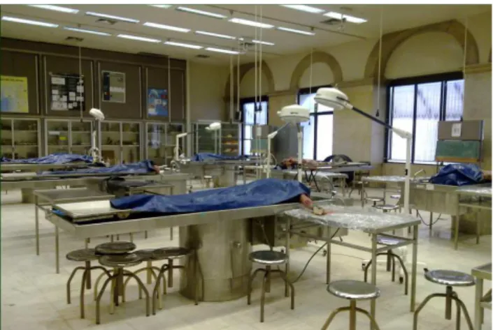
The Department of Anatomy of the New University of Lisbon has been performing routine human cadaveric dis-section, since its foundation in 1973(*1), first for undergrad-uate teaching and research purposes and, more recently, also for the purpose of «Hands-on» postgraduate courses. Simultaneously to these intense teaching activities, with access to cadaveric donations, the Department maintains important activities of scientific research in the domain of human morphological studies and of vascular anatomy, in particular. The Anatomy Department of the New University of Lisbon, presently directed by João Erse de Goyri O’Neill, is well equipped to receive and conserve every cadaveric donation, and the Anatomy dissection room is also well equipped to receive several working stations simultane-ously. (Fig.1)
1 (*) Emeritus Professor J.A. Esperança Pina was the founder of the Faculty of Medical Sciences of Lisbon, where he rebuilt and modern-ized the Lisbon Dissection room in 1973, and later elected President of the International Federation of Associations of Anatomists – I.F.A.A. – (1994/1998). From him, we learned the value of dissection.
Scope of the study: In more than 500 human cadaveric
dissections of arms and forearms, as performed to the pre-sent date in our Department, we find frequent anatomical variations, most of which correspond to classic descriptions and classification, when we review a careful bibliographical research. Last year, during routine cadaveric dissection for teaching purposes, we detected a singular anatomic varia-tion of the extensor muscles of the forearm, which seems previously undescribed.
We presented the preliminary results of this study to the XXII International Symposium of Morphological Sciences,
in Feb. 20121 and prepared the enlarged report of the case
for publication in one of the first issues of the Archives of Anatomy, the official publication of the Portuguese Anatomi-cal Society (AAP/SAP), because it is our strong belief that gross anatomy studies, and gross dissection should be up-dated and reintroduced in modern anatomical studies, both for teaching and research, as for surgical training purposes. (*2)
Much has been written on the subject of anatomical variations of the muscles of the forearm to the fingers. The main anatomical variations belong to the topographic zones I to VII (fingers, hand and wrist), according to the functional classification of C Verdan2,3(*3), commonly in use in
Ortho-paedics, as reviewed by JA Clavero et al.8
The anatomical variations in the forearm compartment (zones VIII-X), though not less frequent, are more rarely
2 (*) Doctors without anatomy are like moles. They work in the dark and
the work of their hands are mounds. (Tiedemann: Heidelberg, 1781– 1861).4
3 (*) C. Verdan’s topographic classification of anatomical zones, as es-tablished in 1981, and modified by M.A.Wehbe (1995),5 is nowadays commonly in use in orthopaedic and reconstructive surgery. The study of anatomical variants of the extensor tendons compartments is of inter-est, not only for the clinical assessment and functional approach, as discussed in the present article, but also and mainly, for the MRI assess-ment and surgical approach of hand and finger injuries, as reported by ZILBER (2004)6 or MEHTA (2009).7
Figure 1 – «The Lisbon Dissection room»: The Anatomy Depart-ment of the New University of Lisbon is well equipped to receive and conserve every cadaveric donation, and the dissection room can receive several working stations simultaneously.
Muscles, in a perfectly defined muscular expansion, bridging obliquely downwards and outwards, between the two main muscular shafts. In our series, this is the first occurrence of such anatomical disposition. Anatomical variations of the extensor tendons to the fingers are frequently detected in the wrist, hand and fingers compartments. The careful analysis of the variants of muscular shafts in the forearm compartment, as commonly reported in the earliest anatomical descriptions will bring renewed light to the functional as-sessment of the extensor mechanism of the human fingers. In this sense, we reviewed the oldest anatomical descriptions, from the 16th
century to the present date.
presented in modern studies. They are more difficult to de-scribe but to our belief, they have the fundamental impor-tance to underlie the functional and clinical variants of the extensor mechanism of fingers in respect to their functional ability. (Fig.2)
MATERIAL AND METHODS
The anatomical specimen in this case-report belongs
to a 73 year old male caucasian cadaver, previously em-balmed through intermittent perfusion of a modified mixture of Theile, and kept in high-freezing chambers, commonly used in our Department, for conservation of donated ca-daveric material.
Careful dissection of the dorsal aspects of the right fore-arm and hand was performed by a group of three students under our supervision, in one of the regular dissection train-ing for teachtrain-ing purposes. The superficial muscular and fas-cial layers of the forearm, hand and fingers were examined and the extensor muscles to the fingers were exposed from their origin in the lateral epicondyle to the isolated tendons in the fingers.
CASE REPORT
In February 2011, during one of the routine human ca-daveric dissections, our attention was caught to the pecu-liar anatomical finding of a variation of the muscle fibres
disposition of the Superficial Extensor Digiti Muscles in the
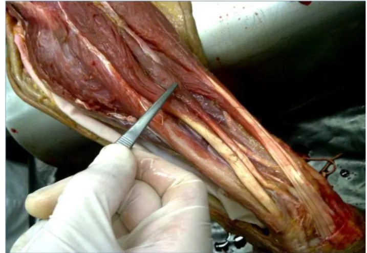
forearm of a human 73 year old male Caucasian cadaver. During the thorough dissection work of the right fore-arm, we clearly identified a thick bundle of muscular fibres,
bridging between the main muscular shafts of the Extensor
Digiti Minimi, and the Extensor Digitorum Communis
Mus-cles. This perfectly defined muscular expansion, of cc. 2 cm
thickness, was obliquely directed downwards and outwards, from the muscular belly of the of the Extensor Digiti Minimi,
to the main muscular shaft of the Extensor Digitorum Com
-munis. (Fig. 3)
The two main shafts of the superficial extensor digiti muscles had a well defined independent trajectory shortly after their common upper insertion in the lateral epicondyle of the umerus, and the muscular bridging occurred in the middle third of the forearm.
Around the upper third of the main muscles, a few other smaller muscle fibre expansions connected the two main muscle bellies. These were better defined, as all the fibrous sheaths and fasciae were dissected, and the main muscular bundles isolated and separated, with dissection tools (Fig. 4) No such muscular disposition was observed in the left forearm of the same cadaver and, in our series of more than 500 human forearm dissections, this is the first occurrence of such anatomical variation.
We cannot consider this as a supernumerary muscle, since no vasculo-nervous bundle was observed in relation to the muscular expansion bridging.
DISCUSSION
In our series of more than 500 human forearm dissec-tions, this is the first occurrence of such anatomical disposi-tion.
Anatomical variations of the extensor tendons to the fingers are frequently detected in the wrist, hand and fin-gers compartments (zones I, II and II of the Verdan classi-fication). We have often found variations in these compart-ments, in our extended series of dissections of forearms and hands in our yearly routine for teaching and research purposes of the Anatomy Department of the New University
Figure 2 – Verdan’s classification of the functional and topographic zones of the extensor muscles of the forearm and hand(Drawing adapted from Clavero’s, on Albinus’s plate (20), of a dorsal view of
the forearm and hand. – (I)= Distal Inter-Phalangeal Joint, drawn in pink; (II)= Middle Phalange , drawn in yellow; (III)= Proximal Inter-Phalangeal Joint, drawn in blue; (IV)= Proximal Phalange, drawn in green; (V)= MCP Joint, drawn in ocre; (VI)= Dorsum of Hand, drawn in yellow; (VII)= Wrist Extensor Compartment, drawn in vio-let; (VIII)= Extrinsic Extensor Tendons, drawn in cyan; and the ad-ditional Wéhbe’s zones: (IX)= Intramuscular Tendon Compartment, drawn in brown; (X)= Muscle belly Compartment, drawn in grey)
Figure 3 – Muscular bridging expansion. Muscular bridging ex-pansion between the main muscular shafts of the Extensor Digiti Minimi and the Extensor Digitorum Communis Muscles, in the right forearm of a 73 year old male caucasian cadaver. (The tweezers point to a clearly defined bundle of muscular fibers connecting the two main muscular bellies)
of Lisbon.9
SR Nayak10 remarks, on this purpose, that ‘the extensor
compartment of the forearm is one of the regions of the hu-man body with frequent variations of its content’. (*4)Most of these variations respect the distribution pattern of the ex-tensor tendons to the fingers, as commonly reported in the
modern anatomical literature.12-17
An interesting variant, frequently found in what regards the extensor apparatus of the fingers, is the presence of
in-tertendinous connections (juncturae tendinum) between the
various extensor tendons, on the dorsum of the hand. Their frequency is so high, that they were commonly depicted in the earliest Anatomy books and drawings, such as
Vesa-lius (1543 - IXth and XIth Plates of the muscles),18 Valverde
(1566 - Libri II, Tabula IX & XV-Fig.XIX),19 Albinus (1747),20 M Verdier (1751),21 or Chaussier (1823).22 (*5)
Modern clinical Anatomy reports often refer the pres-ence of tendinous slips between the extensor tendons on the dorsum of the hand.
HP von Schroeder et al24 classify these Juncturae Tendi
-num of the extensor tendons into three types: Type I
junctu-rae consist of filamentous regions within the intertendinous fascia; Type II consists of much thicker and well-defined connecting bands; and Type III consist of tendon slips from the extensor tendons and were subclassified into «y» or «r» subtypes, depending on shape. In their series of 548
ca-daveric hand dissections, Y Hirai et al25 found that the most
common pattern of intertendinous connections were
clas-4 (*) According to P. Daas (2012),11 the extensor digiti minimi tendon
showed normal anatomy in only 20% of the 100 cases studied.
5 (*) Honoré de Fragonard’s meticulous dissections of whole bodies23 were kept since 1766 to the present days, in France, and his dissections of forearms and hands clearly demonstrate the existence of junctura tendinum between the extensor tendons of the IVth and Vth and of the IIIrd and IVth fingers, at the dorsum of the hand.
sified into Type I in the second intermetacarpal space, into Type III-r in the third metacarpal space, and into type III-y in the fourth intermetacarpal space.
The functional importance of these juncturae would be
to strengthen the frail tendons of the extensor mechanism of the fingers, at the cost of some loss of independence of the IVth finger.26-30
What if the tendinous or muscular slips were more proxi-mal, instead of distal, as is the case in discussion? Wouldn’t
this disposition confer more strength to Vth finger, without
loss of gliding independence of the IVth tendon?
As we expandour review of the anatomical literature,
to older anatomical notes, we discover a nearly forgotten world of accurate and ‘picturesque’ descriptions of Miology, which prove to be quite useful to a modern understanding of the frequent anatomical variations found in cadaveric dissections. We researched the works of some of the most representative authors in Anatomy, from Leonardo da Vinci
(1508)31 to the present date, with JA Esperança Pina.32
With this expanded bibliographic research, one can eas-ily verify that the earliest texts provide the more extensive anatomical descriptions, leading us to a collection of
origi-nal notes on the extensor muscles of the fingers.33
M Lieutaud (1776)34 remarks that the Extensor muscle
of the Vth. Finger should be considered a dependence of the
common Extensor of the fingers. The two tendons have a common origin and a common muscular belly that divides shortly above the extensor retinaculum, to rejoin again, at the metacarpal region, through a tendinous or fibrous slip. Poissonnier (1783) 35 and M Sabatier (1791)36 offer the same description: Both tendons have a common muscular belly until the lower third of the forearm. (…) At the dorsum of the hand, the extensor tendons of the fingers communicate
through thick oblique tendinous slips. Baron Boyer (1815)37
offers the description that most resembles our anatomical variant, as he remarks that the medial margin of the muscu-lar belly of the extensor digiti communis is strongly united to extensor digiti minimi muscle, in the upper half of the fore-arm, and the two muscles remain united in the lower half,
through some bundles of cellular tissue. X. Bichat (1823),38
on the other hand, remarks that the muscular fibres of the Extensor Digiti Minimi are only separated from the muscular fibres of the Extensor Digitorum Communis through fibrous tissue.
None of the quoted works refer the presence of muscu-lar slips between the two muscumuscu-lar bellies in the forearm. Astonishingly, J Cruveilhier (1872)39 (*6) includes, on this
6 (*) Quoting from the original edition of Cruveilhier(Pg.686) :
«Les tendons de l’annulaire et du petit doigt sont si intimement unis entre eux par la bande fibreuse transversale située au-dessus de l’articulation métacarpo-phalangienne, qu’il est bien difficile d’étendre isolément l’un ou l’autre de ces doigts ; C’est là une des grandes difficultés du jeu des instruments de musique, et surtout du piano. Les grands artistes seuls parviennent, par un exercice continuel, qui doit commencer dés l’âge le plus tendre, à obtenir l’indépendance de ces doigts ; encore n’est elle jamais complète.
L’auteur d’un instrument ingénieux destiné à obtenir cette indépendance m’ayant consulté à ce sujet, je lui ai fait connaître les dispositions ana -tomiques qu’il fallait vaincre pour arriver à ce résultat ; son appareil me parait atteindre le but autant que possible.»
Figure 4 –Muscular bridging expansion between the main belly of the Extensor Digiti Minimi and the Extensor Digitorum Communis
Muscles, in the right forearm of a 73 year old male caucasian ca-daver.(The dissection tools are used to deviate the two muscles, to emphasize the presence of the thick bundle of muscular fibres, bridging obliquely between the two main bellies of the muscles. Shouldn’t this disposition be considered a new type of Junctura, the
Junctura Musculorum, in zones VIII and IX of the extensor mecha-nism of the fingers?)
CASO CLÍNICO
purpose, some remarks on musician’s hands injuries that led us to conclude on the modernity and usefulness of our present review, in consideration to repetition strain injuries. His report on the Extensor muscles of the fingers and the
frequent finding of junctura tendinum is stunningly modern
in the sense that he includes several interesting remarks on the functional anatomy of the human fingers and a sur-prising historical note, with the mention of a musician who consulted his advice on building a mechanical device to
exercise on the independence of the 4th finger, to improve
pianist performance. Even though these devices were
com-mon in the 19th century, the anecdote immediately reminded
us of Schumann’s hand injury, as particularly reported by RA Henson & H Urich.40 This article led to an interesting
collection of postscripts,41-44 that we remember reading with
special interest on the British Medical Journal edition of
1978. Jonas Sén45 reported several other cases, of
musi-cian’s repetition strain injuries.
Schumann’s hand injury is the paradigm of repetition strain injuries frequently found in musicians, but also in modern days, in computer and electronic devices users.
CONCLUSIONS
The interest of anatomical studies of the forearm mus-cles has grown in modernity, since the advent of the com-puting era, in the 20th century: Nowadays, every human occupation involves the perfect accuracy of finger motion, from the virtuous pianist, to the manual factory worker in the assembly line, or even the modern intellectual who thrives through long working hours on his laptop keyboard. Even in their leisure times, many people now recur to computer keyboards, either for playing games, or for internet
naviga-tion and social communicanaviga-tion. (*7)
Apart from the obvious and fundamental importance for reconstructive surgery of injured fingers, the profound and meticulous knowledge of the regional anatomy of the fore-arm and hands is fundamental for the better understanding of the functional and clinical management of modern human beings, as repetitive strain injuries of the hand are slowly becoming one of the modern times plagues.
ACKNOWLEDGMENTS
We wish to thank G. Marques, P. Miranda, M. Miranda, B. Mota, first year Anatomy students of the Medical Course of the Faculty of Medical Sciences of the New University of Lisbon. They performed, under our supervision, the careful dissection work that supported the present study.
CONFLICT OF INTERESTS
The authors declare that no conflict of interests was identified. (The main theme of this article was delivered at the XXII International Symposium of Morphological Scienc-es, in São Paulo, Brazil (Feb. 2012), however the abstract and the full text of the paper was totally re-written for sub-mission to Acta Médica Portuguesa.)
FUNDING SOURCES
None stated.
7 (*)in Bettencourt Pires MA, 2013. Hands in Medicine and in Art. In Ed.
C.E.E.C.,Pires L, editora. Science and Humanities – Ways of Seeing the World – Proceedings of the Symposium “Recontextualizing Sciences from the Humanistic Perspective”, Lisboa, Universidade Católica, 2013. (in print)
REFERENCES
1. Bettencourt Pires MA, Pais D, O’Neill JE. An Unusual Variety of the Ex-tensor Digiti Muscles – Case Report , with Notes on Repetition Strain Injuries. In: Abstract Book of the XXII International Symposium of Mor-phological Sciences. São Paulo: XXII ISMS; 2012. p.65.
2. Verdan C. Les anomalies musculo-tendineuses et leur significa-tion en chirurgie de la main. Rev Chir Orthop Reparatrice Appar Mot. 1981;67:221–30.
3. Kleinert HE, Verdan C. Report of the Commitee on Tendon Injuries. J Hand Surg Am. 1983;8:794-8.
4. Turney BW. Anatomy in a Modern Medical Curriculum. Ann R Coll Surg Engl. 2007;89:104–7.
5. Wehbe MA. Anatomy of the extensor mechanism of the hand and wrist. Hand Clin. 1995;11:361-6.
6. Zilber S, Oberlin C. Anatomical variations of the extensor tendons to the fingers over the dorsum of the hand: a study of 50 hands and a review of the literature. Plast Reconstr Surg. 2004;113:214-21.
7. Mehta V, Jyoti A, Suri Rk, Rath G. An assembly of anomalous extensor tendons of the hand – anatomical description and clinical relevance. Acta Medica. 2009;52:27-30.
8. Clavero JA, Golanó P, Fariñas O, Alomar X, Monill Jm, Esplugas M. Extensor mechanism of the fingers: MR imaging-anatomic correlation. Radiographics. 2003;23:593-611.
9. Casal D, Pais D, Toscano T, Bilhim T, Rodrigues L, Figueiredo I, et al. A rare variant of the ulnar artery with important clinical implications: a case report. BMC Res Notes. 2012;5:660.
10. Nayak SR, Krishnamurthy A, Prabhu LV, Rai R, Ranade AV, Madhyastha S. Anatomical variation of radial wrist extensor muscles: a study in ca-davers. Clinics. 2008;63:85-90.
11. Dass P, Prabhu LV, Pai MM, Nayak V, Kumar G, Janardhanan JP. A comprehensive study of the extensor tendons to the medial four digits of the hand. Chang Gung Med J. 2011;34:612-9.
12. von Schroeder HP, Botte MJ. The functional significance of the long ex-tensors and juncturae tendinum in finger extension. J Hand Surg Am. 1993;18:641-7.
13. Cavdar S, Sehirli U. The accessory tendon of the extensor indicis mus-cle. Okajimas Folia Anat Jpn. 1996;73:139-42.
14. Beauchamps R. Our Anatomical Differences. Music and Health, online publication. [consultado em 2012 Dezembro]. Disponível em: http:// www.musicandhealth.co.uk/anatomy.html.
15. Nayak SR, Krishnamurthy A, Pai MM, Prabhu LV, Ramanathan LA, Ganesh Kumar C, et al. Multiple variations of the extensor tendons of the forearm Case Report. Romanian J Morphol Embryol. 2008;49:97–100. 16. Prakash R, Ranade AV, Prabhu LV, Pai MM, Singh G. Múltiples varia-ciones de los músculos extensores del antebrazo en relación con el nervio radial: Reporte de caso y revisión. Int J Morphol. 2008;26:447-9. 17. Wilhelm A. Anatomie der beuge- und strecksehnen. Langenbecks Arch
Chir Suppl Kongressbd. 1992:438-43.
18. Vesalius A. De Humani Corporis Fabrica, Basilea,1543. In: Saunders J, O’Malley D, editors. The Illustrations from the Work of Andreas Vesalius of Brussels. New York: Dover; 1973.
19. Valverde J. Vivae Imagines Partium Corporis Humani. Antuerpiae, Ex. Officina Christophori Plantini. In: Pieter Huys, editors. Nouvelle ed. Paris: Louis Pariente; 2001.p.53-69.
20. Albinus BS. Tabulae sceleti et Musculorum Corporis Humani, Lugduni Batavorum, 1747. In: Hale RB, Coyle T, editors. Albinus on Anatomy, with 80 Original Albinus Plates. New York: Dover; 1978.
21. Verdier M., Abrégé de l’Anatomie du Corps Humain. 2ème ed. Paris: PG Le Mercier; 1751.
22. Chaussier M. Planches Anatomiques, Pl.V. 2ème ed. Paris: Panck-oucke;1823.
CASO CLÍNICO 24. von Schroeder HP, Botte MJ, Gellman H. Anatomy of the juncturae
tend-inum of the hand. J Hand Surg Am. 1990;15:595-602.
25. Hirai Y, Yoshida K, Yamanaka K, Inoue A, Yamaki K, Yoshizuka M. An anatomic study of the extensor tendons of the human hand. J Hand Surg Am. 2001;26:1009-15.
26. von Schroeder HP, Botte MJ. Anatomy of the extensor tendons of the fingers: variations and multiplicity. J Hand Surg Am. 1995;20:27-34. 27. von Schroeder HP, Botte MJ. Functional anatomy of the extensor
ten-dons of the digits. Hand Clin. 1997;13:51-62.
28. Nimbarte AD, Kaz R, Li ZM. Finger joint motion generated by individual extrinsic muscles: a cadaveric study. J Orthop Surg Res. 2008;3:27. 29. Klena JC, Riehlt JT, Beck JD. Anomalous extensor tendons to the long
finger: a cadaveric study of incidence. J Hand Surg Am. 2012;37:938-41.
30. Pinar Y, Gövsa F, Bilge O, Celik S. Accessory tendon slip arising from the extensor carpi ulnaris and its importance for wrist pain. Acta Orthop Traumatol Turc. 2012;46:132-5.
31. Goldsheider L. Leonardo da Vinci – The Artist and the Man. Oxford: Phaidon Press; 1945.
32. Esperança Pina JA. Anatomia Humana da Locomoção. 4ª ed. Lisboa: Lidel; 2010.
33. Gelée Th. L’Anatomie Françoise, en Forme d’Abbrégé. Recueillie des meilleurs autheurs qui ont écrit de cette Science. Rouen: David
Berthe-lin; 1663.
34. Lieutaud M. Anatomie historique et pratique. Nouvelle édition augmen-tée de diverses remarques par M. Portal. Paris: chez Vincent; 1776. 35. Poissonnier PI. Abrégé d’Anatomie à l’Usage des Élèves en Médecine
& Chirurgie. Paris: MC; 1783
36. Sabatier M. Traité Complet d’Anatomie, ou Description de Toutes les Parties du Corps Humain. Tome I: Myologie. 3ème ed. Paris: Théophile Barrois; 1791.
37. Boyer B. Traité Complet D’anatomie, ou Description de toutes les Par-ties du Corps Humain. Tome II: Myologie. 4ème ed. Paris: Migneret Im-primeur; 1815.
38. Bichat X. Traité d’Anatomie Descriptive. Tome II. Paris: Gabon et Com-pagnie Libraires; 1823.
39. Cruveilhier J. Traité d’Anatomie Descriptive. 4ème ed. Paris: Asselin; 1862.
40. Henson RA, Urich H. Schumann’s hand injury. Br Med J. 1978;1:900–3. 41. Ballantyne J. Schumann’s hand injury. Br Med J. 1978;1:1142. 42. Mather H. Schumann’s hand injury. Br Med J. 1978;1:1281. 43. Walker A. Schumann’s hand injury. Br Med J. 1978;1:1420. 44. Henson RA. Schumann’s hand injury. Br Med J. 1978;1:1348. 45. Sén J. Playing the piano: playing with fire? A study of the occupational
hazards of piano playing. A Dissertation submitted for the degree in MA in Music . New York: New York City University Music Department; 1991.
Variação Anatómica Rara de Ausência do Nervo
Ciático: Completamente Substituído Pelos
Nervos Tibial e Fibular Comum
Rare Anatomical Variation of Absence of the Sciatic Nerve:
Completely Substituted by the Tibial and Common Fibular Nerve
Paulo COELHO1, Catarina MELO1, António BERNARDES1
Acta Med Port 2013 May-Jun;26(3):283-286
RESUMO
Introdução: Existem várias publicações referindo variantes anatómicas do nervo ciático, algumas associadas a síndromes clínicos
(como sendo a síndrome do músculo piriforme). Neste contexto, pretendemos apresentar uma variante anatómica rara do nervo ciático.
Casos clínicos: Dois cadáveres leucodérmicos, masculinos, com 74 e 78 anos, falecidos de morte natural, sem patologia do membro
inferior. Em ambos os casos, observou-se ausência do nervo ciático direito, tendo os nervos tibial e fibular comum origem e trajecto independentes, desde a sua origem nas raízes lombo-sagradas até à região poplítea. O nervo ciático contralateral apresentava a anatomia habitual.
Discussão: Analisando a literatura, na Medline, realçamos que apresentamos dois casos raros de ausência do nervo ciático, com
origem e trajecto independentes dos nervos tibial e fibular comum. Esta variante poderá ter implicações clínicas, nomeadamente ser um factor de risco para o insucesso de bloqueios anestésicos poplíteos e para a síndrome do músculo piriforme.
Palavras-chave: Cadáver; Nervo Ciático; Nervo Tibial; Síndrome do Músculo Piriforme; Variação Anatómica.
1. Instituto de Anatomia Normal. Faculdade de Medicina da Universidade de Coimbra. Coimbra. Portugal. Recebido: 28 de Novembro de 2012 - Aceite: 23 de Fevereiro de 2013 | Copyright © Ordem dos Médicos 2013 ABSTRACT
Introduction: There are several reports of sciatic nerve anatomical variations. Some are associated with clinical entities, such as
piri-formis syndrome. We aim to report a rare anatomical variation of this nerve.
Cases report: Two leucodermic, 74 and 78-year-old male subjects, deceased of natural causes, without lower limb relevant medical
history. In both subjects, the right sciatic nerve was absent, with an independent origin and course of the tibial and common fibular nerves. The contralateral sciatic nerve had the common anatomical presentation.
Discussion: After the analysis of the available data indexed in Medline, we conclude that we are reporting two cases of a rare
ana-tomical variation (the absence of sciatic nerve, with an independent origin and course of the tibial and common fibular nerve). This anatomical variation may have clinical importance, as it may be, for example, a risk factor to unsuccessful sciatic nerve popliteal blocks and to the pyriformis syndrome.
Keywords: Cadaver; Genetic Variation; Muscle, Skeletal/abnormalities; Piriformis Muscle Syndrome; Sciatic Nerve/abnormalities;