ORIGIN
AL RESEAR
CH
Corresponding address: Rafael Inácio Barbosa – UFSC, Campus Mato Alto – Rua Pedro João Pereira, nº 150, Araranguá (SC), Brasil – Zip Code: 88905-120 – Email: [email protected] – Financing source: Nothing to declare – Conlict of interest: Nothing to declare – Presentation: Nov. 2016 – Accepted for publication: Mar. 2017 – Approved by the Ethics Committee: 122645/2015.
1Laboratory of Assessment and Rehabilitation of the Locomotor System, Universidade Federal de Santa Catarina (LARAL/UFSC) –
Araranguá (SC), Brazil.
2Laboratory of Clinical Research of the Hand and Upper Limb, Faculdade de Medicina de Ribeirão Preto of Universidade de São Paulo
(LabMão-USP) – Ribeirão Preto, São Paulo, Brazil.
3Graduate program in Rehabilitation Sciences, Universidade Federal de Santa Catarina, Araranguá (SC), Brazil.
ABSTRACT | The objective of this study was to evaluate the efects of a wrist extensor muscles fatigue protocol at the handgrip and lateral pinch strength through dynamometry and surface electromyography (EMG). Forty male individuals were divided into two groups: handgrip and lateral pinch group. The fatigue protocol was based on the 1 Maximal Repetition (1-MR) test, followed by wrist extension movement repeated multiple times with a load of 75% of 1-MR. The volunteers performed hand grip and lateral pinch with a dynamometer. Surface EMG was performed by both groups to analyze the behavior of median frequency (MF) during a fatigue protocol. The muscles extensor
carpi radialis longus and brevis, extensor carpi ulnaris,
extensor digitorum and flexor digitorum superficialis
were analyzed. The strength and EMG evaluations were carried out before and after the fatigue protocol in both groups. The fatigue protocol was efective on hand grip strength reduction (43.5±3.85 kgf in baseline and 36.50±5.1 kgf inal) and lateral pinch strength reduction (10.26±1.01 kgf in baseline and 8.54±0.86 kgf inal) (p<0.05, 95% CI). At the EMG analysis, using median frequency, an ulnaris carpal extensor muscle fatigue at the handgrip group was evidenced. The indings indicate that wrist extensors fatigue can decrease the strength in functional activities such as handgrip, resulting in upper limb dysfunctions.
Keywords | Grip; Pinch Strenght; Fatigue; Eletromiography; Dinamometry.
100
RESUMO | O objetivo deste estudo foi avaliar os efeitos de um protocolo de fadiga dos músculos extensores de punho na força de preensão e da pinça lateral através da dinamometria e eletromiograia de superfície (EMG). Foram selecionados 40 indivíduos do sexo masculino, divididos em dois grupos: preensão ou pinça lateral. O protocolo de fadiga foi baseado no teste de 1 Repetição Máxima (1-RM), seguido da realização do movimento de extensão de punho repetidas vezes com carga de 75% da 1-RM. Os voluntários realizaram as tarefas de preensão ou pinça lateral associadas à dinamometria. A EMG foi realizada para ambos os grupos, analisando o comportamento, segundo o protocolo, pela frequência mediana (FM) do extensor radial do carpo (ERC), do extensor ulnar do carpo (EUC) e do lexor supericial dos dedos (FD). A dinamometria de preensão ou pinça lateral e a EMG foram realizadas antes e após o protocolo de fadiga para ambos os grupos. O protocolo de fadiga foi eicaz na diminuição da força de preensão palmar (43,5±3,85 kgf inicial e 36,50±5,1 kgf inal) e da pinça lateral (10,26±1,01 kgf inicial e 8,54±0,86 kgf inal), bem como na diminuição da FM, sugerindo uma condição de fadiga do EUC no grupo preensão. Os achados do presente estudo possibilitam relacionar a fadiga dos extensores de punho à diminuição de força em atividades funcionais, como a preensão, o que pode implicar em disfunções musculoesqueléticas do membro superior.
Descritores | Força da Mão; Força de Pinça; Fadiga; Eletromiograia; Dinamometria.
Fatigue of the wrist extensor muscles decreases
palmar grip strength
Fadiga dos músculos extensores do punho diminui a força de preensão palmar
La fatiga de los músculos extensores de muñeca disminuye la fuerza de prensión palmar
Vitor Kinoshita Souza1,3, Adrian Freitas Claudino¹, Heloyse Uliam Kuriki1,3, Alexandre Marcio Marcolino1,2,3,
RESUMEN | En este estudio se evalúa los resultados de un protocolo de fatiga de los músculos extensores de muñeca en la fuerza de presión y de pinza empleando la dinamometría y la electromiografía de superficie (EMG). Se eligieron a cuarenta hombres, los cuales fueron divididos en dos grupos: el de presión y el de pinza lateral. Se basó el protocolo de fatiga en la prueba de 1 Repetición Máxima (1RM), y se realizó el movimiento de extensión de muñeca muchas veces con carga del 75% de la 1RM. Los participantes realizaron la tarea de prensión o de pinza lateral asociada a la de dinamometría. La EMG fue realizada por ambos grupos, en los que se evaluó el comportamiento ante el protocolo de la frecuencia mediana (Fm) del extensor radial del carpo (ERC), del extensor cubital del carpo (ECC) y del flexor superficial
de los dedos (FD). Se realizaron la dinamometría de presión o de pinza lateral y la EMG antes y después del protocolo de fatiga en ambos grupos. El protocolo de fatiga fue eficaz en la disminución de la fuerza de presión palmar (43,5±3,85 kgfinicial y 36,50±5,1 kgffinal) y de pinza lateral (10,26±1,01 kgfinicial y 8,54±0,86 kgffinal), así como en la disminución de la Fm, lo que demuestra una condición de fatiga del ECC en el grupo de presión. Los resultados de este estudio permiten relacionar la fatiga de los extensores de muñeca a la disminución de fuerza en las actividades funcionales, lo que puede causar trastornos musculoesqueléticos del miembro superior.
Palabras clave | Fuerza de la Mano; Fuerza de Pinza; Fatiga; Electromiografía; Dinamometría.
INTRODUCTION
In recent years, trauma-orthopedic injuries of the upper limb have been featuring in the scientiic literature with the increased incidence of traumatic injuries1,2 and also of diferent pathologies related to musculoskeletal overload3,4. In the evaluation of results of patients treated for upper limb disorders, various clinical parameters are used, among which we highlight the lateral pinch and palmar grip strength5,6. Individuals with palmar grip and lateral pinch dysfunction present deicit in daily life activities, thus the return of strength to this body segment is necessary7,8.
Sensory capacity combined with agility are essential to the daily performance of the distal segment movements of the upper limb. Muscles in this region are of the utmost importance because they enable the practice of movements, such as the lateral pinch and the grip, therefore, a set of muscle actions is necessary such as the wrist extensor muscles activation associated with contraction of inger lexors9-11.
Bawa et al.12 describe the synergy that exists between the wrist extensor muscle and inger lexor muscle. Extensor muscles act on stabilization of the wrist in extension during activities of grip and pinch. Studies that investigate this synergistic relationship in functional activities are important for the understanding of pathophysiology and also to outline prevention and treatment protocols in musculoskeletal dysfunctions such as lateral epicondylitis5,13.
Caporrino et al.14, in a populational study of the palmar grip strength with Jamar® dynamometer for the Brazilian population, deined that the average of palmar grip strength in the dominant side is 44.2±8.9 kgf for male individuals and 31.6±7.5 kg for female individuals. With the same goal, Araujo et al.15 conducted a populational study of pinch strength with pinch gauge™ dynamometer and concluded that the strength of lateral pinch in the male gender presents an average of 9.9±1.9 kgf and 6.7±1.4 kgf in the female gender. Studies related to normative data for grip and pinch are presented in diferent countries and serve as a reference in clinical practice in order to compare possible changes of grip strength and pinch, arising from diferent lesions of the upper limb6,16. Tang et al.17 describe that changes in biomechanical parameters of the wrist can be an important cause of diseases and dysfunctions. Alizadehkhaiyat et al.18 describe the fatigue as a possible etiology of wrist and elbow dysfunctions.
results in adenosine diphosphate (ADP), which justiies the increase of its concentration during muscle fatigue19. he electromyographic (EMG) assessment seeks to investigate parameters involving muscle action such as fatigue, conduction velocity, diagnosis of muscular diseases and the muscle recruitment pattern20-22. Median frequency (MF) is the most reliable parameter for measuring the changes of EMG spectra, since this parameter is less sensitive to noises and more sensitive to physiological changes related to muscular fatigue such as the accumulation of lactate and extracellular potassium. his metabolic accumulation leads to reduction in the conduction velocity of the action potential, resulting in a decline in the MF values as the muscle becomes fatigued23-25. Studies demonstrate the diiculty in assessing the EMG of the forearm muscles due to proximity of the extensor and lexor compartments26,27. Rota et al.28 describe the inluence of fatigue in muscular performance of tennis athletes, and report the importance of surface electromyography in the evaluation of activation and fatigue in the muscles of the upper limb in this population.
In this context, this study aimed to evaluate the pattern of surface EMG in a fatigue protocol of the wrist extensor muscles and the relationship of this protocol in the alteration of grip strength and lateral pinch.
METHODOLOGY
he study was characterized as randomized controlled, with a proposal for evaluation of individuals before and after the fatigue protocol of the wrist extensor muscles, having as outcomes the palmar grip strength, the lateral pinch and the electromyographic activity in diferent groups. It was approved by the Research Ethics Committee at Universidade Federal de Santa Catarina (UFSC) – 122645/2015 and all subjects recruited signed an informed consent form.
SUBJECTS
Forty male individuals were volunteers and included in the research, aged between 18 and 25 years, no regular physical activity practitioners, who were subdivided into two groups of 20 individuals: Handgrip Group or Lateral Pinch Group.
History of nerve damage associated with bone or joint complex multiple lesions, the presence of central nervous system injury, rheumatic diseases, leprosy and any disease in the upper limbs were considered criteria for non-recruitment.
Randomization to assemble groups was based on a sequence of random numbers generated by the Excel® program.
Evaluations were carried out in LARAL (Laboratory for Evaluation and Rehabilitation of the Locomotor System – UFSC/Araranguá) and divided into two phases: on the irst day, maximal repetition (1-MR) was calculated for the wrist extensor muscles in the dominant upper limb and, after a week, the pre-fatigue evaluation was carried out (EMG associated with task of grip or lateral pinch, depending on the group), the extensor fatigue protocol and the post-fatigue evaluation (EMG associated with grip or lateral pinch task).
PROCEDURES
Fatigue Protocol
Participants were told not to perform moderate or vigorous physical activity 24 hours before data collection.
he load used for the fatigue protocol was based on the 1-MR test, which consisted in the biggest load the volunteer has achieved the full wrist extension, starting from full lexion.
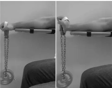
he fatigue protocol consisted in the performance of lexion and extension movements of the wrist in the maximum range, with the elbow in extension (Figure 1), with 75% of 1-MR load, preset to every volunteer, associated with the use of a metronome with frequency of 50 bpm, being interrupted when the volunteer was unable to maintain the frequency in two consecutive or three alternate movements or failed to perform the maximum range29,30.
Figure 1. Positioning of the upper limb and resistance to implementation of the fatigue protocol of the wrist extensor muscles.
Grip or lateral pinch task associated with EMG
he tasks performed by the volunteers were: palmar grip using Jamar® dynamometer (Handgrip Group) or lateral pinch with the Pinch Gauge® (Lateral Pinch Group). he individual’s positioning for evaluation followed the recommendation of ASHT (American Society of Hand herapy), which is followed by SBTM (Brazilian Society of herapy of the Hand and Upper Limb)31. Individuals were positioned comfortably on a chair without armrests, feet fully supported on the loor, the arm parallel to the body, elbow at 90º and forearm in neutral position. Evaluations occurred before and after the proposed fatigue protocol.
Muscle electrical signal was captured by the electromyograph Miotool 400 (Miotec®, Porto Alegre, RS, Brazil), interfaced with the Miograph® software (Miotec®), with a 14-bit resolution analog-to-digital converter (A/D), acquisition ampliied in 2000 Hz and 100dB common-mode rejection, with 10-500 Hz bandpass ilter. Electrodes with bipolar contact Ag/AgCl (Silver/silver chloride) and 20 mm distance between poles – Medtrace® were positioned in the muscles: extensor carpi radialis (ECR) – longus and brevis –, extensor carpi ulnaris (ECU) and lexor digitorum supericialis (FD)32 (Figure 2). hree maximum voluntary isometric contractions (MVIC) maintained by six seconds were requested, with one minute rest interval between each task for both groups. he evaluations occurred before and after the fatigue protocol, with the reference electrode positioned over the acromion of the dominant member.
Figure 2: Positioning of electrodes for EMG signal capture of extensor carpi ulnaris (ECU), extensor carpi radialis (ECR) and lexor digitorum supericialis (FDS) muscles during the task of palmar grip strength and lateral pinch.
Data Analysis
he electromyography tracings were processed in the frequency domain, extracting the median frequency (MF) in Hertz; the time of exhaustion of fatigue protocol is calculated in seconds, and the measures of grip strength and lateral pinch are measured in Kgf.
Statistical analysis was performed by the program GraphPad Prism™, version 6.0. Data normality was checked by the Shapiro-Wilk test, applying the One Way ANOVA test with Tukey’s post hoc analysis, considering the signiicance of 5%.
RESULTS
Table 1 presents the anthropometric data and the age of the sample, which were homogeneous between the groups.
Table 1.Initial characteristics of the groups
Grip (n=20) Lateral pinch (n=20)
Age (SD) – years 22.32 (±3.58) 21.94 (±1.73)
Dominance (R/L) 17/3 18/2
BMI (SD) – kg/m² 24.75 (±2.15) 25.11 (±3.02)
1-MR (SD) – kg 12.35 (±2.32) 12.62 (±2.49)
BMI: Body mass index; MR: Maximal repetition.
In the assessment of grip strength and lateral pinch, we can observe that there were signiicant diferences when comparing assessments before and after fatigue protocol of the wrist extensor muscles, evidencing decreased strength in both groups (Figure 3).
50
40
30
20
10
0
Grip before fatigue Grip after fatigue Lateral pinch before fatigue Lateral pinch after fatigue
Dynamometry
Kg
F
Figure 3. Average values (standard deviation) of force in kilogram-force of groups grip and lateral pinch in the moments before and after fatigue. *p< 0.05 - CI 95% grip before strength versus grip after strength
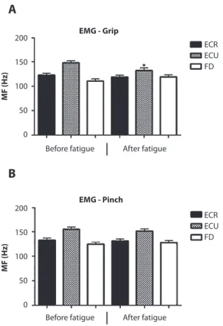
On the analysis of electromyography tracings from MF values, we can observe that it occurred more often in the ECU for both groups, in the initial assessment when compared with other muscles. It is worth mentioning that only the ECU of the Handgrip Group presented a signiicant decrease in the MF values when compared with initial and inal assessment (Figure 4).
EMG - Grip
Before fatigue After fatigue
ECR ECU FD 200
150
100
50
0
A
MF (H
z)
200
150
100
50
0
ECR ECU FD
B
EMG - Pinch
MF (H
z)
Before fatigue After fatigue
Figure 4. A – Average values of the median frequency (MF) of extensor carpi radialis (ECR), extensor carpi ulnaris (ECU) and lexor digitorum supericialis (FDS) muscles for the grip group. B – Average values of the median frequency (MF) of extensor carpi radialis (ECR), extensor carpi ulnaris (ECU) and lexor digitorum supericialis (FDS) muscles for the lateral pinch group, * p< 0.05 – CI 95% versus its respective before fatigue.
DISCUSSION
he assessment of palmar grip strength lateral pinch is widely used to evaluate upper limb disabilities as well as its ability to work. Manual tasks, such as palmar grip and lateral pinch, are study objects since the hand combines two diferent functions: strength and dexterity. In addition, palmar grip strength is predictive of future disabilities, morbidity and mortality, not only in older adults, but also in middle-aged and young people6,33-35, which strengthens the importance of studies in this area. he aim of this study was to evaluate the immediate efects of a fatigue protocol in the wrist extensor muscles and their relationship with the palmar grip strength and lateral pinch in diferent groups. Individuals accepted well the protocol, and no complaint occurred regarding the time of assessment. he protocol has proven to be efective in decreasing the strength of inger lexors, highlighting the synergistic efect between the wrist extensors and lexors of ingers. Corroborating this study, Danna-Dos Santos et al.36 refer that fatigue changes muscle strength and coordination of the hand muscles.
Diferent studies seek to observe the biomechanical properties in the synergistic relationship between the wrist extensors and inger lexors in musculoskeletal dysfunctions. Studies sought to evaluate the function of the forearm muscles in professional tennis players with or without epicondylitis; among the indings, the authors report the need to evaluate activation and fatigue in wrist extensor muscle in an attempt to identify players more susceptible to development of lateral epicondylitis18,37. his study sought to identify the relation of fatigue of the extensors in the changes of grip strength and lateral pinch in normal individuals, since the indings regarding grip indicate the need for future studies analyzing the pattern in individuals with elbow lateral epicondylitis, commonly observed in the population assisted by physical therapy.
he electromyographic assessment has been used for the evaluation of forearm muscles, in search of a recruitment pattern. Even with a wide variety of studies, we can observe that there is still no consensus on the positioning of electrodes and the inluence of diferent wrist angles in the palmar grip task32,34,38, 39.
neuromuscular activation, due to increased recruitment of motor units to compensate for the saturation of ibers that are already fatigued, avoiding the immediate failure of the system24. Our electromyographic indings showed a decrease of the ECU median frequency in both groups, with statistical diference for the Handgrip Group after the fatigue protocol. In the Handgrip Group evaluation, only the ECU presented decline in the MF, which suggests its fatigue after the protocol in week 2, when comparing before and after assessments of the wrist extensors fatigue.
Decline in the MF is also observed in the study of Da Silva et al.25 in muscles of the lumbar region, when the authors sought to compare the parameters of fatigue in young and older people with chronic low back pain. Larivière et al.40 showed the same change on the MF in the gluteus maximus muscle when they analyzed the speciicity of an exercise about the “Roman Chair” in healthy people or people with low back pain. Da Silva et al.41 evaluated the reproducibility of MF in muscle fatigue located in the quadriceps venters and concluded that the MF is a reproducible and sensitive parameter for the characterization of muscle fatigue. Accordingly, Rota et al.28 describe the inluence of muscle fatigue of the pectoralis major muscle and the muscles of the forearm in decreasing the performance of tennis athletes.
his study demonstrates the importance of electromyographic evaluation of the wrist extensor muscles associated with the palmar grip strength or lateral pinch as indicative of muscle fatigue. herefore, we suggest the incorporation of these analyses in clinical routines involving the evaluation and treatment of patients with upper limb dysfunctions, more speciically, of the wrist extensor muscles and inger lexors of individuals who perform resisted repeated movements of the wrist.
Among the study limitations, we considered the small follow-up time of volunteers, suggesting an efect in the short term and the need for comparison with other models associated with trauma-orthopedic pathologies in the upper limb.
CONCLUSION
In the sample and in the model used, we can conclude that the fatigue protocol of wrist extensor muscles was efective in decreasing palmar grip strength and lateral
pinch. In addition, the extensor carpi ulnaris muscle showed a decrease in the median frequency, suggesting fatigue of the muscle. he indings suggest that further studies with this methodology can bring contributions regarding synergistic assessment or agonist/antagonist relationship of these muscle groups, relating to the etiology and treatment of musculoskeletal dysfunctions of the forearms and wrist.
REFERENCES
1. Avery DM, Rodner CM, Edgar CM. Sports-related wrist and hand injuries: a review. J Orthop Surg Res. 2016;11:99. doi: 10.1186/s13018-016-0432-8
2. Rubin G, Peleg K, Givon A; Israel Trauma Group, Rozen N. Upper extremity fractures among hospitalized road traic accident adult. Am J Emerg Med. 2015;33:250-3. doi: 10.1016/j. ajem.2014.11.048.
3. Henderson CJ, Kobayashi KM. Ulnar-sided wrist pain in the athlete. Orthop Clin North Am. 2016;47(4):789-98. doi: 10.1016/j.ocl.2016.05.017.
4. Dimitrios S. Exercise for tendinopathy. World J Methodol. 2015;5(2):51-4. doi: 10.5662/wjm.v5.i2.51
5. Chourasia AO, Buhr KA, Rabago DP, Kijowski R, Irwin CB, et al. Efect of lateral epicondylosis on grip force development. J Hand Ther. 2012;25(1):27-37. doi: 10.1016/j.jht.2011.09.003 6. Fain E, Weatherford C. Comparative study of millennials’
(age 20-34 years) grip and lateral pinch with the norms. J Hand Ther. 2016;29(4):1-5. doi: 10.1016/j.jht.2015.12.006. 7. Vinjamuri R, Mao ZH, Sclabassi R, Sun M. Limitations of
surface EMG signals of extrinsic muscles in predicting postures of human hand. Conf Proc IEEE Eng Med Biol Soc. 2006;1:5491-4.. DOI: 10.1109/IEMBS.2006.260329
8. Krischak A, Krasteva F, Schneider D, Gulkin F, Gebhard M. Physiotherapy after volar plating of wrist fractures is efective using a home exercise program. Arch Phys Med Rehabil. 2009;90(4):537-44.
9. Hoozemans MJM, Van Dieën JH. Prediction of handgrip forces using surface EMG of forearm muscles. J Electrrmyogr Kinesiol. 2005;15(4):358-66. DOI: 10.1016/j.jelekin.2004.09.001 10. Finneran A, O’sullivan L. Efects of grip type and wrist posture
on forearm EMG activity, endurance time and movement accuracy. Int J Industr Ergon. 2013;43(1):91-9. http://dx.doi. org/10.1016/j.ergon.2012.11.012
11. Shimose R, Matsunaga A, Muro M. Efect of submaximal isometric wrist extension training on grip strength. Eur J Appl Physiol. 2011;111(3):557-65. DOI: 10.1007/s00421-010-1675-4 12. Bawa P, Chalmers GR, Jones KE, Segaard K, Walsh MI.
Control of the wrist joint in humans. Eur J Appl Physiol. 2000;83(2):116-27.
14. Caporrino FA, Faloppa F, Santos JBG, Réssio C, Soares FHC, et al. Estudo populacional da força de preensão palmar com dinamômetro Jamar®. Rev Bras Ortop. 1998;33(2):150-4. 15. Araújo MP, Pola MP, Caporrino FA, Faloppa F, Albertoni M.
Estudo populacional das forças das pinças polpa-a-polpa, trípode e lateral. Rev Bras Ortop. 2002;37(11/12):496-504. 16. Steiber N. Strong or weak handgrip? normative reference
values for the german population across the life course stratiied by sex, age, and body height. PLoS ONE. 2016;11(10):e0163917.
17. Tang JB, Ryu J, Han J, Omokawa S,Kish V, Wearden S. Biomechanical changes of the wrist lexor and extensor tendons following loss of scaphoid integrity. J Orthop Res. 1997;15(1):69-75. doi: 10.1002/jor.1100150111
18. Alizadehkhaiyat O, Fisher AC, Kemp GJ, Vishwanathan K, Frostick SP. Upper limb muscle imbalance in tennis elbow: a functional and electromyographic assessment. J Orthop Res. 2007;25:1651-7. doi: 10.1002/jor.20458
19. Allen DG, Lamb GD, Westerblad H. Skeletal muscle fatigue: cellular mechanisms. Physiol Rev. 2008;88:287-332. doi: 10.1152/physrev.00015.2007
20. Reaz MBI, Hussain MS, Mohd-yasin F. Techniques of EMG signal analysis: detection, processing, classiication and applications. Biol Proced Online. 2006;8(1):11-35. doi: 10.1251/ bpo115
21. Pinter IJ, Bobbert MF, Soest AJKV, Smeets JBJ. Isometric torque–angle relationships of the elbow lexors and extensors in the transverse plane. J Electromyogr Kinesiol. 2010;20(5):923-31. doi: 10.1016/j.jelekin.2010.05.001
22. Keir PJ, Brow MM. Force, frequency and gripping alter upper extremity muscle activity during a cyclic push task. Ergonomics. 2012;55(7):813-24. doi: 10.1080/00140139.2012.668947 23. Bonato P, Roy SH, Knalitz M, De Luca CJ.. Time-frequency
parameters of the surface myoelectric signal for assessing muscle fatigue during cyclic dynamic contractions. IEEE Trans Biomed Eng. 2001 Jul;48(7):745-53. doi: 10.1109/10.930899 24. De luca CJ. The use of surface electromyography in
biomechanics. Journal Applied Biomechanics. 1997;13:135-163. 25. Da silva RA et al. Back muscle fatigue of younger and older
adults with and without chronic low back pain using two protocols: A case-control study. Journal of Electromyography and Kinesiology. 2015;25(6):928-936
26. Mogk JPM, Keir PJ. The efects of posture on forearm muscle loading during gripping. Ergonomics. 2003;46(9):956-975. 27. Kong Y, Hallbeck MS, Jung M. Crosstalk efect on surface
electromyogram of the forearm lexors during a static grip task. Journal of Electromyography and Kinesiology. 2010;20:1223-1229.
28. Rota S, Morel B, Saboul D, Rogowski I, Hautier C. Inluence of fatigue on upper limb muscle activity and performance
in tennis. Journal of Electromyography and Kinesiology. 2014;24:90-97.
29. Bruniera CAV, Rogério FRPG, Rodacki ALF. Stabilometric response during single-leg stance after lower limb muscle fatigue. Brazilian Journal of Physical Therapy. 2013;17(5):464-469
30. Leal junior EC, Lopes-martins RA, Dalan F et al. Efect of 655-nm low-level laser therapy on exercise-induced skeletal musclefatigue in humans. Photomed Laser Surg. 2008;26:419-424.
31. Mathiowetz V, Wiemer DM, Federman SM. Grip and pinch strength:norms for 6- to 19-year-olds. The American Journal of Occupational Therapy. 1986;40(10):705-711.
32. Marcolino AM, Barbosa RI, Rodrigues E, Tamanini G, Colombari F, Fonseca MC. Inluence of volar and dorsal static orthoses in diferent wrist positions on muscle activation and grip strength in healthy subjects. Hand Therapy. 2014;19:114-125. 33. Sayer, AA et al. Grip strength and mortality: a biomarker of
ageing? The Lancet. 2015;386:226-227
34. Tanaka DM, Ferreira, Aline Miranda, Colombari F, Barbosa RI, Marcolino AM, Mazzer N, Fonseca MC. Muscles co-activation and wrist position during sustained grip in healthy subjects. Revista do Instituto de Ciências da Saúde. 2014;32:194-197. 35. Roberts HC, Denison HJ, Martin HJ, Patel HP, Syddall H,
Cooper C, Sayer AA. A review of the measurement of grip strength in clinical and epidemiological studies: towards a standardised approach. Age and Ageing. 2011;40:423-429. 36. Danna-Dos Santos A, Poston B, Jesunathadas M, Bobich
LR, Hamm TM, Santello M. Inluence of Fatigue on Hand Muscle Coordination and EMG-EMG Coherence During Three-Digit Grasping. Journal of Neurophysiology. 2010;104(6):3576-3587.
37. Alizadehkhaiyat O, Frostick SP. Electromyographic assessment of forearm muscle function in tennis players with and without Lateral Epicondylitis. Journal of Electromyography and Kinesiology. 2015;25:876-886
38. Ferreira AM, Fonseca MCR, Tanaka DM, Barbosa RI, Marcolino AM, Elui VMC, Mazzer N. Should we think about wrist extensor after lexor tendon repair? SAGE Open Medicine. 2013;1:1-6 39. Coldham F, Lewis J, Lee H. The Reliability of One vs. Three
Grip Trials in Symptomatic and Asymptomatic Subjects. Journal of Hand Therapy. 2006;19:318-327.
40. Larivière C, Da silva RA, Arsenault AB, Nadeau S, Plamondon A, Vadeboncoeur R. Speciicity of a back muscle roman chair exercise in healthy and back pain subjects. Medicine & Science in Sports & Exercise. 2011;43:157-64.