Chromosome mapping of 5S rRNA genes differentiates Brazilian
populations of
Leporellus vittatus
(Anostomidae, Characiformes)
Cecilia Teixeira de Aguilar
1and Pedro Manoel Galetti Junior
2 1Departamento de Genética, Instituto de Biologia, Universidade Federal do Rio de Janeiro,
Rio de Janeiro, RJ, Brazil.
2
Departamento de Genética e Evolução, Universidade Federal de São Carlos, São Carlos, SP, Brazil.
Abstract
Among the anostomid fishes, the genusLeporellus is represented by only three species: L. nattereri, endemic of the Amazon River,L. retropinnis, endemic of the Piracicaba River, and L. vittatus, widely distributed in rivers from Peru, Colombia, Guianas, and different major hydrographic basins of Brazil. A cytogenetic study carried out on specimens ofLeporellus vittatus from three major Brazilian hydrographic basins evidenced a karyotype of 54 metacentric and submetacentric chromosomes. C-banding analysis revealed the presence of large pericentromeric heterochromatic
segments in all chromosomes and a telomeric block coincident with the NOR sites. Ag, CMA3or MM staining, and
FISH with ribosomal probes located the 45S ribosomal genes on the terminal region of the long arm of the 12th
chro-mosome pair of all populations. Nevertheless, in the specimens from the Paraná and São Francisco Basins the 5S rDNA clusters were interstitially located by FISH on the long arm of the 2nd
chromosome pair, while in the specimens from the Tocantins-Araguaia Basin these sites were observed on the long arm of the 9th
chromosome pair and on the short arm of the 17th
chromosome pair. These data suggest that the species currently namedLeporellus vittatus may
comprise a complex of cryptic species.
Key words:karyotype, C-bands, 45S rDNA, 5S rDNA.
Received: August 30, 2006; Accepted:July 10, 2007.
Introduction
The Anostomidae family comprises 12 genera of typ-ically Neotropical fishes, occurring from Central America to South America (Garavello and Britski, 2003). According to the current taxonomic classification, based on morpho-logical characters, the genusLeporellusis the smallest one of this family, represented by only three species: L. nattereri, endemic of the Amazon River (Northern Brazil), L. retropinnis, endemic of the Piracicaba River (Southeast-ern Brazil), andL. vittatus, widely distributed in rivers from Peru, Colombia, Guianas, and three major Brazilian hydro-graphic basins: Paraná, São Francisco and Tocantins-Ara-guaia (Fowler, 1950; Garavello and Britski, 2003).
Nevertheless, there is little agreement as to the limits of the genus. The wide geographical distribution of L. vittatushas raised questions about the cospecificity of local populations of this species. As the major drainage basins of Brazil began to develop during the Tertiary (Buerlen, 1970)
and several teleost fish species of the families Cichlidae, Characidae, and Curimatidae have already been described as endemic of distinct hydrographic basins (Kullander, 1983; Menezes, 1988; Vari, 1988), it is possible that, due to the limited gene flow, some populations have genetically diverged, although the conservative morphology prevents the detection of such differentiation. Therefore, the species known asLeporellus vittatusmay be actually representing a complex of species.
Classical population models of chromosome evolu-tion have postulated that small and/or restricted popula-tions may show a higher karyotypic diversity than migra-tory and/or large populations, which seem to retain more conservative karyotypes, at least at the macrostructure level (Lande, 1979).
Previous cytogenetic studies have determined the diploid number, the constitutive heterochromatin distribu-tion pattern (C-bands), and the locadistribu-tion of the nucleolus or-ganizer regions (NORs) by silver-staining on specimens of just one population of L. vittatus from the Paraná Basin (Galettiet al., 1991).
In view of these considerations, the aim of the present study was to investigate the possible geographical variation
www.sbg.org.br
Send correspondence to Cecilia Teixeira de Aguilar. Departamento de Genética, Instituto de Biologia, Universidade Federal do Rio de Janeiro, Bloco A, Cidade Universitária, Ilha do Fundão, 21941-590 Rio de Janeiro, RJ, Brazil. E-mail: [email protected].
of Leporellus vittatus through cytogenetic comparisons. Samples collected in three major Brazilian hydrographic basins were cytogenetically analyzed and Giemsa karyo-types, C-banding patterns, and ribosomal DNA sites (rDNA) studied by silver staining, base-specific fluoro-chromes (chromomycin A3 or mithramycin, and
4’,6-diamidino-2-phenylindole) and fluorescencein situ hybrid-ization were investigated in order to find potential chromo-some markers.
Material and Methods
Sampling sites
Samples of Leporellus vittatus representing three distinct populations of major Brazilian hydrographic bas-ins were collected. Seventeen specimens from the Mogi-Guaçu River, Pirassununga, São Paulo State (21° 9’ S and 47° 4’ W), Paraná Basin, Southeastern Brazil; four from the São Francisco River, Três Marias, Minas Gerais State (18° 2’ S and 45° 2’ W), São Francisco Basin, Southeast-ern Brazil; and three from the Araguaia River, Barra do Garças, Mato Grosso State (15° 9’ S and 52° 3’ W), Araguaia-Tocantins Basin, Central Brazil were analyzed (Figure 1).
Chromosome staining techniques
Mitotic chromosomes were obtained from kidney cel-lular suspensions through the air-drying technique (Ber-tolloet al., 1978) or, alternatively, by short term solid tissue culture (Fenocchioet al., 1991). Giemsa karyotypes were established for the three populations. The constitutive heterochromatin distribution pattern was investigated by barium hydroxide treatment (Sumner, 1972). The nucleolus organizer regions (Ag-NORs) were observed by colloidal silver-staining (Howell and Black, 1980). Fluorochrome staining with the GC-specific chromomycin A3(CMA3) or
mithramycin (MM), and the AT-specific 4’,6-diamidino-2-phenylindole (DAPI) was carried as described by Schmid (1980) and Schweizer (1978), respectively.
Fluorescencein situhybridization
Fluorescencein situ hybridization (FISH) was per-formed basically according to Pinkelet al.(1986), using a cocktail of 18S and 28S cloned fragments of the rDNA of Xenopus laevis (Cortadas and Pavon, 1982), and a 5S rDNA probe of the fishLeporinus elongatus, obtained by PCR (polymerase chain reaction) as described in Martins and Galetti (1999). The probes were labeled with bio-tin-16-dUTP by nick translation. The metaphase chromo-some slides were incubated with RNase (40 µg/mL) for
1.5 h at 37 °C in a moist chamber. The chromosomal DNA was denatured for 5 min at 70 °C in a solution of 70% formamide in 2 x SSC. After that, 40µL of hybridization
mixture (1 µg of denatured probe, 50% formamide, and
10% dextran sulphate in 2 x SSC) were applied to the slides under a glass coverslip. The hybridization was performed overnight at 37 °C in a moist chamber. The slides were then washed three times at 37 °C, once in a solution of 50% formamide in 2 x SSC, and twice in 2 x SSC, for 15 min each. The probes were detected by avidin-FITC conjugate. The signal was enhanced by biotinylated anti-avidin and avidin-FITC. Afterwards, the chromosomes were counter-stained with 70µL of propidium iodide (100µg/mL) and
the slides were mounted with 25 µL of the anti-fading
Vectashield®Mounting Medium.
Results
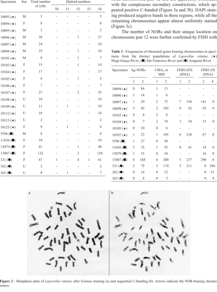
A total of 24 specimens were cytogenetically ana-lyzed. Specimens from all populations had a modal diploid number of 54, composed of metacentric and submeta-centric chromosomes (FN = 108) (Table 1 and Figure 2a).
The different applied staining techniques produced a pattern common to specimens of both sexes from all the populations. C-banding analysis revealed the presence of large pericentromeric heterochromatic segments in all chromosomes and a block at the terminal region of a mid-dle-sized chromosome pair (Figure 2b).
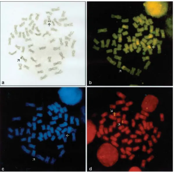
Silver nitrate and chromomycin A3 or mithramycin
staining revealed the presence of two NOR-bearing
mosomes in this species (Table 2). The NOR sites were lo-cated near the telomere of the long arm of the medium-sized chromosome pair 12 and are apparently coincident with the conspicuous secondary constrictions, which ap-peared positive C-banded (Figure 3a and 3b). DAPI stain-ing produced negative bands in these regions, while all the remaining chromosomes appear almost uniformly stained (Figure 3c).
The number of NORs and their unique location on chromosome pair 12 were further confirmed by FISH with
Table 1- Diploid numbers found inLeporellus vittatus: populations from (♦) Mogi-Guaçu River, (■) São Francisco River and (●) Araguaia River.
Specimens Sex Total number of cells
Diploid numbers
50 51 52 53 54
10093 (♦) M 5 - - - - 5
10094 (♦) F 8 - - - - 8
10095 (♦) M 3 - - - - 3
10096 (♦) M 38 - - 1 - 37
10097 (♦) M 24 - - - - 24
10098 (♦) M 25 - - 1 - 24
10102 (♦) M 8 - - 1 - 7
10103 (♦) F 15 - - 1 - 14
10104 (♦) F 17 - - - - 17
10105 (♦) F 6 - - 1 - 5
10106 (♦) F 7 - - - - 7
10107 (♦) F 27 - - 2 - 25
10108 (♦) U 10 - - - - 10
10109 (♦) U 11 - 1 - - 10
10112 (♦) U 16 - - - - 16
10113 (♦) U 2 - - - - 2
10125 (♦) F 9 - 1 - - 8
5596 (■) M 8 - - - - 8
11636 (■) F 10 - - - - 10
13079 (■) F 41 - - 1 - 40
13087 (■) F 132 - - 2 - 130
331 (●) F 67 1 - 4 1 61
362 (●) U 2 - - - - 2
363 (●) U 8 - 1 - - 7
Figure 2- Metaphase plate ofLeporellus vittatusafter Giemsa staining (a) and sequential C-banding (b). Arrows indicate the NOR-bearing chromo-somes.
Table 2- Frequencies of ribosomal genes bearing chromosomes in speci-mens from the distinct populations of Leporellus vittatus: (♦)
Mogi-Guaçu River, (■) São Francisco River and (●) Araguaia River.
Specimens Ag-NORs CMA3or MM
FISH (45S rDNA)
FISH (5S rDNA)
1 2 1 2 1 2 2 4
10094 (♦) 0 84 1 13 - - -
-10096 (♦) 1 14 1 6 - - -
-10097 (♦) 1 29 2 75 7 156 141 0
10098 (♦) 3 45 2 105 8 92 92 0
10103 (♦) 0 4 2 8 - - -
-10104 (♦) 0 7 2 74 3 54 15 0
10105 (♦) 0 10 0 6 - - -
-10107 (♦) 1 23 1 105 6 210 67 0
5596 (■) 1 27 0 30 - - -
-11636 (■) 3 35 1 35 0 41 14 0
13079 (■) 1 33 0 34 - - 16 0
13087 (■) 4 168 0 200 5 237 290 0
331 (●) 2 75 3 174 5 211 0 288
362 (●) 0 14 0 12 - - 0 33
biotinylated 18S and 28S rDNA (45S rDNA) probes (Fig-ure 3d).
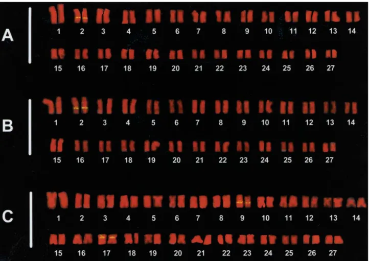
The 5S ribosomal genes were detected by FISH on chromosome pairs distinct from the ones bearing the 45S rDNA clusters. In the specimens from the Paraná and São Francisco Basins the 5S rDNA clusters were interstitially located on the long arm of the 2ndchromosome pair (Figure
4a and b). The specimens from the Tocantins-Araguaia Ba-sin showed two chromosome pairs bearing these sites. In this population the 5S rDNA sites were observed on the long arm of the 9thchromosome pair and on the short arm of
the 17thchromosome pair (Figure 4c).
Discussion
According to Buerlen (1970), the major drainage bas-ins of Brazil began to develop during the Tertiary. Al-though the precise time of formation of each hydrographic system cannot be determined, several teleost fish species of the families Cichlidae, Characidae, and Curimatidae have
been described as endemic of distinct hydrographic basins, reinforcing the supposed vicariant events (Kullander, 1983; Menezes, 1988; Vari, 1988).
The cytogenetic markers studied here included differ-ent banding techniques, fluorochrome staining, and in situ hybridization with ribosomal probes, allowing a careful in-vestigation of the constitutive heterochromatin and, partic-ularly, of the ribosomal sites, which are chromosomal regions often described as variable in fishes.
Although the three studied populations ofLeporellus vittatus share the same karyotypic structure already de-scribed by Galettiet al.(1991) for the population from the Paraná Basin and no differences could be detected in the heterochromatin distribution pattern and in the number and location of the 45S rDNA sites, interpopulation differences were evidenced concerning the number and location of the 5S rDNA clusters.
The association between heterochromatin and NOR sites observed in all the three studied populations seems to
be a common feature in fishes and has also been described for many other fish species (Galettiet al., 1991; Rossiet al., 1996; Aguilar and Galetti, 1997; Martins and Galetti, 1997). This NOR-associated heterochromatin, positive stained with CMA3, is thus GC-rich as originally reported
by Amemiya and Gold (1986). DAPI staining, an AT-specific fluorochrome, confirmed the GC-rich nature of this heterochromatin, since negative bands coincident to the NOR sites were observed.
The lack of large chromosomal differences among the populations is not surprising since the karyotypic macro-structure ofL. vittatus seems to be conserved among the Anostomidae (Galetti et al., 1981; Martins and Galetti, 1997, 1998) and other related Characiformes families, such as Curimatidae (Venere and Galetti, 1989; Feldberget al., 1992), Parodontidae (Jesus and Moreira-Filho, 2000), and Prochilodontidae (Pauls and Bertollo, 1990), suggesting that the karyotype with 2n = 54, FN = 108, is ancient among the Characiformes.
Nevertheless, subtle changes in the chromosomal microstructure involving distinct rearrangements in the ri-bosomal regions seem to have occurred during the evolu-tionary diversification of the Anostomidae and may be
strictly related to species differentiation. Intrapopulational chromosomal polymorphisms involving the number of 45S rDNA sites have already been reported in Leporinus friderici andLeporinus trifasciatus (Galettiet al., 1991; Galettiet al., 1995a,b), despite the conserved chromosome structure (2n = 54, FN = 108). In the present study we iden-tified differences in the number and location of the 5S rDNA clusters among distinct populations ofLeporellus vittatusfrom major Brazilian hydrographic basins.
In Neotropical characiform fishes, the 5S rDNA clus-ters are generally distributed in an inclus-terstitial position in two autosomal chromosome pairs and are usually not syn-tenic to the 45S rDNA sites, suggesting that this could be a common condition for the 5S rRNA gene organization in the genome of these fishes (Martins and Galetti, 1999, 2000, 2001; Born and Bertollo, 2000).
However, variations concerning the number and loca-tion of 5S rDNA sites have already been described for many other fish species. Multiple sites have been described for some salmonid species that show up to eight sites lo-cated on distinct autosomal chromosome pairs (Fujiwaraet al., 1998), and forAstyanax scabripinnis, a characid fish, that presents eight 5S rDNA sites located on four distinct
Figure 4- Karyotypes ofLeporellus vittatus: FISH with 5S ribosomal probe in specimens from Mogi-Guaçu (a), São Francisco (b) and Araguaia (c)
chromosome pairs (Ferroet al., 2000). The 5S rDNA clus-ters have also been located on the X or Y sex chromosomes of some salmonid species (Moranet al., 1996; Iturraet al., 2001; Steinet al., 2001), and on the Y-chromosome of the males ofChionodraco hamatus, an Antarctic fish (Mazzei et al., 2004). Frequently, differences in the number and po-sition of 5S rDNA sites have been reported as good chro-mosome markers to discriminate closely related fish species, such as in some families of the orders Mugili-formes and PerciMugili-formes (Gornunget al., 2001; Molina and Galetti, 2002; Rossiet al., 2005). Accordingly, the inter-population differences observed in the present study con-cerning the number and location of the 5S rDNA clusters between Paraná/São Francisco and Tocantins-Araguaia populations ofL. vittatussuggest that the species currently named Leporellus vittatus may comprise a complex of cryptic species.
The recent discovery that, during the construction of the Furnas hydroelectric power dam in the upper Paraná River Basin in the early 1960s, the Piumhi River drainage outflow was diverted into the headwaters of the São Fran-cisco River Basin has raised questioning about the current São Francisco watershed ichthyofauna structure (Moreira-Filho and Buckup, 2005). As this transposition event al-lowed the entire fish fauna of the Piumhi River and associ-ated swamps, lakes, and tributaries to intermingle with the fish fauna of the São Francisco Basin, it may have contrib-uted to the chromosomal stability described by many au-thors for Paraná and São Francisco Neotropical fish popu-lations, as also described in the present study. Therefore, further studies comparing the different populations ofL. vittatusand other anostomid species through molecular ap-proaches, such as DNA sequencing of mitochondrial genes, may probably give a more precise answer to the present question – IsLeporellus vittatusa complex of cryptic spe-cies?
Acknowledgments
This work was supported by Coordenação de Aper-feiçoamento de Pessoal de Nível Superior (CAPES), Con-selho Nacional de Desenvolvimento Científico e Tecnoló-gico (CNPq) and Fundação Universitária José Bonifácio (FUJB).
References
Aguilar CT and Galetti Jr PM (1997) Chromosomal studies in South Atlantic serranids (Pisces, Perciformes). Cytobios 89:105-114.
Amemiya CT and Gold JR (1986) Chromomycin A3stains
nucle-olus organizer regions of fish chromosomes. Copeia 1:226-231.
Bertollo LAC, Takahashi CS and Moreira-Filho O (1978) Cyto-taxonomy considerations on Hoplias lacerdae (Pisces,
Erithrynidae). Braz J Genet 1:103-120.
Born GG and Bertollo LAC (2000) An XX/XY sex chromosome system in a fish species,Hoplias malabaricus, with a poly-morphic NOR-bearing X chromosome. Chrom Res 8:111-118.
Buerlen K (1970) Geologie von Brasilien. Gebrüder Borntraeger, Berlin, 444 pp.
Cortadas J and Pavon MC (1982) The organization of ribosomal genes in vertebrates. EMBO J 1:1075-1080.
Feldberg E, Porto JIR and Bertollo LAC (1992) Karyotype evolu-tion in Curimatidae (Teleostei, Characiformes) of Amazon region. I. Studies on the genera Curimata,Psectrogaster, SteindachnerinaandCurimatella. Braz J Genet 15:369-383.
Fenocchio AS, Venere PC, Cesar ACG, Dias AL and Bertollo LAC (1991) Short term culture from solid tissues of fishes. Caryologia 44:161-166.
Ferro DAD, Neo DM, Moreira-Filho O and Bertollo LAC (2000) Nucleolar organizing regions, 18S and 5S rDNA in
Astyanax scabripinnis (Pisces, Characidae): Populations
distribution and functional diversity. Genetica 110:55-62. Fowler HW (1950) Os Peixes de Água Doce do Brasil. In:
Arqui-vos de Zoologia do Estado de São Paulo, v. VI. Depar-tamento de Zoologia da Secretaria da Agricultura, Indústria e Comércio, São Paulo, pp 205-404.
Fujiwara A, Abe S, Yamaha E, Yamazaki F and Yoshida MC (1998) Chromosomal localization and heterochromatin as-sociation of ribosomal RNA gene loci and silver-stained nu-cleolar organizer regions in salmonid fishes. Chrom Res 6:463-471.
Galetti Jr PM, Foresti F, Bertollo LAC and Moreira-Filho O (1981) Karyotypic similarity in three genera (Leporinus, LeporellusandSchizodon) of the family Anostomidae (Pis-ces, Teleostei). Braz J Genet 4:11-15.
Galetti Jr PM, Mestriner CA, Venere PC and Foresti F (1991) Heterochromatin and karyotype reorganization in fish of the family Anostomidae (Characiformes). Cytogenet Cell Genet 56:116-121.
Galetti Jr PM, Mestriner CA, Monaco PJ and Rasch EM (1995a) Post-zygotic modifications and intra- and inter-individual nucleolar organizing region variations in fish: Report of a case involvingLeporinus friderici. Chrom Res 3:285-290.
Galetti Jr PM, Lima NRW and Venere PC (1995b) A mono-phyletic ZW sex chromosome system in Leporinus
(Anostomidae, Characiformes). Cytologia 60:375-382. Garavello JC and Britski HA (2003) Family Anostomidae
(Head-standers). In: Reis RE, Kullander SO and Ferraris Jr CJ (eds) Check List of the Freshwater Fishes of South and Central America. 1st ed. EDIPUCRS, Porto Alegre, pp 71-84. Gornung E, Cordisco CA, Rossi AR, Innocentiis DS, Crosetti D
and Sola L (2001) Chromosomal evolution in Mugilidae: Karyotype characterization ofLiza saliensand comparative localization of major and minor ribosomal genes in the six Mediterranean mullets. Mar Biol 139:55-60.
Howell WM and Black DA (1980) Controlled silver-staining of nucleolus organizer regions with a protective colloidal de-veloper: A 1-step method. Experientia 36:1014-1015. Iturra P, Lam N, De La Fuente M, Vergara N and Medrano JF
(2001) Characterization of sex chromosomes in rainbow trout and coho salmon using fluorescencein situ
Jesus CM and Moreira-Filho O (2000) Karyotypes of three spe-cies ofParodon(Teleostei, Parodontidae). Ichthyol Explor Freshwaters 11:75-80.
Kullander SO (1983) A revision of the South American cichlid ge-nusCichlasoma(Teleostei, Cichlidae). Swedish Museum of
Natural History, Sweden, 296 pp.
Lande R (1979) Effective deme sizes during long-term evolution estimated from rates of chromosomal rearrangement. Evolu-tion 33:234-251.
Martins C and Galetti Jr PM (1997) Narrow chromosome diver-sity in fish of the genus Schizodon (Characiformes, Anostomidae). Cytobios 92:139-147.
Martins C and Galetti Jr PM (1998) Karyotype similarity between two sympatric Schizodon fish species (Anostomidae,
Characiformes) from the Paraguay River basin. Genet Mol Biol 21:355-360.
Martins C and Galetti Jr PM (1999) Chromosomal stability of 5S rDNA genes in Leporinus fish (Anostomidae,
Characiformes). Chrom Res 7:363-367.
Martins C and Galetti Jr PM (2000). Conservative distribution of 5S rDNA loci inSchizodon(Pisces, Anostomidae) chromo-somes. Chrom Res 8:353-355.
Martins C and Galetti Jr PM (2001) Two 5S rDNA arrays in Neo-tropical fish species: Is it a general rule for fishes? Genetica 111:439-446.
Mazzei F, Ghigliotti L, Bonillo C, Coutanceau JP, Ozouf-Costaz C and Pisano E (2004) Chromosomal patterns of major and 5S ribosomal DNA in six icefish species (Perciformes, Notothenioidei,Channichthyidae). Polar Biol 28:47-55. Menezes NA (1988) Implications of the distribution patterns of
the species of Oligosarcus (Teleostei, Characidae) from
Central and Southern South America. In: Vanzoline PE and Ronald Heyer W (eds) Proceedings of a Workshop on Neo-tropical Distribution Patterns. Academia Brasileira de Ciên-cias, Rio de Janeiro, pp 295-304.
Molina WF and Galetti Jr PM (2002) Robertsonian rearrange-ments in the reef fish Chromis (Perciformes,
Pomacentridae) involving chromosomes bearing 5S rRNA genes. Genet Mol Biol 25:373-377.
Moran P, Martinez JL, Garcia-Vazquez E and Pendas AM (1996) Sex chromosome linkage of 5S rDNA in rainbow trout (Oncorhynchus mykiss). Cytogenet Cell Genet 75:145-150. Moreira-Filho O and Buckup PA (2005) A poorly known case of watershed transposition between the São Francisco and up-per Paraná River basins. Neotrop Ichthyol 3:449-452. Pauls E and Bertollo LAC (1990) Distribution of supernumerary
chromosome system and aspects of karyotypic evolution in the genusProchilodus(Pisces, Prochilodontidae). Genetica 81:117-123.
Pinkel D, Straume T and Gray JW (1986) Cytogenetic analysis us-ing quantitative, high sensitivity, fluorescence hybridiza-tion. Proc Nat Acad Sci USA 83:2934-2938.
Rossi AN, Crossetti D, Gornung E and Sola L (1996). Cytogenetic analysis of global populations ofMugil cephalus(striped
mullet) by different staining techniques and fluorescentin situhybridization. Heredity 76:77-82.
Rossi AR, Gornung E, Sola L and Nirchio M (2005) Comparative molecular cytogenetic analysis of two congeneric species,
Mugil curemaandM. liza(Pisces, Mugiliformes),
character-ized by significant karyotype diversity. Genetica 125:27-32. Schmid M (1980) Chromosome banding in Amphibia IV.
Differ-entiation of GC- and AT-rich chromosome regions inAnura. Chromosoma 77:83-103.
Schweizer D, Ambros P and Andrle M (1978) Modification of DAPI banding on human chromosomes by pre-staining with a DNA binding oligopeptide antibiotic distamycin A. Expl Cell Res 111:327-332.
Stein J, Phillips RB and Devlin RH (2001) Identification of the Y chromosome in chinook salmon (Oncorhynchus tshawytscha). Cytogenet Cell Genet 92:108-110.
Sumner AT (1972) A simple technique for demonstrating centromeric heterocromatin. Expl Cell Res 75:304-306. Vari RP (1988) The Curimatidae, a lowland Neotropical fish
fam-ily (Pisces, Characiformes): Distribution, endemism and phylogenetic biogeography. Proceedings of a Workshop on Neotropical Distribution Patterns, pp 313-348.
Venere PC and Galetti Jr PM (1989) Chromosome evolution and phylogenetic relationships of some Neotropical Characiformes of the family Curimatidae. Brazil J Genet 12:17-25.
Associate Editor: Lurdes Foresti de Almeida-Toledo