Nauplius
THE JOURNAL OF THE BRAZILIAN CRUSTACEAN SOCIETY
e-ISSN 2358-2936
www.scielo.br/nau www.crustacea.org.br
Intraspecific variation in megalopae
of
Clibanarius antillensis
(Anomura,
Diogenidae) among western Atlantic
populations
Sergio Cházaro-Olvera
1Rafael Robles
2Jesús Montoya-Mendoza
3Josué Abraham Herrera-López
11 Laboratorio de Crustáceos, Facultad de Estudios Superiores Iztacala, Universidad
Nacional Autónoma de México, Av. de los Barrios No.1, Los Reyes Iztacala, C.P. 54090, Tlalnepantla, Estado de México, México.
SCO E-mail: [email protected] JAHL E-mail: [email protected]
2 Facultad de Ciencias Químico Biológicas, Universidad Autónoma de Campeche.
Humberto Lanz Cárdenas y Fracc. Ecológico Ambiental Siglo XXIII, Colonia Exhacienda Kalá, CP. 24085, San Francisco de Campeche. Campeche, México.
RR E-mail: [email protected]
3 Laboratorio de Investigación Acuícola Aplicada, Tecnológico Nacional de México,
Instituto Tecnológico de Boca del Río (ITBOCA), Carretera Veracruz-Córdoba Km 12, Boca del Río, Boca del Río, CP. 94290. Veracruz, México
JMM E-mail: [email protected]
ZOOBANK: http://zoobank.org/
urn:lsid:zoobank.org:pub:ABE11F35-05BA-4D8D-B606-F656DEAD4168
ABSTRACT
The objective of this study was to describe the morphology of Clibanarius antillensis Stimpson, 1859 megalopae collected in the vicinity of Isla Sacrificios, Mexico, and compare it to previous descriptions originated from Brazilian and Panamanian specimens raised in laboratory conditions. We found four meristic differences between the Brazilian and the Panamanian and Mexican populations with the Brazilian population: the outer flagellum of antennule in the Panamanian and Mexican populations has more aesthetascs on the second, third, and fourth segments of the outer flagellum (0, 6, 5, 3, 0) than the Brazilian population (0, 4, 4, 2, 0); the maxilla has more setae on the scaphognathite in the Brazilian specimens (70) than in the Mexican and Panamanian specimens (49-62); the crista dentata of the third maxilliped is formed by only three denticles in the Brazilian specimens, while 4 or 6 denticles form the same structure in the Panamanian and Mexican populations; there are fewer number of setae on the endopod of the uropod in the Brazilian population. The differences may be explained by intraspecific variation.
KEY WORDS
Crustacea, Anomura, interspecific variation, megalopa stage, taxonomy. CORRESPONDING AUTHOR
Sergio Cházaro-Olvera
SUBMITTED 06 August 2018 ACCEPTED 23 October 2018 PUBLISHED 03 December 2018
DOI 10.1590/2358-2936e2018031
All content of the journal, except where identified, is licensed under a
orcid.org/0000-0002-7598-7300
orcid.org/0000-0003-0212-1211 orcid.org/0000-0003-0531-5557
Cházaro-Olvera et al. Morphological variations on megalopa of C. antillensis
INTRODUCTION
The genus Clibanarius Dana 1852 is represented by 60 species worldwide (Lemaitre and McLaughlin, 2018); four of these species can be found in the Gulf of Mexico, Clibanarius antillensis Stimpson, 1859, C. sclopetarius Herbst 1796, C. tricolor (Gibbes, 1850), and
C. vittatus (Bosc, 1802) (Felder et al., 2009). These four species have also been reported in Veracruz (Álvarez
et al., 2011). Recent plankton studies performed in the Parque Nacional Sistema Arrecifal Veracruzano (PNSAV), on the southwestern Gulf of Mexico, have found megalopae of eight species of hermit crabs of the family Diogenidae: Calcinus tibicen (Herbst, 1791), Clibanarius antillensis, C. sclopetarius, C. vittatus,
Dardanus insignis (de Saussure, 1858), Paguristes sericeus A. Milne Edwards, 1880, P. spinipes A. Milne-Edwards, 1880, and Petrochirus diogenes (Linnaeus, 1758) (Cházaro-Olvera et al., 2013).
Bartilotti et al. (2008) described the complete larval development of two species of Clibanarius. While doing so, the authors reviewed morphological larval characters among species of the genus. In general, larval development within the genus is very homogenous, consisting of four or five zoeal stages and one megalopa. However, morphometry of the larval stages varies within the genus and species can be identified based on specific larval characters (Siddiqui et al., 1991; 1993; Bartilotti et al., 2008). Although morphology of larval characters are believed to be species-specific, some morphological variations have been found among larval developmental stages described under laboratory conditions of C. antillensis
from Brazil (Brossi-Garcia and Hebling, 1983) and Panama (Siddiqui et al., 1991); these morphological variations are believed to be the result of geographic differences and/or differing laboratory conditions (Siddiqui et al., 1991; Bartilotti et al., 2008).
The distribution of the hermit crab C. antillensis
extends throughout the western Atlantic including Bermuda, Eastern Florida, Gulf of Mexico, Caribbean Sea, and Brazil (Felder et al., 2009). Within-species larval variations in decapod crustaceans are not uncommon in the marine environment. In particular, latitudinal variations have been explained by increased seasonality and unpredictability of primary productivity: a macro-ecological gradient is formed, and it modifies the number of larval instars or the morphology in the
same stage of development in some marine crustaceans (Anger, 2001; Oliphant et al., 2013). The objective of this study was to compare the morphology of the megalopa of C. antillensis collected at the PNSAV to those reared in laboratory conditions from both Brazilian and Panamanian populations.
MATERIAL AND METHODS
The PNSAV is a coral reef system located in the northwest sector of the Bay of Campeche, in the polygon delimited by 19°00’00’’–19°16’00’’N and 95°45’00’’–96°12’00’’W. It is formed by 23 coral reefs distributed in northern and southern groups, which are separated by the mouth of the Jamapa River (Granados
et al., 2007) (Fig. 1).
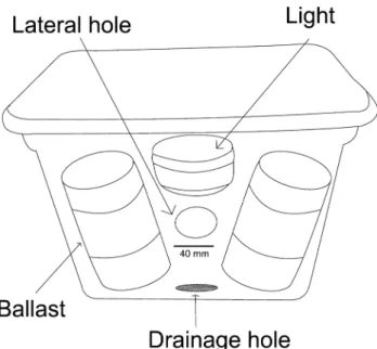
Megalopae were collected using light traps placed off the northwest of Isla Sacrificios for three consecutive nights, on June 2015. These collecting cycles were initiated at 21:00h and stopped at 01:00h on the next day during the new moon phase. The temperature recorded was almost invariable (28 to 29 °C), and salinity was 35 psu. All traps were constructed with plastic boxes that were 0.4 m long, 0.25 m wide and 0.30 m high, with 40 mm diameter inlet holes in the upper part of each side of the box; the box contained a white light of 38 lumens (Fig. 2). Trap contents were collected manually and placed in 500 ml plastic bottles at the end of the sampling period. All samples were preserved in 70% ethanol.
Samples were sorted by using a Motic SMZ-168 microscope at the Crustacean Laboratory, FES Iztacala, UNAM. Identification of the larvae followed current literature (Lang and Young, 1977; Brossi-Garcia, 1987; Sidiqui et al., 1991; Bartilotti et al., 2008; Cházaro-Olvera
Clark et al. (1998), while setal classification followed that proposed by Garm (2004). Images of the larvae were taken using a Leica DM750 microscope equipped with an Omax 14MP USB 3.0 digital camera. The illustrations were made with the Corel Draw V.12 program.
RESULTS
We found 2,795 larvae of the genus Clibanarius: 968 of these were identified as C. antillensis, 656 as C. sclopetarius, and 1171 as C. vittatus (Tab. 1). We were able to differentiate the species C. antillensis from C. sclopetarius and C. vittatus considering the total length, number of aestethascs in the external flagellum of the antennule, number of segments of the antenna, number of segments of the palp of the mandible and the number of plumose marginal setae in the endopod and exopod of uropods (Tab. 2).
Table 1. Abundance of Clibanarius species in the sampling area.
Species Sampling time Total
21:00 22:00 23:00 00:00 01:00
Clibanarius
antillensis 216 443 291 13 5 968 Clibanarius
sclopetarius 327 211 86 25 7 656 Clibanarius
vittatus 542 364 242 16 7 1171
Total 1085 1018 619 54 19 2795
Figure 1. Sampling station northwest off Sacrificios Island (*), Sistema Arrecifal Veracruzano.
Cházaro-Olvera et al. Morphological variations on megalopa of C. antillensis
We found intrapopulation variation for seven characters of the megalopa of C. antillensis, size (2.0–2.3mm TL), number of setae on the scaphognathite of maxilla (50–55), number of denticles of crista dentata (4–6), and number of setae on the protopod (0–4), endopod (11–14), and exopod (19–21) of uropods (Tab. 2). When comparing the same characters to other populations of
C. antillensis, we also found inter-population differences, which are described below in the remarks section.
Clibanarius antillensis Stimpson, 1859
Megalopa
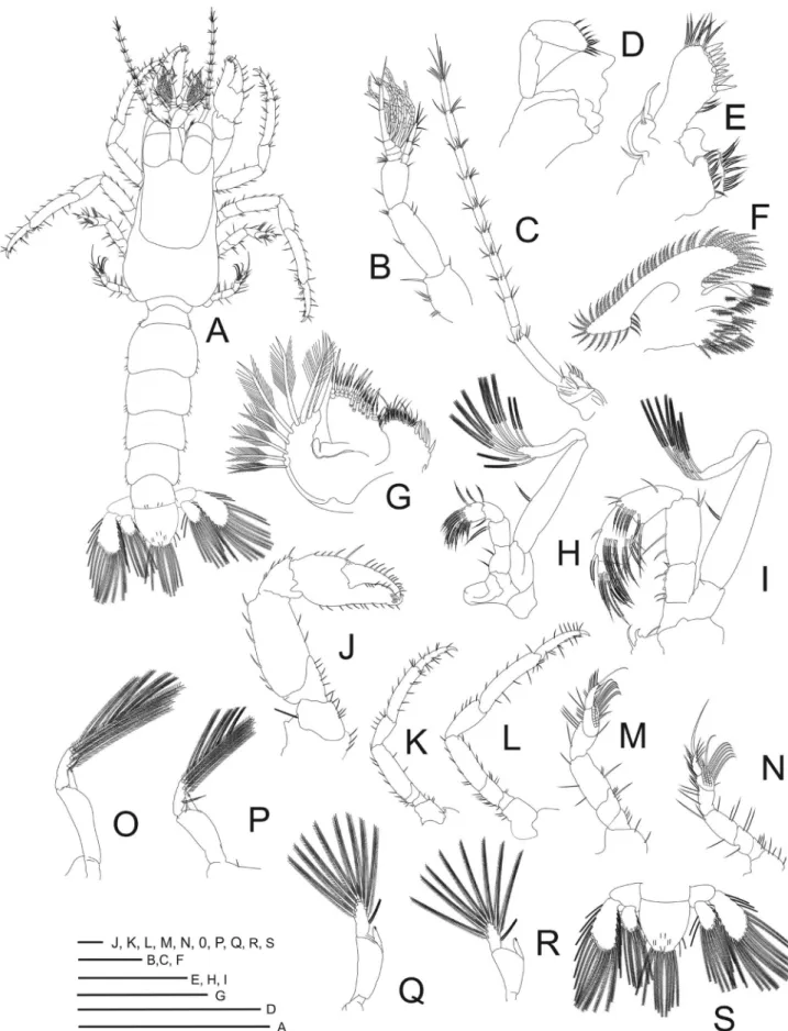
Size (Fig. 2A). Carapace length = 0.9–1.1 mm; carapace width = 0.7‒0.81 mm; total length = 2.0–2.3 mm; n = 96.
Carapace (Fig. 3A). Longer than broad, with few scattered setae; rostrum triangular; ocular peduncles bulbous, reaching half of distal segment of antennular peduncle.
Antennule (Fig. 3B). Biramous. Peduncle 3-segmented; basal segment with 6 simple setae, 1 strong seta at ventrodistal angle; penultimate segment with 3 subterminal setae, 2 simple terminal setae; basal segment with 2 subterminal, 4 short terminal setae. Endopod 3-segmented, with 1, 4, 6 (3 subterminal, 3 terminal) setae, respectively. Exopod with 5 segments, aesthetascs as 0, 6, 5, 3, 0, last segment with 3 subterminal and 1 long terminal setae.
Antenna(Fig. 3C). Basal segment not delineated. Second segment with angular process ending on tip, third segment with 5 simple marginal setae. Flagellum with 10 segments, with setae from proximal to distal segment: 0, 3, 4, 5, 4, 5, 5, 6, 5, 9 (6 subterminal and 3 terminal). Exopod reduced, with 5 plumose marginal setae.
Mandible (Fig. 3D). Reduced, simple; palp 2-segmented, 9 marginal serrate setae.
Maxillule (Fig. 3E). Coxal endite with 17 serrate setae. Basal endite with 3 plumose intermediate, 12 cuspidate, 7 serrate setae. Endopod with well-developed internal lobe, with 1 simple long terminal seta, external lobe recurved.
Maxilla(Fig. 3F). Coxal endite with 2 plumose subterminal, 4 terminal setae. Basal endite with 10 plumose setae at distal lobe and 5 at proximal lobe. Endopod narrow, significantly twisted, not lobed, without setae. Scaphognathite elongate, proximal free lobe of protopod, with 55 to 60 plumose marginal setae. Maxilliped 1 (Fig. 3G). Coxal endite with 5 cuspidate, 10 plumose submarginal setae. Basal endite with 5 cuspidate, 11 marginal plumose setae. Endopod 1-segmented and narrow. Exopod 1-segmented, with 9 plumose marginal setae.
Maxilliped 2(Fig. 3H). Endopod 4-segmented, with 1 or 2, 2, 7, 5 or 6 plumose setae. Exopod 2-segmented, 8 or 9 plumose terminal setae, 1 or 2 plumose setae on inner margin.
Maxilliped3(Fig. 3I). Endopod 5-segmented; crista dentata on proximal segment with 4 to 6 denticles, 2 subterminal, 5 simple terminal setae, second segment with 2 simple subterminal setae, 2 simple terminal setae, third segment with 11 subterminal setae, 5 terminal setae, 5 marginal setae, fourth segment with 13 plumose setae and fifth segment with 7 plumose setae. Exopod 2-segmented, with 1 simple marginal seta, 8 plumose terminal setae.
First pereiopod (chelipeds) (Fig. 3J). Chelae equal, with segments smooth; coxa with 3 simple setae; basis with 6 simple setae; ischium with 5 simple setae; merus, longest segment, with 8 simple setae; carpus with 6
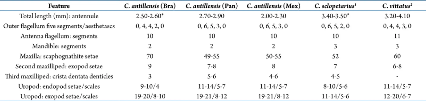
Table 2. Comparison of relevant megalopae features of Clibanarius antillensis, collected from a Mexican population to those obtained in laboratory conditions from Brazilian (Bra) (Brossi-Garcia and Hebling, 1983) and Panamanian (Pan) populations (Siddiqui et al., 1991). The same features are compared to C. sclopetarius and C. vittatus collected in the same area (1, Brossi-Garcia, 1987; 2, Lang
and Young, 1977; * obtained from the figure).
Feature C. antillensis (Bra) C. antillensis (Pan) C. antillensis (Mex) C. sclopetarius1 C. vittatus2
Total length (mm): antennule 2.50-2.60* 2.70-2.90 2.00-2.30 3.40-3.50* 3.20-4.10 Outer flagellum five segments/aesthetascs 0, 4, 4, 2, 0 0, 6, 5, 3, 0 0, 6, 5, 3, 0 0, 6, 5, 2, 0 0, 4, 4, 3, 0
Antenna flagellum: segments 10 10 10 10 11
Mandible: segments 2 2 2 3 3
Maxilla: scaphognathite setae 70 49-55 50-55 52 60
Second maxilliped: exopod setae 9 7-8 8 7 6-8
Third maxilliped: crista dentata denticles 3 5-6 4-6 4-5 ‒
Cházaro-Olvera et al. Morphological variations on megalopa of C. antillensis
simple setae; propodus with 16 simple setae, distal upper extremity corneous; dactyl length about half palm including the propodal prolongation, with 16 simple setae and distal extremity corneous.
Second pereiopod (Fig. 3K). Coxa with 2 simple setae; basis smaller with 3 simple setae; ischium with 4 simple setae; merus, longest segment, with 9 simple setae; carpus less than half the ischium length, with 5 simple setae; propodus longer than carpus, with 15 simple setae and; dactylus very stout, apically curved with corneous tip, with 11 simple setae and 3 spines.
Third pereiopod (Fig. 3L). Coxa with 3 simple setae; basis with 3 simple setae; ischium with 11 simple setae; merus longer than ischium and with 10 simple setae; carpus with 5 simple setae; propodus longer than carpus, with 14 simple setae and 3 stout cuspidate seta; dactylus very stout, apically curved and corneous, with 16 simple setae and 4 spines.
Fourth pereiopod (Fig. 3M). Coxa with 2 simple setae; basis with 4 simple setae; ischium with 4 simple setae; merus with 6 simple setae; carpus with 5 simple setae; propodus, with 5 simple setae and 17 pseudochaetae scales forming protopodal plate; dactyl with 1 long papposerrate seta, 7–9 simple setae and 3 teeth on distal extremity.
Fifth pereiopod (Fig. 3N). Coxa with 4 simple setae; basis with 4 simple setae; ischium with 3 simple setae; merus with 6 simple setae; carpus with 5 simple setae; propodus, with 6–7 long papposerrate, 10–11 simple setae and 23 pseudochaetae scales; dactyl with 7 simple setae, 1 long papposerrate distal and 5 pseudochaetae scales.
Pleon (Fig. 3A). Pleonites 2–5 with rounded posterolateral angles, lateral margins with 3 simple short setae; pleonite 6 with 4 simple setae at posterior dorsal margin and 2 simple setae at posterolateral angles.
Pleopods(Figs. 3O–R). Pleopod developed on pleonites 2–5, decreasing in size distally. Endopod unsegmented, with pair of hooks at apical margin. Exopods 2-segmented with 9 plumose setae.
Uropods(Fig. 3S). Segment protopodal with 0–3 simple setae. Endopod with 1 or 2 simple setae on dorsal surface, 11–14 plumose marginal setae and 5–7 corneal scales; Exopods with 0–4 short plumose setae on dorsal surface, 19–21 plumose marginal setae and 8–12 corneous scales.
Telson (Fig. 3S). Shape rounded posterior margin, with 9 posterior plumose marginal setae, 2 pairs of
submarginal short plumose setae, 4 pairs of short simple setae on dorsal surface.
Remarks. We found four meristic differences among the Brazilian (Brossi-Garcia and Hebling, 1983) the Panamanian (Siddiqui et al., 1991) and Mexican populations (Tab. 1). The antennule in the Panamanian and Mexican populations had more aesthetascs on the second, third and fourth segments (0. 6, 5, 3, 0) with respect to the Brazilian population (0, 4, 4, 2, 0). There were fewer plumose setae on the scaphognathite in the Panamanian and Mexican populations (49–55) with respect to the Brazilian population (70 plumose setae). In the second maxilliped, the difference was of 1 seta, whereas in maxilliped 3, there were 1–3 fewer denticles in the Brazilian population on the crista dentata. The exopod of the uropod had fewer (10) marginal plumose setae in the Brazilian population.
DISCUSSION
We identified hundreds of larvae of C. antillensis,
C. sclopetarius, and C. vittatus and the observed key characters of the megalopa of C. sclopetarius and C. vittatus corresponded to those observed in the literature (Lang and Young, 1977; Brossi-Garcia, 1987). Thus there was no doubt concerning the identity of the megalopae of C. antillensis collected; such variations in morphology of the megalopae must be explained.
We found that the Panamanian and Mexican populations were morphologically more similar in the number of aesthetascs on the antennule, setae on the scaphognathite, second maxilliped, number of denticles on the crista dentate and number of setae on the exopod of the uropod.
There are two potential explanations for such differences: either these differences are the result of intraspecific variations, probably following a latitudinal gradient or there are two cryptic taxa, currently unrecognized: one distributed mainly in Brazil and another distributed in the Caribbean, Mexico and the USA.
and salinity. This flexibility is an evolutionary and ecological strategy which enhances survival and allows the distribution of the larvae to be extended (Welch and Epifanio, 1995; Hartnoll, 2001; Thatje and Bacardit, 2000; Negreiros-Fransozo et al., 2008). In this respect, Tirmizi and Siddiqui (1980), McLaughlin and Gore (1988) and Siddiqui et al. (1991) found differences in the setation and spinationof Pagurus kulkarnii
Sankolli, 1962, Pagurus hirsutiusculus (Dana, 1851) and C. antilliensis. Furthermore, the morphological variability increases in the later larval or decapodid stages. For example, in Crangon crangon (Linnaeus, 1758), in the first decapodid stage, at least two forms can be distinguished, differing primarily in the developmental state of the antennae, the maxillipeds, the pereiopods, and the pleopods (Linck, 1995). Brossi-Garcia and Hebling (1983) found more setae on the scaphognathite of the maxilla of the Brazilian population; this could be explained by the availability of food affecting the life cycle of many benthic marine invertebrates, with significant implications for their survival, growth and metamorphosis (Calado and Leal, 2015; Pechenik and Tyrell, 2015). It has been found that the morphology of the feeding structures is more developed in the megalopa and juveniles, and the mouthparts present an increasing number of setae compared to previous developmental stages, which indicates that these stages are more able to capture food and they are capable of processing solid food available in their benthonic environment (Abrunhosa
et al., 2006). The ability to process small food particles increases because of the increment in the size of the foregut and the number of setae on the mouthparts (Abrunhosa and Melo, 2008). The phytoplankton is most abundant in high latitudes and in upwelling zones along the equator and near coastlines. However, phytoplankton is scarce in the subtropical regions, where nutrient levels are low and drop off in the summer (NASA, 2018).
For the second potential explanation, we were considering genetically comparing different populations of C. antillensis since it has been shown that cryptic species do occur among species of this genus. Furthermore, Negri et al. (2014) found morphological and genetic differences strong enough to resurrect C. symmetricus (Randall, 1840). This species was reported for many years under the name C. vittatus because of their close morphological similarities. The authors
defined the genetic divergences between C. vittatus
and C. symmetricus, identifying that the latter species may occur more broadly in the Caribbean, the Antilles, or the southern Gulf of Mexico. Similar to C. vittatus,
C. antillensis has a broad range in the western Atlantic, from the southeastern coast of the United States, and from Bermuda to Brazil (Felder et al., 2009), although the distribution is not continuous, so differences might be found. While the present study was in review, we were not aware that Nishikawa (2017) already preformed a robust and representative sampling with two molecular markers (16S and COI) and found no genetic structure among different western Atlantic populations of C. antillensis, including populations from Mexico, Panama and Brazil. Thus it appears that differences observed in the megalopa stage among geographically separated populations are the result of interpopulation variation.
ACKNOWLEDGEMENTS
We appreciate the support to the national sabbatical program 2018 of CONACYT. We also thank the authorities of SEMARNAT, CONANP and CONAPESCA-DGOPA (SAGARPA) for the permits granted (PPF/DGOPA-051/15). RR wishes to acknowledge PRODEP-SEP, Mexico, through the program “Apoyo a la Incorporación de NPTC (Ago/1/2018 –Jul/31/2019)”. We are also thankful to F.L. Mantelatto and two anonymous reviewers for suggestions made to an earlier version of this paper.
Disclosure Statement
No potential conflict of interest was reported by the authors.
REFERENCES
Abrunhosa, F. and Melo, M. 2008. Development and functional morphology of the foreguts of larvae and postlarvae of three crustacean decapods. Brazilian Journal of Biology, 68: 221–228.
Abrunhosa, F.A; Melo, M.; Lima, J.F. and Abrunhosa J. 2006. Developmental morphology of mouthparts and foregut of the larvae and postlarvae of Lepidophthalmus siriboia Felder and Rodrigues, 1993 (Decapoda: Callianassidae). Acta Amazonica, 36: 335–342.
Cházaro-Olvera et al. Morphological variations on megalopa of C. antillensis
La biodiversidad en Veracruz. Estudio de estado. Diversidad de especies: conocimiento actual. México, D.F., CONABIO.
Anger, K. 2001. The biology of decapod crustacean larvae. Rotterdam, Balkema Publishers. 300p.
Bartilotti, C.; Calado, R. and dos Santos, A. 2008. Complete larval development of the hermit crabs Clibanarius aequabilis and
Clibanarius erythropus (Decapoda: Anomura: Diogenidae), under laboratory conditions, with a revision of the larval features of genus Clibanarius. Helgoland Marine Research, 62: 103–121.
Bosc, L.A.G. 1802. Histoire Naturelle des Crustacés, contenant leur description et leurs meours: avec figures dessinées d’après nature. Vol. 2. Paris, De Guilleminet, 226p.
Brossi-Garcia, A.L. and Hebling, N.J. 1983. Desenvolvimento pós-embrionário de Clibanarius antillensis Stimpson, 1859 (Crustacea, Diogenidae), em laboratório. Boletim de Zoologia, 6: 89–111.
Brossi-Garcia, A.L. 1987. Morphology of the larval stages of
Clibanarius sclopetarius (Herbst, 1796) (Decapoda, Diogenidae) reared in the laboratory. Crustaceana, 52: 251–275.
Calado, R. and Leal, M.C. 2015. Trophic ecology of benthic marine invertebrates with bi-phasic life cycles: what are we still missing? Advances in Marine Biology, 71: 1–70.
Cházaro-Olvera, S.; Winfield, I.; Ortiz, M.; Cházaro-Martínez, E.; Vázquez-López, H. and Horta-Puga, G.J. 2013. Morphology of megalopae from Diogenidae family (Decapoda, Anomura) in Veracruz, south-western Gulf of Mexico: identification keys to genera and species. American Journal of Life Sciences, 1: 261–266.
Clark, P.F.; Calazans, D.K. and Pohle, G.W. 1998. Accuracy and standardization of brachyuran larval descriptions. Invertebrate Reproduction and Development, 33: 127–144.
Dana, J.D.1851. Conspectus Crustaceorum quæ in Orbis Terrarum circumnavigatione, Carolo Wilkes e classe Reipublicæ Fœderatæ Duce, lexit et descripsit. Proceedings of the Academy of Natural Sciences of Philadelphia, 5: 267–272.
Dana, J.D. 1852. Crustacea. Part I. United States Exploring Expedition. During the years 1838, 1839, 1840, 1841, 1842. Under the command of Charles Wilkes, U.S.N. Vol. 13. Philadelphia, C. Sherman, 685p.
Felder, D.L.; Álvarez, F.; Goy, J.W. and Lemaitre, R. 2009. Decapoda (Crustacea) of the Gulf of Mexico, with comments on the Amphionidacea. P. 1019–1104. In: D.L. Felder and D.K. Camp (eds), Gulf of Mexico Origin, Waters and Biota. Texas, Texas A&M University Press.
Gibbes, L.R. 1850. On the carcinological collections of the cabinets of natural history in the United States. Proceedings of the American Association for the Advancement of Science, 3: 167–201.
Garm, A. 2004. Revising the definition of the crustacean seta and setal classification systems based on examinations of the mouthpart setae of seven species of decapods. Zoological Journal of the Linnean Society, 142: 233–252.
Granados, B.A.; Abarca, A. L. G and Vargas, H.J.M. 2007. Investigaciones Científicas en el Sistema Arrecifal Veracruzano. Campeche, Universidad Autónoma de Campeche, 304p.
Hartnoll, R.G. 2001. Growth in Crustacea-twenty years on. Hydrobiologia, 449: 111–122.
Herbst, J.F.W. 1782-1804. Versuch einer Naturgeschichte der Krabben und Krebse nebst einer systematischen Beschreibung ihrer verschiedenen Artenm 1 [1782–1790]: 1–274, 2 [1791– 1796]: 1–226, 3 [1799–1804]: 1–216; 72 pls. Berlin and Stralsund.
Lang, W.H. and Young A.M.1977. The larval development of
Clibanarius vittatus (Bosc) (Crustacea: Decapoda: Diogenidae) reared in the laboratory. Biology Bulletin, 152: 84–104.
Lemaitre, R. and McLaughlin, P. 2018. World Paguroidea and Lomisoidea database. Clibanarius Dana, 1852. Accessed through: World Register of Marine Species at: http://www. marinespecies.org/aphia.php?p=taxdetails&id=106841 on 2018-05-17.
Linck, B.M. 1995. Einfluß von Temperatur und Salzgehalt auf die Larven der Nordseegarnele Crangon crangon. M.Sc. Thesis, Universität Oldenburg, Germany, 174 pp. [Unpublished]
Linnaeus, C. 1758. Systema Naturae per regna tria naturae, secundum classes, ordines, genera, species, cum characteribus, differentiis, synonymis, locis. Editio decima, reformata. Laurentius Salvius: Holmiae II, 824 pp.
McLaughlin, P.A. and Gore, R.H. 1988. Studies on the provenzanoi
and other pagurid groups: I. The larval stages of Pagurus maclaughlinae Garcia-Gómez, 1982 (Decapoda: Anomura: Paguridae) reared under laboratory conditions. Journal of Crustacean Biology, 8: 262–282.
Milne Edwards, A. 1880. Reports on the results of dredging, under the supervision of Alexander Agassiz, in the Gulf of Mexico and in the Caribbean Sea, 1877, ’78, ’79 by the United States Coast Survey Steamer “Blake”. VIII.-Études préliminaires sur les Crustacés. Bulletin of the Museum of Comparative Zoology at Harvard College, 8: 1–68, 2 plates.
NASA, 2018. Image by Jesse Allen & Robert Simmon, based on MODIS data from the GSFC Ocean color team. Average chlorophyll concentration in the global oceans from July 2002–May 2010 https://earthobservatory.nasa.gov/ Features/Phytoplankton/page4.php
Negreiros-Fransozo, M.L.; Fransozo, A. and Hirose G.L. 2008. The megalopa and early juvenile development of Hepatus pudibundus (Crustacea: Brachyura: Aethroidea) reared from neuston samples. Revista brasileira de Zoologia, 25: 608–616.
Negri. M.; Lemaitre, R. and Mantelatto, F.L. 2014. Molecular and morphological resurrection of Clibanarius symmetricus
(Randall, 1840), a cryptic species hiding under the name for the “thinstripe” hermit crab C. vittatus (Bosc, 1802) (Decapoda: Anomura: Diogenidae). Journal of Crustacean Biology, 34: 848–861.
Nishikawa, K.S. 2017. Variabilidade genética e morfológica do ermitão Clibanarius antillensis Stimpson, 1859 ao longo de sua distribuição. Universidade de São Paulo, Faculdade de Filosofia, Ciência e Letras de Ribeirão Preto, Ribeirão Preto, Brazil, Trabalho de Conclusão de Curso. 72p. [Unpublished]
Oliphant, A.; Hauton, C. and Thatje, S. 2013. The implications of temperature-mediated plasticity in larval instar number for development within a marine invertebrate, the shrimp
Palaemonetes varians. PloS One, 8: e75785.
Randall, J.W. 1840. Catalogue of the Crustacea brought by Thomas Nuttall and J. K. Townsend from the West Coast of North America and the Sandwich Islands, with descriptions of such species as are apparently new, among which are included several species of different localities, previously existing in the Collection of the Academy. Journal of the Academy of Natural Sciences of Philadelphia, 8, 106–147, pls. 3–7.
Sankolli, K.N. 1962. On a new species of hermit crab Pagurus kulkarni sp. nov. (Anomura: Paguridae). Journal of Zoological Society India, 13: 136–142.
Saussure, H. de. 1858. Mémoire sur divers Crustacés nouveaux des Antilles et du Mexique. Mémoires de la Société de Physique et d’Histoire Naturelle de Genève, 14: 417–496, pls. 1-6.
Siddiqui, F.A.; McLaughlin, P.A. and Crain, J.A. 1991. Larval development of Clibanarius antillensis Stimpson, 1859 (Crustacea: Anomura: Diogenidae) reared under laboratory conditions: A comparison between Panamanian and Brazilian populations. Journal of Natural History, 25: 917–932.
Siddiqui, F.A.; McLaughlin, P.A. and Crain, J.A. 1993. Larval development of the hermit crab Clibanarius albidigitus
(Crustacea: Anomura: Diogenidae) reared under laboratory conditions. Marine Biology, 116: 603–613.
Stimpson, W. 1859. Notes on North American Crustacea, 1.
Annals of the Lyceum of Natural History of New York, 7 (1862) (2): 49–94, pl. 1. [p. 1–47 on separate published March 1859; dated 1860 by Stimpson (1871: 119); republished with original pagination in 1860 in complete volume].
Thatje, S. and Bacardit R. 2000. Morphological variability in larval stages of Nauticarismagellanica (A. Milne Edwards, 1891) (Decapoda: Caridea: Hippolytidae) from South American waters. Bulletin of Marine Science, 66: 375–398.
Tirmizi, N.M. and Siddiqui, F.A. 1980. Notes on the laboratory reared larvae of Pagurus kulkarnii Sankolli (Decapoda, Paguridae). Crustaceana, 38: 155–168.