Contextual Region of Interest Based
Medical Image Compression using
Contextual Listless SPIHT Algorithm for
Brain Images
*
Mrs. S.Sridevi1, Dr.V.R.Vijayakumar2
1. Dept. of Computer Science & Engg, Sethu Institute of Technology, India Email:[email protected], ph:91-8760182838
2. Dept. of Electronics and Communication Engg, Anna University, India Email:[email protected]
Abstract- Medical Imaging plays a major role in medical diagnosis. Storing these medical images and transmitting them is quite challenging. Due to the extensive use of medical images like CT and MR scan, the application of digital imaging technology in the medical domain has grown rapidly. These medical imagery are stored for a longer period for the continuous monitoring of the patients. So, the medical images need to be compressed to reduce the storage cost and for transmission without any loss. In this paper, a context based method called Contextual Listless Set Partitioning in Hierarchical Trees (CLSPIHT) algorithm for brain images is proposed to overcome this challenge. Here, the region containing the most inportant information for diagnosis purpose is referred as contextual region of interest. In this method, the Contextual Region of Interest(CROI) is encoded separately with a low compression rate ie, with high bpp and the Back Ground region(BG) is encoded with low bpp. Finally, the two regions are merged together to construct the output image. Our experimental results show that the proposed Contextual Listless SPIHT (CLSPIHT) yields very good image quality without any diagnostic loss. Compression performance parameters (Mean Square Error, Peak Signal to Noice Ratio, and Coefficient of Correlation) are improved by our method and it is compared with the other existing methods of JPEG2000,and the ROI based methods such as CSPIHT and CVQ on magnetic resonance images. As a result, it is found that our proposed algorithm gives better results and using this method, we can overcome the limitations in storage and transmission of medical images.
Keywords: CLSPIHT, CROI, BG,MSE,CR,PSNR, CoC, MR images I.INTRODUCTION
Nowadays, Magnetic Resonance(MR) Images play a vital role in diagnosing various diseases. The medical images have to be stored and retrieved for frequent analysis. Medical image compression techniques play a major role in storing and transmitting the medical images in various applications such as telemedicine, health care , medical sciences etc. All the important information needed for diagnosis will be preserved only if the compression techniques are effective. The main aim of compression techniques is to maintain all the necessary information needed for diagnosing the digital images. There are many imaging devices that continue to produce large amount of data per patient which needs storage and transformation for continuous monitoring.
Many compression algorithms[1] produce high compression rates with affordable loss of quality but physician may not accept any loss in diagnostically important regions of images. The region which contains the more important information for diagnosis is referred to as region of interest[7,10,13]. The main challenge in compressing medical images is to compress the ROI region without any loss of important information.
In our work, we propose a new technique based on ROI . Normally ROI is selected based on wavelet based compression techniques. There are several ROI based methods presented by researchers. Most popular method is based on SPIHT[3,4,7,10] and the other methods are based on bit plane coding techniques[2,8]. The proposed Contextual Listless Set Partitioning in Hierarchical Trees [CLSPIHT] method gives excellent performance with comparable efficiency and a high quality of reconstructed image.
The Contextual LSPIHT allows certain parts of an image which contains important information to be coded with high bit rate compared to the background which contains the patient’s information. The Contextual Regions and the background regions are encoded separately with high bit rate and low bit rate respectively. Then the two encoded images are merged to get a compressed image.
II.RELATED WORKS
encoding. First, it needs to encode the ROI shape and if the shape is arbitrary then coding will consume more no of bits which minimizes the efficiency of coding.
Another method for ROI is the Maxshift method[6,11], which lacks in flexibility in scaling value and overflow of bitstream. Contextual SPIHT (CSPIHT) algorithm for ultrasound image compression method is introduced by Ansari and Anand[5]. This method also encodes the CROI and the BG information separately.
Embedded Block coding is another ROI based method, introduced by Tauman[8]. The main advantage of this method is to get a target compression ratio and need not perform multiple compression. But this method suffers from very high computational complexity.
Recently, S.M.Hosseini et al[9], proposed a technique based on Contextual vector quantization for CT image compression. In our work, we propose a similar contextual based technique for the compression of Magnetic Resonance Images which uses Listless SPIHT instead of CSPIHT.
III. Overview of ROI Coding Techniques
A. Scaling Method by JPEG2K
In JPEG2K, ROI coding is the scaling method in which the wavelet coefficients belonging to the contextual regions are shifted upward. Moreover this method allows the use of arbitrary scaling value. But it has some drawbacks. First, the shape information of the ROI needs to be encoded. Second, if arbitrary ROI shapes are selected then the coding will consume more number of bits which in turn decreases the overall efficiency of coding.
B. Maxshift
This method (defined in JPEG 2K-part 1) uses the arbitrary scaling value without the need for transmitting the shape information to the decoder. Here, the mapping is based on the wavelet filters[1] which maps the ROI from the spatial domain to the wavelet domain. Wavelet coefficients [12]that are not part of ROI are scaled down. The main advantage of this method is that encoding of arbitrarily shaped ROI is possible without the shape information.
C.Contextual SPIHT
Mostly, DWT[1,17] is applied on the whole image in conventional image compression methods, but in the contextual based coding, different thresholds are applied to the wavelet coefficients of each band to get better compression ratio. CSPIHT uses different compression rates to wavelet coefficients in different CROI. Segmentation approach is used to separate CROI and not the BG.
D. Contextual Vector Quantization
This method is also based on Contextual SPIHT method. Here instead of SPIHT, Vector Quantization [14]is used to encode the image parts. The main aim of this approach is to separate CROI and background (BG) and then encode both regions using CVG. Here the CROI is encoded with high bit rate and the BG is encoded with low bit rate. Finally, the two images are merged to get the compressed image.
IV. PROPOSED CONTEXTUAL LISTLESS SPIHT
Initially, the LL sub band coefficients are marked as Mark=’1’ and others are assigned the value as Mark=’0’. In the sorting pass, the significant coefficients are marked as mark=2, and encoded by sign and magnitude bits. In the refinement pass, all the coefficients which are marked as mark=3 is refined.
Here five markers are being used for significant or insignificant sets and to know the state of the nodes of trees. Each tree node which lies in the LL sub band is marked initially as Mark=5, and the others are marked as mark=0. In the Scanning pass, if a tree that lies in the present spatial resolution level are tested for significance and if it is type A and is coded by one significant bit and the set is being partitioned into four children and a grand descendant. The first four descendants are checked for the significance and the output is coded by sign and the magnitude. The pixel states are updated accordingly.
The grand descendants are processed for the significant test and it they found significant, four new type A with offspring as root node is being formed with the Mark=’5’ and root node is reassigned to the value Mark=’4’, otherwise it is type B with Mark=’6’. If there are no type A nodes in the spatial region, next spatial region is checked for significance and marked as Mark=’7’. The type B is marked as mark=8, if it does not lie in the present spatial region. In the next spatial region, the mark=7 is changed to mark=5 and mark=8 is to mark=6 and again checked for significance.
In this way, all the coefficients corresponding to the same spatial resolution level are encoded and the undesired coefficients are discarded. If the desired bit rate and compression ratio is achieved, then the encoding can be terminated.
A. Region Separation
Segmentation approach[15] is applied to separate the contextual region and the background images. For the segmentation of regions, techniques like region growing[19], edge detection are used. In our approach, region growing method is used to separate the CROI and the background regions. The main aim of this region growing technique is to group spatially connected pixels which lies within a small range. By this method, the original image is divided into some predefined regions. Based on some criterion, the pixels may be merged together to form a larger regions. From this, we need a seed point. From that seed point, the neighboring pixels are checked with region growing condition. That is, pixels which have similar attributes are appended to form a larger region. Based on the condition, the growing algorithm appends the similar pixels. Normally, similarity can be measured in terms of the difference between the pixel’s intensity value and the mean of region corresponding to that particular pixel. The process is recursively repeated until no spatially connected pixels meet the condition. The procedure is repeated for all the pixels to include any one of the regions based on the condition.
B. Proposed Contextual Listless SPIHT (CLSPIHT) algorithm
1. Load the input image
2. In the preprocessing, Gaussian Filtering is applied to eliminate noise if any 3. Using a region growing method, the CROI and BG are separated
4. CROI region is encoded using CLSPIHT with high bpp 5. BG region is encoded using CLSPIHT with low bpp
6. Merge the two encoded regions (CROI and BG) to get the compressed image 7. Decode the compressed image to get the reconstructed image
8. The image is correlated with the original image, if the correlation is not satisfactory, bit rates are reassigned to CROI and BG and the process is repeated till the desired bpp and CR is achieved.
C. Performance Metrics
The efficiency of the compression algorithm is measured in terms of performance measuring parameters[18] such as Compression Ratio (CR), Peak Signal Noise Ratio (PSNR), bit rate (bpp), mean square error (MSE) and Correlation coeeficient (CoC)[5,9].
1. Peak Signal to Noise Ratio (PSNR)
dB
MSE
PSNR
=
2 10
255
log
10
(1)2. Compression Ratio
CR=
image
ed
uncompress
the
of
Size
image
compressed
the
of
Size
3. 2 1 1
)
,
(
)
,
(
.
1
= = ∧−
=
M x N yy
x
f
y
x
f
N
M
MSE
(3)4.
= = ∧ = = = = ∧=
M x N y M x N y M x N yy
x
f
y
x
f
y
x
f
y
x
f
CoC
1 1 2 1 1 2 1 1)
,
(
)
,
(
)
,
(
).
,
(
(4)V.EXPERIMENTAL RESULTS AND DISCUSSIONS
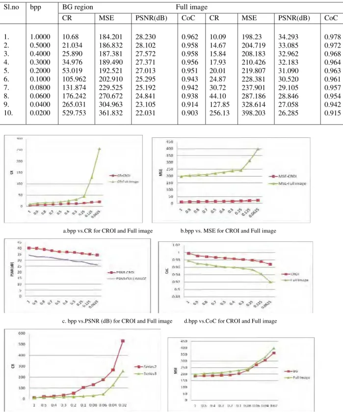
The proposed method is implemented and tested using the image processing software tool, MATLAB 7.9. We considered magnetic resonance brain images for the test purpose. The performance parameters bpp, Compression ratio (CR), Mean square error (MSE), Peak Signal to Noise Ratio (PSNR) and Correlation Coefficients (CoC) are given in Table.I & II. The parameters are obtained separately for CROI region and the entire image area. Figure 5. Show the original image and the extracted CROI region, BG and the reconstructed image is obtained by merging the two extracted regions.
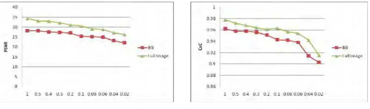
The compression parameters for several compression ratios for CROI region and the entire image is listed in Table I. and the comparison of bpp with MSE,CR,PSNR are analysed and plotted in Figure 2(a-d). Table II. Shows the same performance parameters of compression for background (BG) image and the complete image and plotted in Figure 2.(e-h).
Table I. CLSPIHT- CROI Region and the full image parameters
Table III. shows, our proposed methods outperforms all the existing methods such as Maxshift,CSPIHT,CVQ interms of PSNR and CoC and also plotted in Figure 3. To show our proposed method is even better than the general compresson methods of JPEG2000, SPIHT, it is compared in terms of same PSNR and CoC and it is plotted in Figure 4.
We considered 10 different bitrates for CROI and BG, and achieved better compression quality compared to the existing methods. Our proposed method’s advantage over all the listed method is more evident as the compression ratio increases. It is clearly stated in the Table III and Table IV and also plotted in Figure.3 & 4. Our proposed method keeps all the important information needed for the diagnostic purposes with out any loss in the quality.
Sl.no bpp CROI region Full image
CR MSE PSNR(dB) CoC CR MSE PSNR(dB) CoC
Table II. CLSPIHT- BG region and the full image parameters.
a.bpp vs.CR for CROI and Full image b.bpp vs. MSE for CROI and Full image
c. bpp vs.PSNR (dB) for CROI and Full image d.bpp vs.CoC for CROI and Full image
e.bpp vs CR for BG and Full image f.bpp vs. MSE for BG and Full image
Sl.no bpp BG region Full image
CR MSE PSNR(dB) CoC CR MSE PSNR(dB) CoC
1. 2. 3. 4. 5. 6. 7. 8. 9. 10.
1.0000 0.5000 0.4000 0.3000 0.2000 0.1000 0.0800 0.0600 0.0400 0.0200
10.68 21.034 25.890 34.976 53.019 105.962 131.874 176.242 265.031 529.753
184.201 186.832 187.381 189.490 192.521 202.910 229.525 270.672 304.963 361.832
28.230 28.102 27.572 27.371 27.013 25.295 25.192 24.841 23.105 22.031
0.962 0.958 0.958 0.956 0.951 0.943 0.942 0.938 0.914 0.903
10.09 14.67 15.84 17.93 20.01 24.87 30.72 44.10 127.85 256.13
198.23 204.719 208.183 210.426 219.807 228.381 237.901 287.186 328.614 398.203
34.293 33.085 32.962 32.183 31.090 30.520 29.105 28.846 27.058 26.285
g. bpp vs.PSNR (dB) for BG and Full image h.bpp vs.CoC for BG and Full image
Figure 2.Comparison of compression parameters – bpp vs.MSE,CR,PSNR and CoC for both CROI and BG (CROI and complete image, BG and Complete Iimage) for 10 different bit rates (bpp).
Table III. Comparison of Maxshift, CSPIHT, CVQ and our proposed method (CLSPIHT), in terms of PSNR (dB) and CoC.
Table IV. Comparison of JPEG2000,SPIHT, and the proposed CLSPIHT, in terms of PSNR (dB) and CoC.
bpp PSNR (dB) CoC
JPEG2000 SPIHT CLSPIHT JPEG2000 SPIHT CLSPIHT
1.0000 0.5000 0.2500 0.1250 0.0625
34.45 31.73 27.41 26.01 23.47
36.49 33.10 29.73 28.05 26.14
42.79 40.13 39.72 37.95 37.26
0.998 0.996 0.991 0.989 0.985
0.999 0.997 0.994 0.989 0.983
0.999 0.998 0.999 0.999 0.998
a.bpp vs.PSNR (dB) for Maxshift, CSPIHT, b.bpp vs.CoC for Maxshift,CSPIHT, CVQ and our proposed CLSPIHT CVQ and our proposed CLSPIHT
Figure 3.Comparison of bpp vs.PSNR (dB) and CoC for Maxshift,CSPIHT, CVQ and our proposed method (CLSPIHT).
bpp CR PSNR(dB) CoC
Maxshift CSPIHT CVQ CLSPIHT Maxshift CSPIHT CVQ CLSPIHT
1.0000 0.5000 0.2500 0.1250 0.0625
10.57 34.97 68.01 127.85 256.13
40.73 37.28 32.19 29.81 25.04
40.27 36.62 33.81 29.95 27.74
41.32 39.04 38.26 36.39 34.68
42.79 40.13 39.72 37.95 37.26
0.997 0.996 0.995 0.993 0.989
0.998 0.996 0.993 0.992 0.991
0.999 0.997 0.997 0.995 0.996
c.bpp vs.PSNR (dB) for JPEG2000,SPIHT, d.bpp vs.CoC for JPEG2000,SPIHT,
and our proposed CLSPIHT and our proposed CLSPIHT Figure 4. Comparison of bpp vs.PSNR (dB) and CoC for JPEG2000, SPIHT and our proposed CLSPIHT
a.Original Image b. CROI region c. BG region d. Reconstructed image (CROI & BG)
Figure.5 Original image and the separation of CROI and BG, final reconstructed image using our proposed CLSPIHT.
VI.CONCLUSIONS
In this work, we have proposed Contextual Listless SPIHT (CLSPIHT) which uses region of interest concept. Our main idea is to separate the contextual region of interest which contains the most important information for diagnosis and the back ground image which consists of patient information. Here, CROI region is encoded separately and the BG regions are encoded separately using Contextual Listless SPIHT. Finally, the two extracted regions are merged together to get the reconstructed image. The results obtained in this method are listed in Table I and Table II and it clearly show the better performance in terms of PSNR, CR, MSE and CoC for different bit rates. The performance parameters bpp and the PSNR comparative results of different methods are compared and our method show the improved performance. From the comparison with the traditional methods such as JPEG2000 and SPIHT, our method provides better compression performance in terms of PSNR and CoC as well as good visual quality of reconstructed image. So, we conclude that our proposed CLSPIHT method is a good selection for minimizing the storage cost and the transmission time of medical images.
ACKNOWLEDGEMENT
The authors wish to thank Dr. Amba Bhavani, Amba Clinic and Dr. Devaki from Devaki Cancer Institute, Madurai for the provision of medical image data sets used in this study.
REFERENCES
[1] S.Grgic,M.Grgic, B.Zovko-cihlar,Performance analysis of image compression using wavelets, IEEE Trans.Ind.Electron. 48(2001) 682-695.
[2] J.Askelof,M.Carlander,C.Christopoulos, Region of interest coding in JPEG2000, Signal Process,image Commun,17 (2002) 105-111
[3] A.said and W.Pearlman, A new, fast and efficient image codec based on set partitioning, IEEE Trans.Circuits Syst.VideoTechnol..6,pp.243-250,.(1996)
[4] Martin, K. Lukac, R. Plataniotis, K.N., SPIHT-Based Coding of the Shape and Texture of Arbitrarily Shaped Visual Objects, IEEE Transactions on Circuits and Systems for Video Technology, 16(10), pp. 1196-1208,(2006)
[5] M.A. Ansari,R.S.Anand, Context based medical image compression for ultrasound images with contextual set partitioning in hierarchial trees algorithm, Adv.Eng.Software 40, 487-496, (2009)
[6] P.G.Tahoces,J.R.Varela,M.J.Lado, Image compression:Maxshift ROI encoding options in JPEG2000, Comput.Vision Image Understanding ,109 139-145, (2008)
[7] M.A.Ansari,R.S.Anans, A novel ROI based algorithm with DCT, wavelet transform and set partitioning in hierarchical trees for medical image compression, Int,J.Sci.Computing (IJSC), 2(1), 7-22, (2008)
1170,(2000)
[9] Seyed Morteza Hosseini,Ahmad-Reza Naghsh-Nilchi, Medical ultrasound image compression using Contextual Vector quantization, Computers in Biology and Medicine http://dx.doi.org/10.1016/j.compbiomed.2012.04.006, (2012) [10] Park K.Park Hw. Region of interest coding based on set partitioning in hierarchical trees, IEEE Trans circuit syst video
tecno,12(2):106-113, (2002)
[11] Xu Yan, etal. The coding technique of image with multiple ROIs using standard Maxshift method. In: The 30th annual conference
of the IEEE industrial Electronics society, Busan, Korea,p.2077-80, (2004)
[12] K.Sayood, Introduction to data compression, second Ed. Morgan Kaufman publishers, San Francico, USA, (2000)
[13] Penedo.M,W.A.Pearlman,P.G.Tahoces,M.Souto and J.J.Vidal, Region based wavelet coding methods for digital mammography, IEEE Trans.Med.Imag.,22:1288-1296,DOI:10.1109/TMI.2003.817812, (2003)
[14] T.C.Lu,C.Y.chang,A Survey of VQ codebook generation, J.Inf.Hiding Multimed.signal proc.(JIHMSP) ,1(3), 190-203,(2010) [15] Rongchang Zhao, Yide Ma, A region segmentation method for region-oriented image compression, Neurocomputing (Elsevier),
85,45-52,(2012)
[16] A.P.Bradley,F.W.M.Stentiford, JPEG 2000 and region of interest coding in: Digital Image Computing:Techniques and Applications (DICTA’02), Melbourne, Australia, January, pp.303-308, (2002)
[17] Shih-Chung B.Lo, Huai Li and Matthew T.Freedman, Optimization of wavelet decomposition for image compression and feature preservation, IEEE Trans. on Medical Imaging, vol.22(9), 1141-1151.
[18] T.H.Oh.R.Besar,Image Quality measures of compressed medical images, in: 4th National Conference on Telecommunication
Technology Proceedings, Shah Alam,Malaysia,(2003)
[19] R.C.Conzalez,R.E.woods,Digital Image Processing, 2nd edn.,Prentice Hall, New Jersey,(2002)
[20] F. W. Wheeler and W. A. Pearlman, “SPIHT image compression without lists”, IEEE conference on acoustics, speech and signal processing (ICASSP2000), vol. 4, pp. 2047-2050,(2000)